MODULE 9
Expansion and Airways
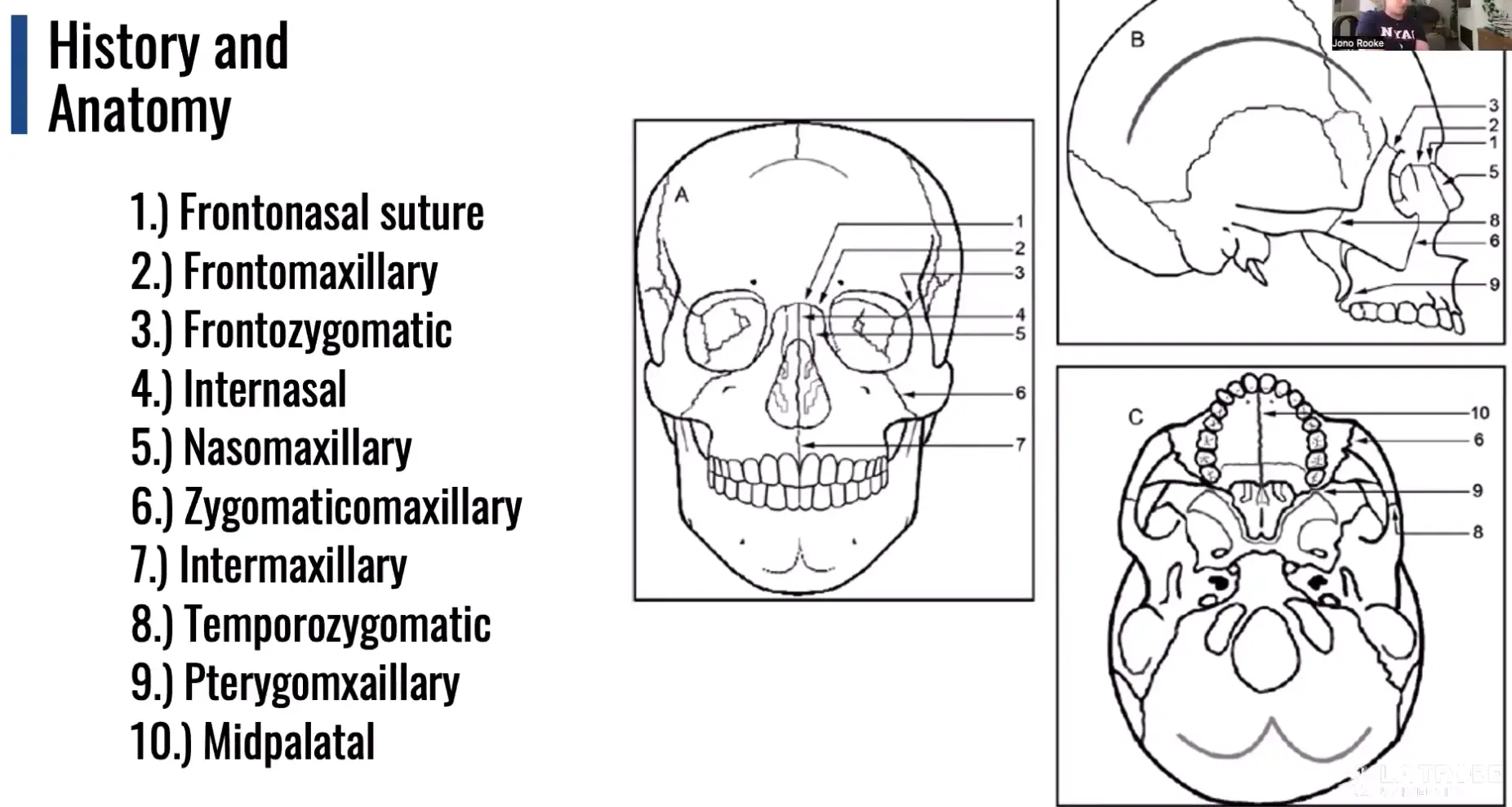
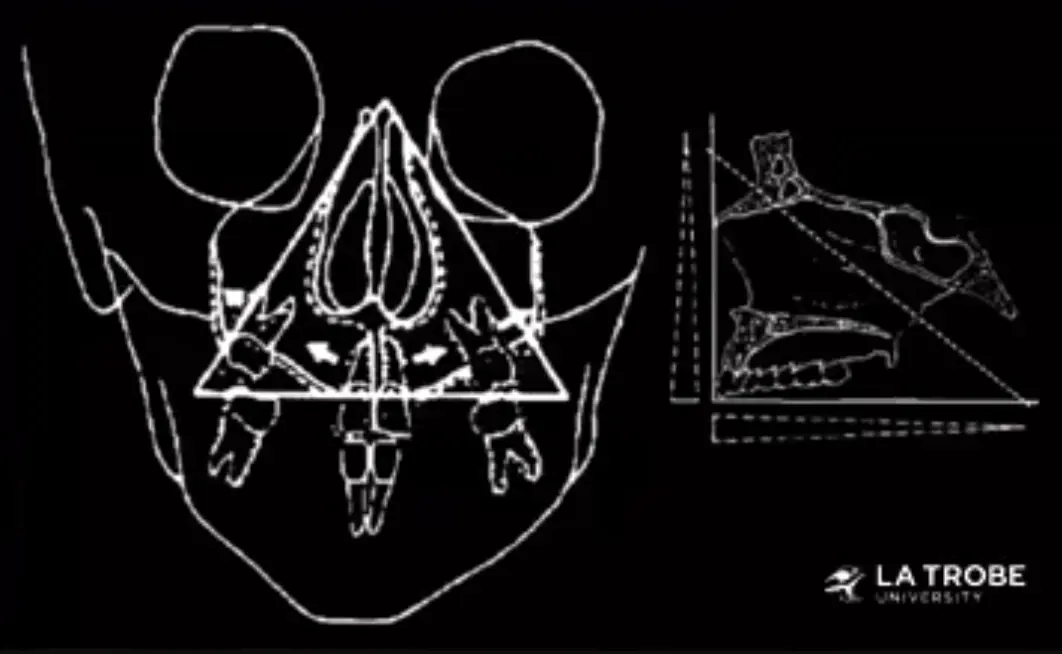
- maximum is always at the level of inferior turbinates with a variable degree of expansion superiorly
Boundaries / anatomy of the airways
- hard tissue boundaries
- upper and lower incisors
- anteriorly piri form rim
- superiorly cranial base
- posteriorly cervical vertebrae
- inferiorsly hyoid bone
- laterally: width of palate, middle cranial fossa, distance between ascending rami
- soft tissue
- pharyngeal muscles
- tongue
- soft palate
- turbinates
- pharyngeal tonsils
- adenoids, nares
Hard tissue growth
- anterior cranial base increases in length via growth at sphenoepthmoidal synchondrosis until age of 7
- increases in posterior cranial base relate to growth at sphenooccipital synchondrosis until age 13
- anterior cranial base carries nasomaxillary complex forward, at same time midface bones are displaced anteriorly and inferiorly
- mandible elongates, displaced downward and forward with deposition of bone on posterior and superior borders of the ramus, increasing height of rami and increasing distance between ascending rami
- resorption of anterior border of ramus increases corpus length (oropharyngeal length)
Soft tissue growth
- skeletal boundaries of airway increasing, lymphatic tissues are shrinking
- decrease in tonsils/adenoids result in enormous increase in size of upper airway in adolescence/childhood
- these changes in growth exceed any ortho/orthopaedic effects on airway shape or size
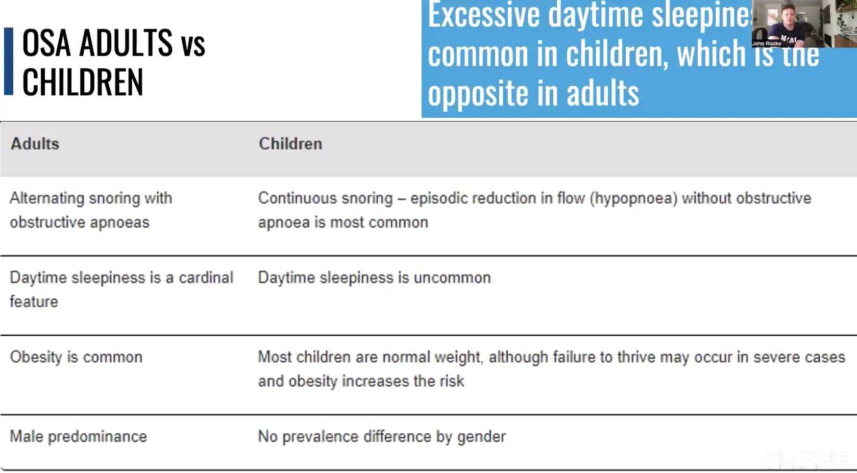
Childhood sleep disordered breathing
- significant problem for proportion of children
- ranges from loud snoring to obstructive sleep apnoea
- childhood OSA often remits without any intervention during growth phases
- certain craniofacial, genetic syndromes and obesity = higher prevalence of OSA /SDB
How is sleep disordered breathing treated
- adenoidectomy
- half had normalised by 7months
- can spontaneously resolve over time
- dentist perform clinical risk assessment for OSA and refer at risk patients to appropriate physician for definitive diagnosis of OSA
- examples
- snoring
- sleep related behaviours
- daytime sleepiness
- difficulty concentrating
- formal diagnosis of ADHD
- mouth breathing during sleep
- medications
- developmental delays
- bed wetting that is not age appropriate
- hard to wake up in the morning
- morning headaches
- difficult breathing during sleep
- pauses in breathing during sleep
- fall asleep quickly
- nasal obstruction
Developing airway
Consequences of OSA / SDB
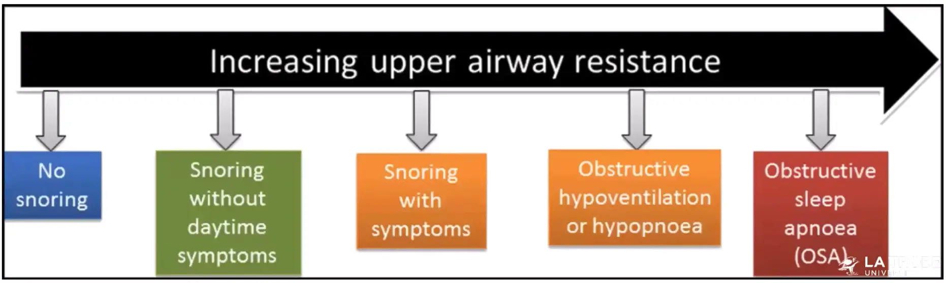
- true measure is going to be an overnight sleep study
- consequences in children
- cardiovascular
- increased BP during sleep
- ventricular wall thickening
- increase risk of life long hypertension
- neurocognitive / behavioural
- memory impairment
- attention deficit
- daytime behavioural difficulties
- learning and behaviour impairments
- cardiovascular
- msot common cause of childhood OSA is enlargement of the tonsils and adenoids
- allergic rhinitis
- obesity - up to 50% overweight children, compared to 1-5% of normal weight children
- more common in
- hypotonia
- micrognathia
- cleft palate
- down syndrome
- achondroplasia
Maxillary expansion and expanders
3 types
- rapid maxillary expansion RME
- slow maxillary expansion SME
- surgically assisted maxillary expansion SAME
- for adults or fused sutures
Types of expanders
- removeable or fixed
- rate of activation should not exceed one turn a week in 0.25mm / turn screw
- fixed
- bonded
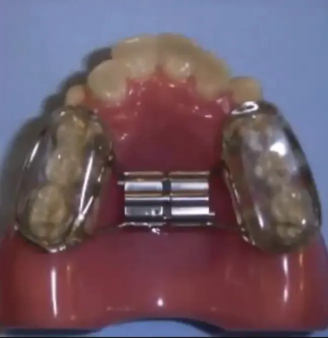
- flat plane occlusal splint wiht haas appliance
- recomended for AOB, perio compromised, TMD symptoms
- very hard to clean
- banded
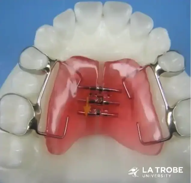
- Haas type with acrylic palatal flanges
- Hyrax: expansion screw and metal framework without acrylic palatal coverage
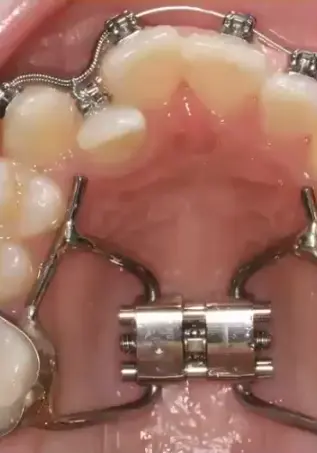
- other types
- minne
- fan shaped
- quad helix (bottom right)
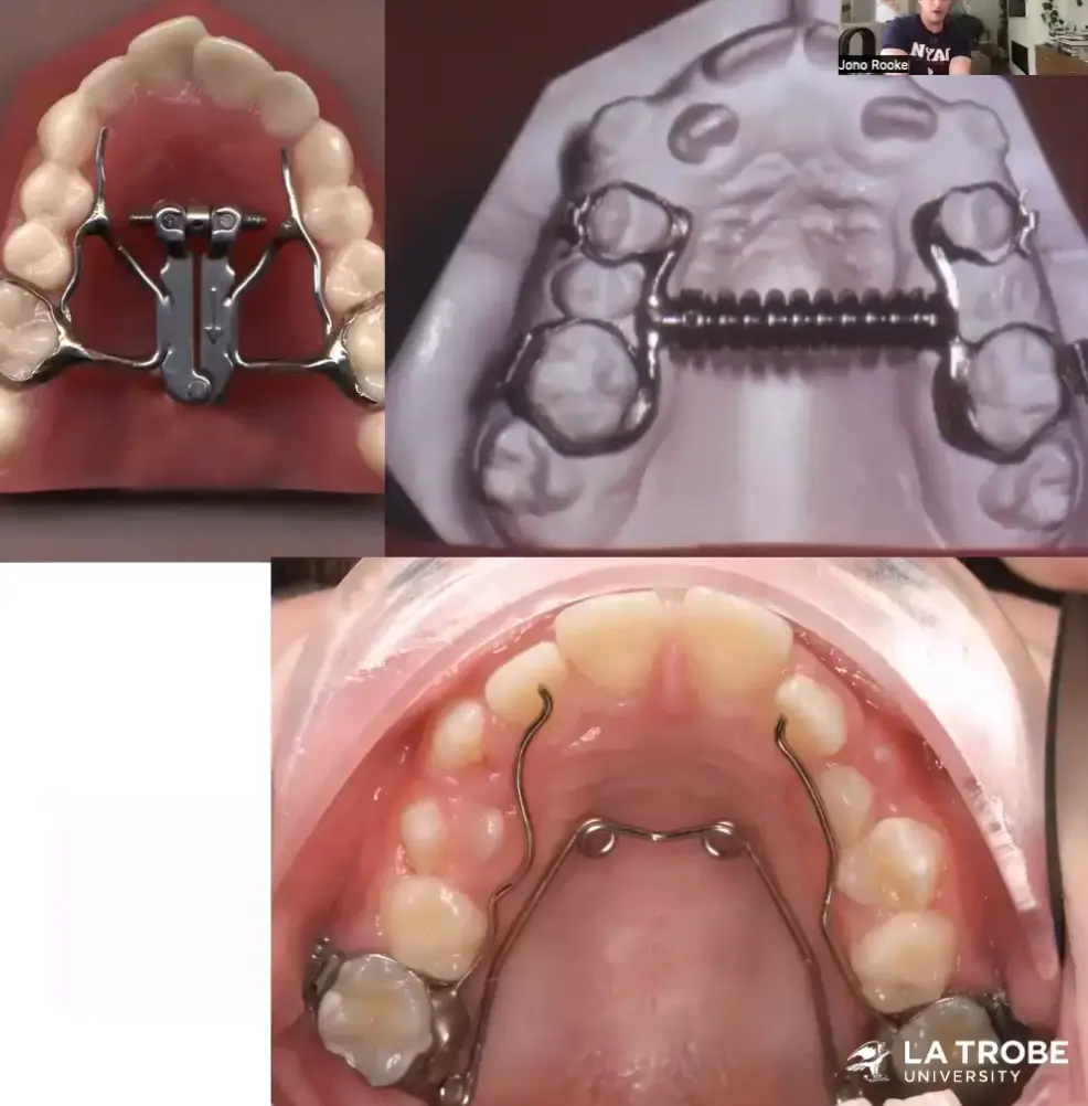
- bonded
Expansion effects on airway
- assessment
- study casts - not useful
- ceph studies - PA head films, occlusal radio, lat cephs - hard to determine airway
- 3d imaging - MRI CT CBCT computational fluid dynamics
- are airway measurements using cbct accurate
- large amont of inaccuracy in airway measurement from CBCT
- positioning or patient, swallowing, breathing during scan etc.
- low inter and intra examiner reliability
- assessment of airway patency
- rhinoscopy or nasoendoscopy
- rhinomanometry and acoustic rhinometry have been proven to be useful to provide objective assessment of airflow through nose and to measure nasal cavity measurements
- AR, CT MRI are good correlation
RME effect on airway
- increase nasal cavity width, volume, and minimal cross sectional area
- palatal disjunction causes separation of inferior turbinates as lateral walls are carried away from each other
- displaces palate downward, result in opening of posterior nasal aperture, allowing easier passage of air
- anterior tongue position has been found in some studies, which could improve airflow
- once RME results in increased nasal airway patency and reduced nasal airway resistance (NAR), airway flow increases, craniocervical angulation consequently reduced
- some studies show mandibular forward repositioning in all patients except CIII
- palatal flattening when present may contribute to lowering of nasal floor and improvement of nasal permeability
- greater impact on anterior region of nasal cavity, perhaps due to greatest site of separation at the incisal level
- nasal resistance after RME significantly lower than prior
- oropharyngeal no changes
- 12yo prospective cohort study for OSA in children RME, hypoxic events lowered, o2 sat up
Quality of life
- ten children SDB longitudinal study for RME,
- might aid in quality of life for kids with narrow maxilla
Airway patency and breathing mode
- RME may permanently change functional matrix of upper airway since it results in increase in nasal airflow and thoracic capacity
- resistance after RME drops to predominantly nasal breathing subjects values
- breathing mode is unconscious motor function, change from predominantly oral to nasal is difficult to achieve
RME SUMMARY
- not recommended alone if main purpose is to improve nasal breathing
- only for skeletal crossbite
Jaws Dr Sandra Kahn
Harvold et al 1982
- 16 three year old rhesus monkeys
- occluded nostrils of 8 of them
- observed facial growth over 3 years by taking and analysing serial cephalograms
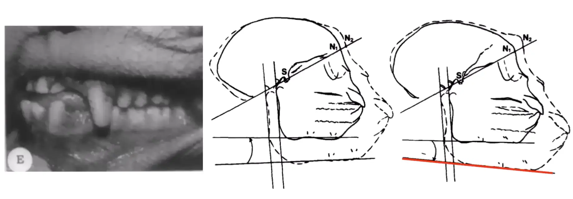
- no data presented within study, magnitude of chance
- statement only explaining significant different between groups
- steep mandibular plane angle and increased face height can be viewed as a result of induced neuromuscular imbalance between neck and facial muscles in the experimental monkeys
- critique
- difficult to apply findings to humans
- definitely wouldn't be accepted on peer review
- rhesus monkeys are obligate nasal breathers
Linder aronson et al 1986, Woodside et al 1991
- facial growth following adenectomies
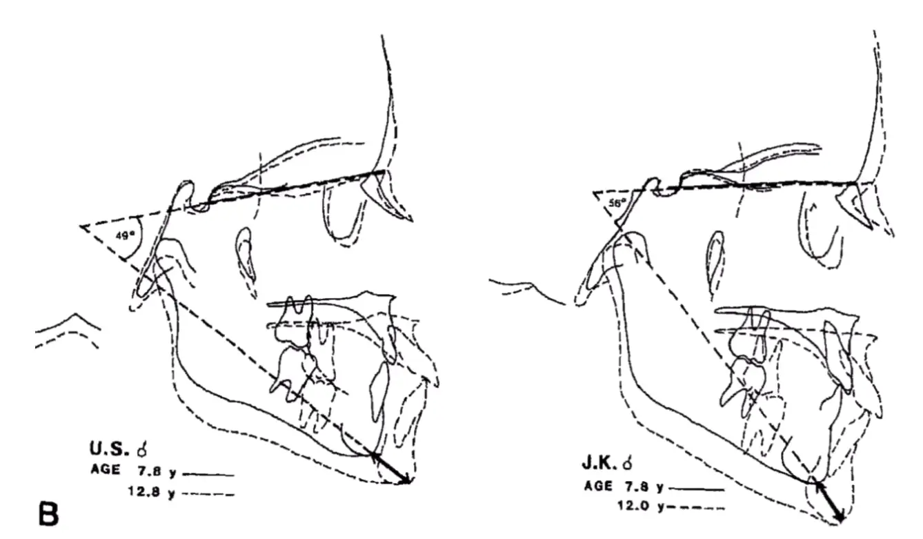
- severe nasopharyngeal obstruction
- 48 changed from mouth to nasal breathing
- results 1986
- girls post op had more horizontal growth (wide CI though)
- difference not found in boys
- growth direction more variable for adenectomy group
- actual difference is 3.1 degree in mandibular plane error (within standard variation)
- results 1991
- mandibular length 3.8mm and 2.5mm
- effect size small, large 95% CI, uncertainty in data
- critique
- only children with severe nasopharyngeal obstruction
- ceph differences minimal and not clinically significant
- sample had severe attrition bias
- genders analysed separately
Conclusion
- minimal evidence mode of breathing influences the craniofacial skeletal and facial growth
Neanderthal
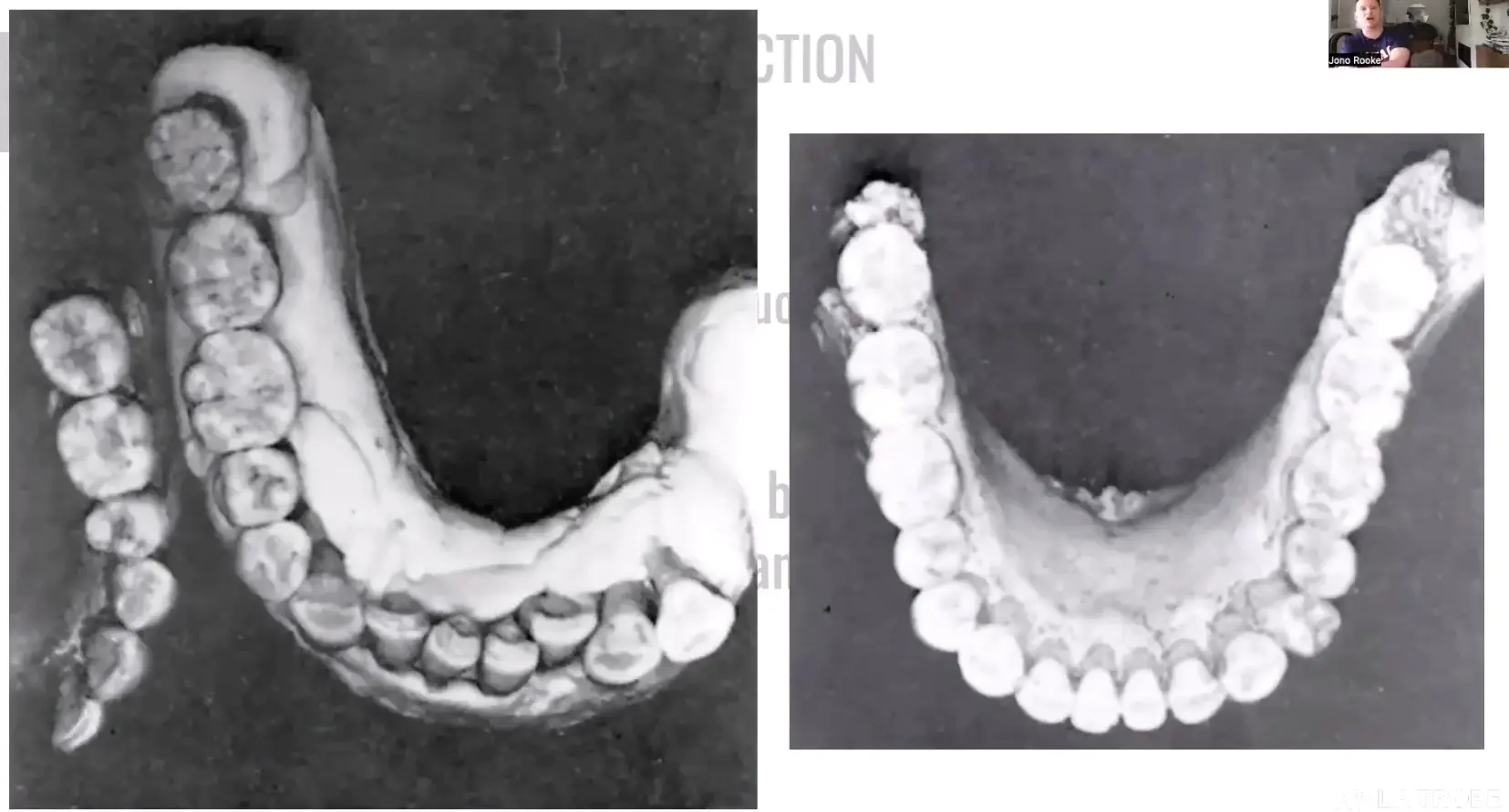
Magdalenian girl
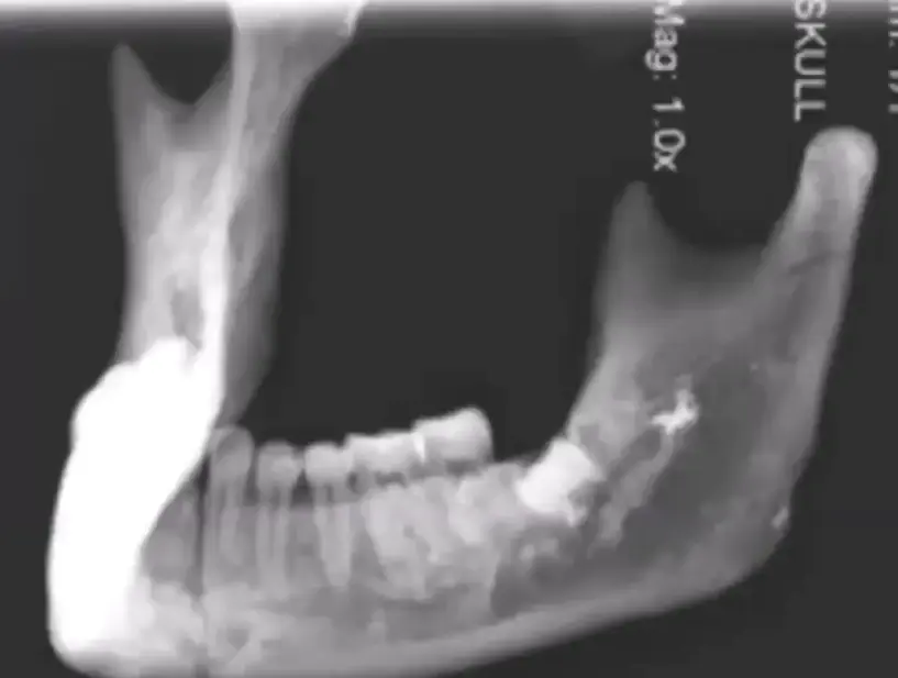
- earliest case of impacted wisdom teeth 13-15k year old skeleton