Dentoalveolar Trauma
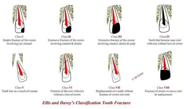
Ellis classification
- class 1 - fracture little or no dentine
- class 2 - fracture dentine but no pulp exposure
- class 3 - extensive crown fracture with pulp exposure
- class 4 - tooth becomes non vital with/without loss of crown structure
- class 5 - tooth lost as result of trauma
- class 6 - roto fracture
- class 7 - tooth displacement with out crown or root fracture
- class 8 - fracture of crown en masse and its replacement
- class 9 - injury to primary dentition
Mechanism of injury
- energy of impact
- resilience of impacting object
- shape of impacting object
- direction of impacting force
Examination and diagnosis
- when where how did the injury occur
- previous injuries
- amnesia, unconsciousness, drowsiness, vomiting, headache
- spontaneous tooth pain
- teeth reactive to thermal changes, sweet sour foods
- teeth tender to touch or during eating
- any change in bite
- Check for extraoral wounds; palpate facial skeleton
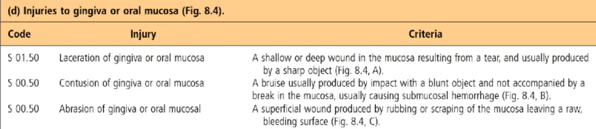
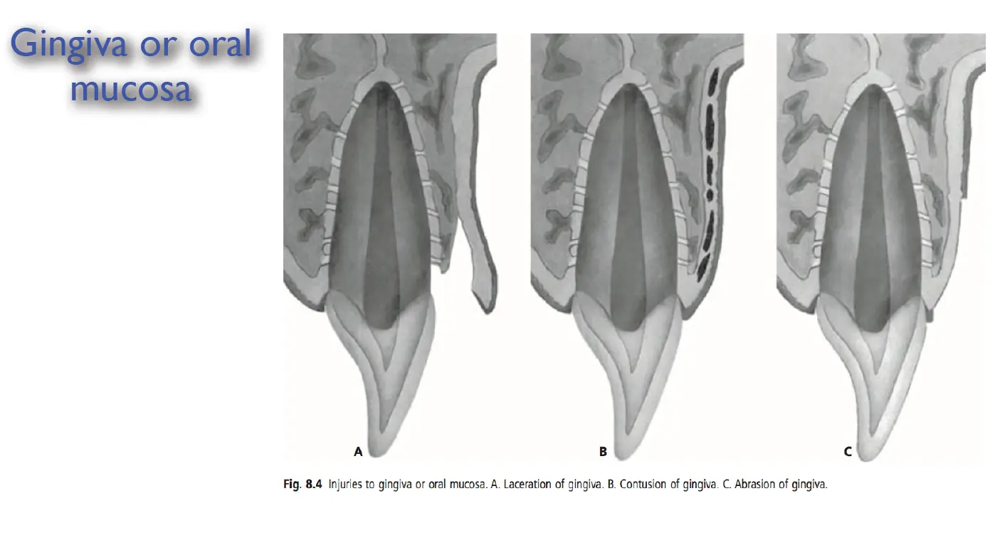
- Record soft tissue injuries to oral mucosa and gingiva
- Check crowns of teeth for fractures, pulp exposures, colour changes
- Check displacement of teeth
- Occlusal disturbances
- Abnormal mobility of teeth or alveolar fragments
- Tenderness of teeth to percussion and changes in percussion note
- Pulp sensibility testing
Radiographic examination
- reveals the stage of root development
- discloses injuries affecting the root
- mandibular fracture
- maxillofacial injuries
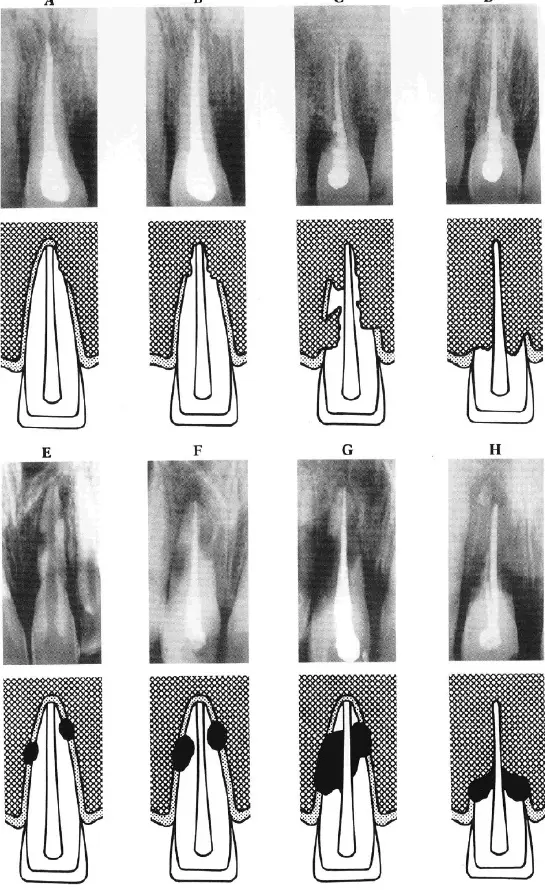
Dental resorption
- resorptive process similar to bone
- differences between cells resorbing dentine and bone
- dentinoclasts
- morphologically similar to osteoclasts
- fewer nuclei
- very small clear zones
Resorption types relating to trauma
- external
- surface
- inflammatory root
- replacement (ankylosis)
- cervical
- internal
- internal inflammatory
- internal surface and tunnelling (seen in root fractures)
Surface resorption
- pathogenesis
- result of complex localised injury to PDL
- mechanical injury/bacterial contamination
- macrophages remove debris -> resorption cavity
- repair with cementum
- diagnosis
- more resorption lacunae superficial, cant be seen radiographically
- deeper cavity result in altered root outline
- clinical management
- none, self limiting
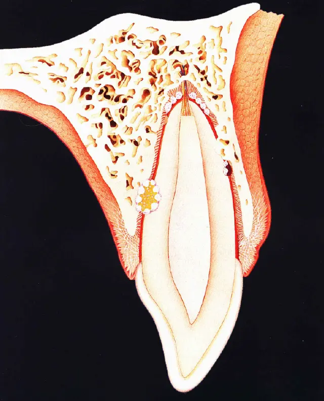
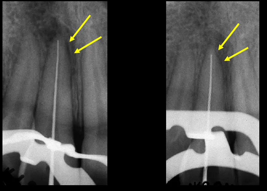
Inflammatory root resorption
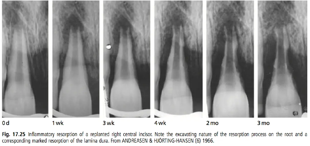
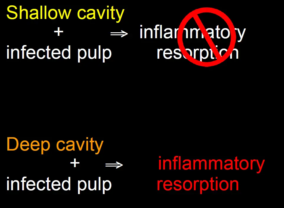
- pathogenesis
- bacteria in pulp release toxins
- toxin diffsue through tubule to PDL
- stimulate osteoclasts on root surface
- inflammatory root resorption
- resorption can be rapid
- within few months
- aggressive after avulsion injuries in 6-10yo (wider dentinal tubules and thin protective cementum)
- resorption slower in older age groups
- diagnosis
- radiographically
- radiolucent bowl shaped cavitations along root surface
- corresponding radiolucency in adjacent bone
- can detect as early as 2 weeks after replantation
- clinically
- tender to percussion
- dull tone (vs ankylosis)
- radiographically
- management
- endo + endo medication
Replacement resorption (Ankylosis)
- histology
- fusion of alveolar bone to root surface
- pathogenesis
- extensive damage to PDL
- bone repair dominates
- replacement resorption
- 2 directions
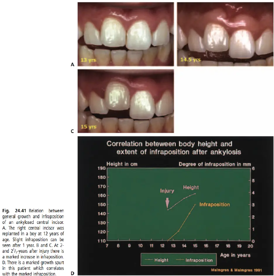
- progressive replacement resorption
- tooth becomes part of continuous bone remodelling
- transient replacement resorption
- replacement resorption exposes dentinal tubules + infected root canal
- replacement + inflammatory resorption
- progressive replacement resorption
- diagnosis
- percussion test - raised note
- decreased tooth mobility - perio test
- radiograph - disappearance of PDL space
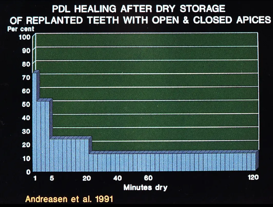
- anderson et al 1984
- <10%
- 10 -20%
-
20%
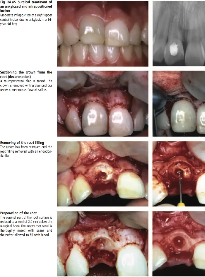
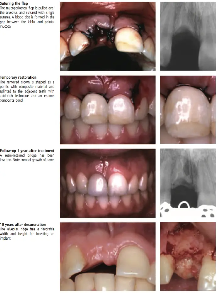
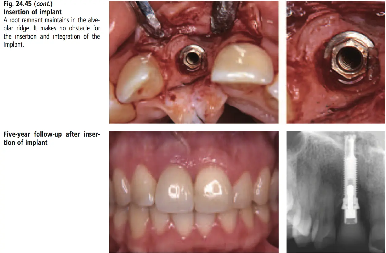
- clinical management
- no treatment
- young patients
- disturb normal growth of alveolar process
- exo
Cervical resorption
- pathogenesis
- unknown
- traumatic conditions ? suggested role of bacteria
- cervical area
- unknown
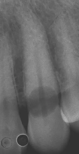
- diagnosis
- radiographically
- single lacuna in cervical area
- alveolar bone
- trace pulp chamber
- pink spot
- radiographically
- clinical management
- exposure resorption lacuna
- orthodontic or surgical
- removal of resorptive tissue
- surgical curettage
- trichloroacetic acid (90% solution)
- exposure resorption lacuna
Internal inflammatory resorption
- pathogenesis
- inflammatory signal
- collateral blood supply?
- cervical area
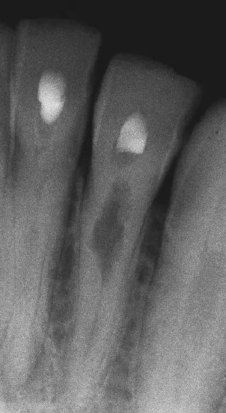
- diagnosis
- radiolucency, oval shaped enlargement
- root surface resorption
- management
- removal of residual pulp tissue
- chemo mechanical preparation
- medication
- root filling
- thermoplastiticise GP technique
- external communication
- long term CH or surgical
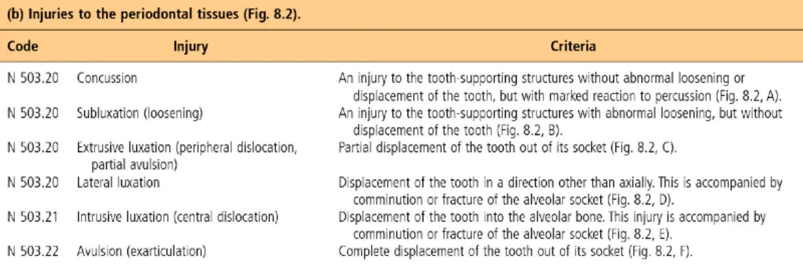
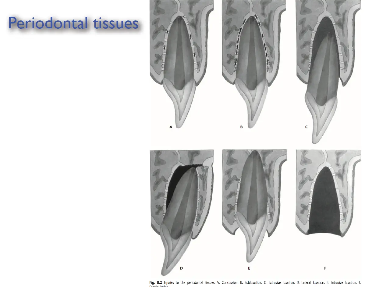
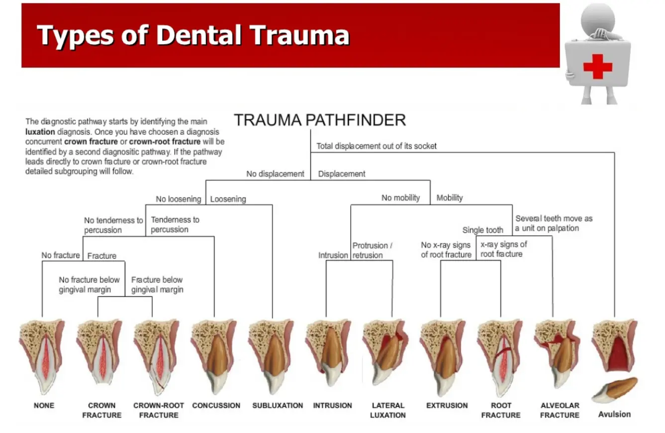
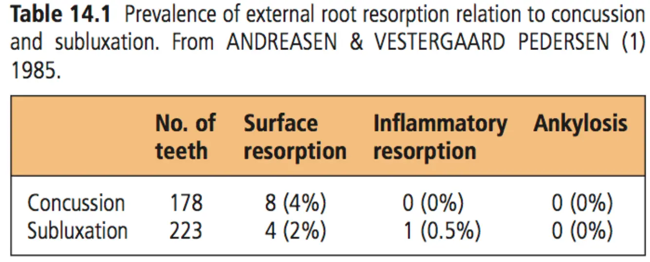
Concussion/Subluxation
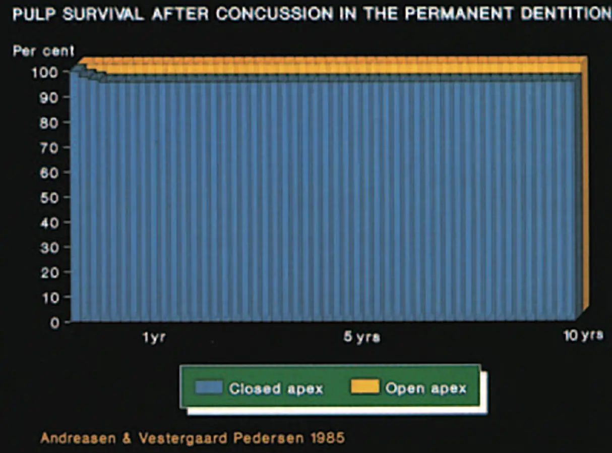
- concussion
- minor damage to PDL, no gingival bleeding
- tooth is TTpercussion, no loosening or displacement
- no radiographic changes
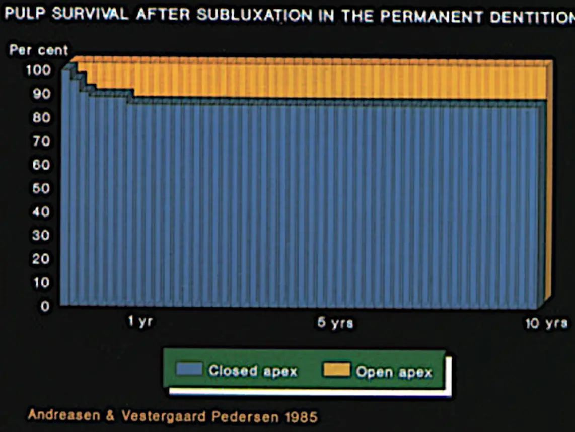
- Subluxation
- rupture PDL fibres, some mobility, may have gingival bleeding
- TTpercussion
- possible widened PDL on PA film
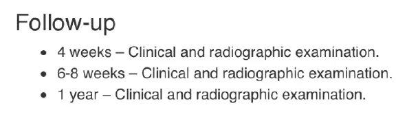
- management
- relief of occlusal interference may be necessary
- incase of severe loosening or multiple tooth injuries, teeth may be splinted (2 weeks)
- soft diet 14 days otherwise
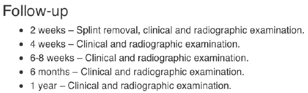
- CONCUSSION FOLLOW UP
- SUBLUX FOLLOW UP
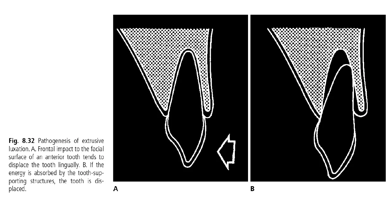
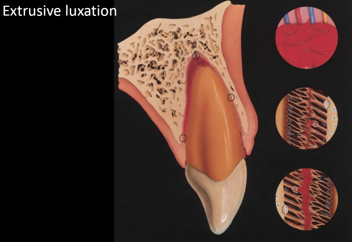
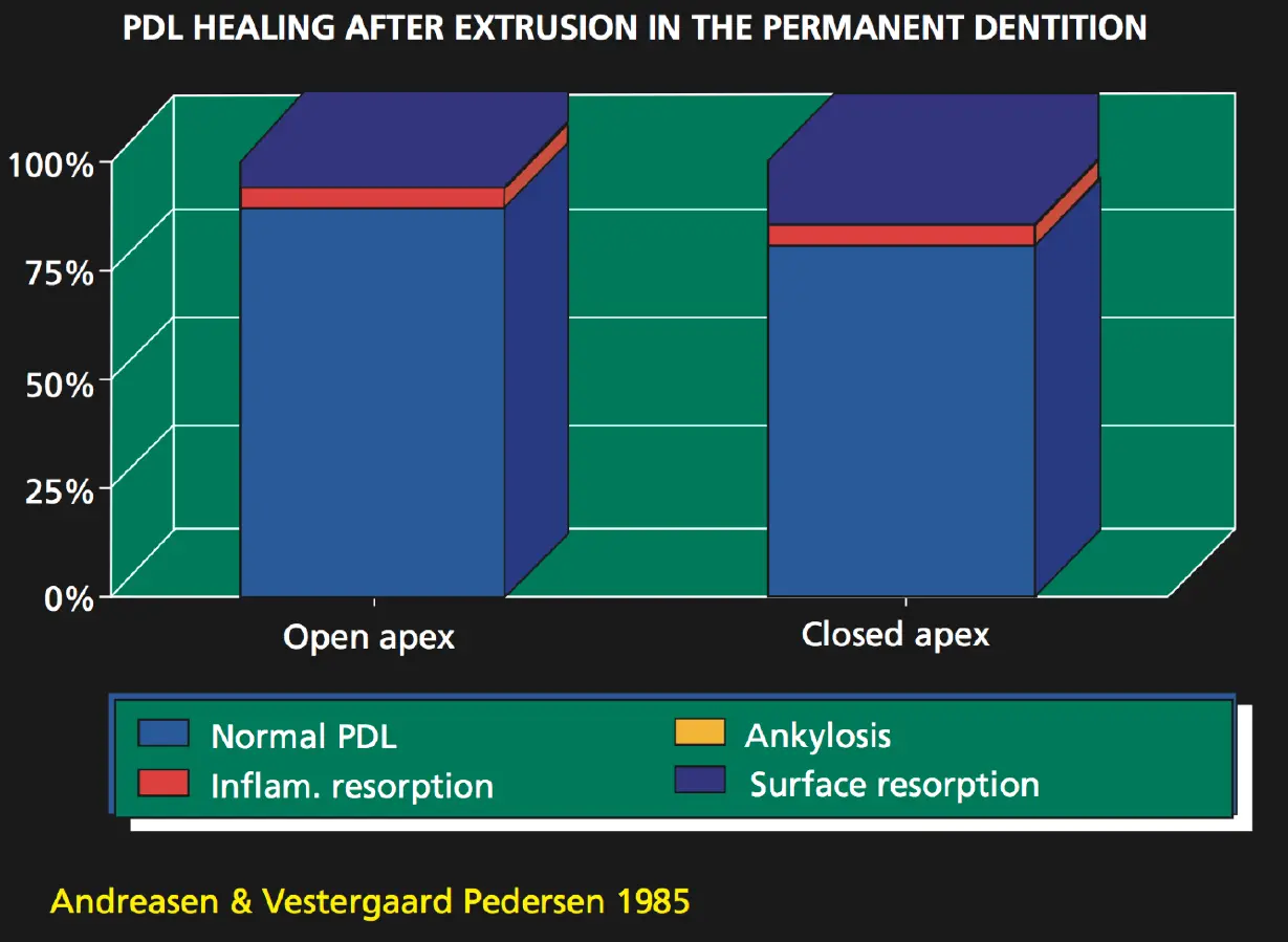
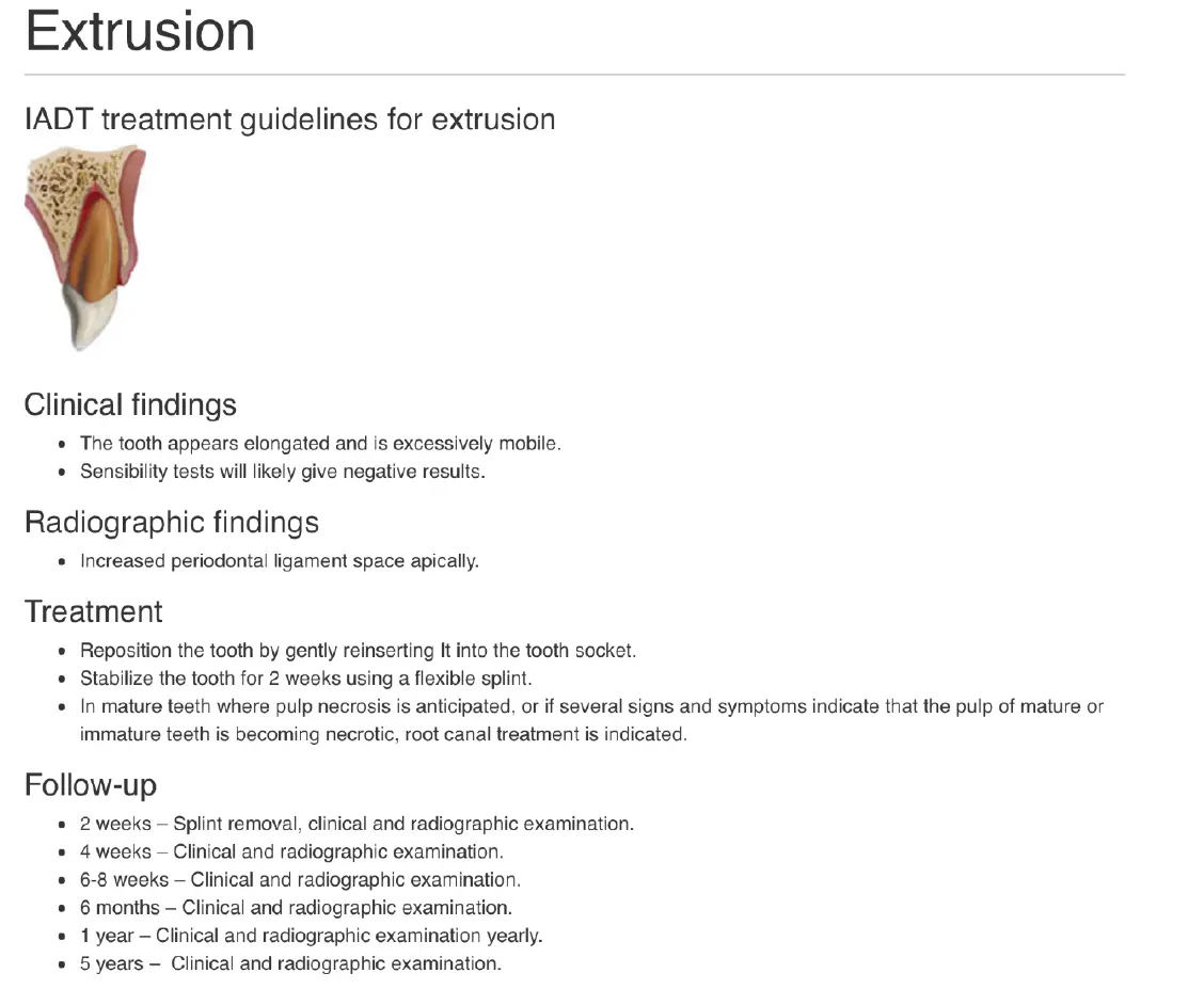
**Extrusive Luxation
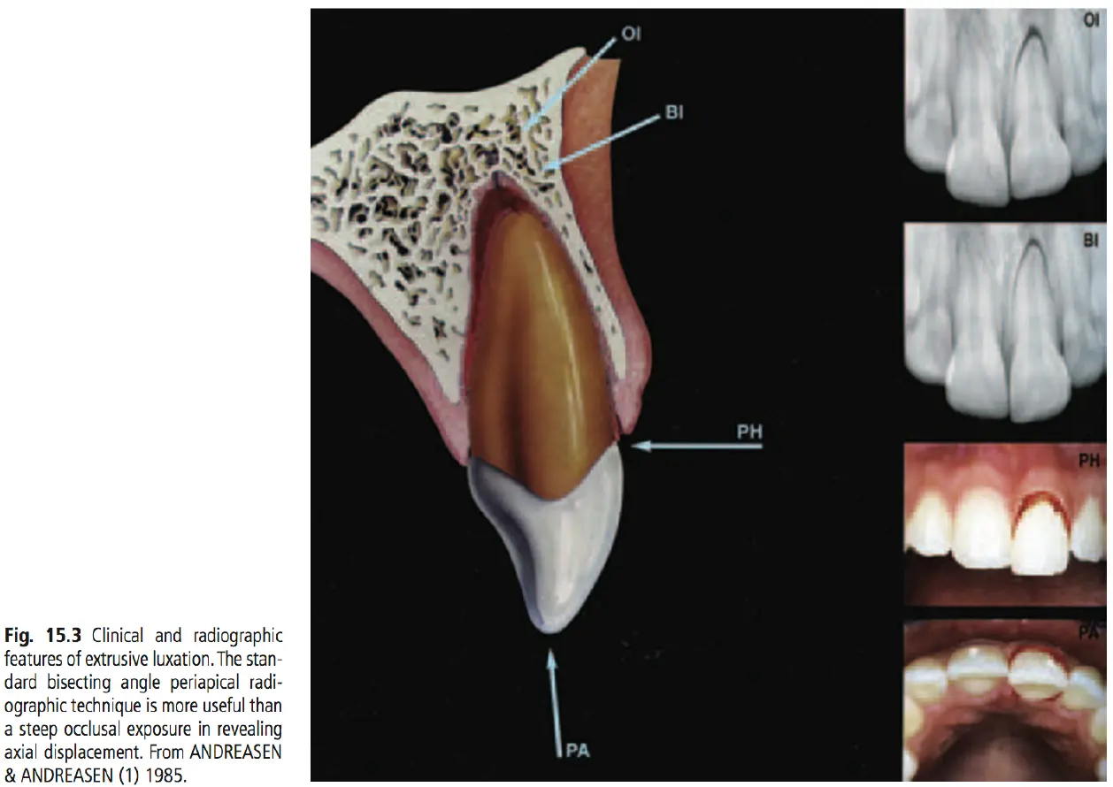
Clinical and radiographic findings
- extruded teeth appear elongated often with lingual deviation
- always bleeding from PDL
- expanding PDL space apically on PA
Management
- gently reposition using finger pressure
- splint tooth for 2 weeks
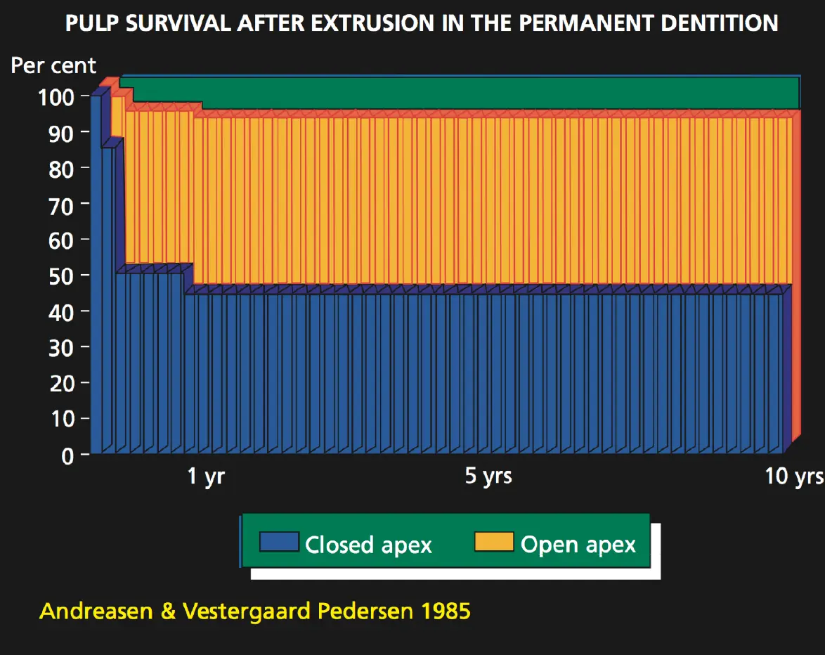
- endo in cases where pulp necrosis anticipated/signs and symptoms indicate becoming necrotic
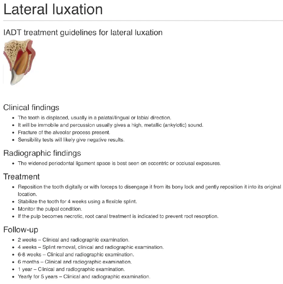
lateral luxation
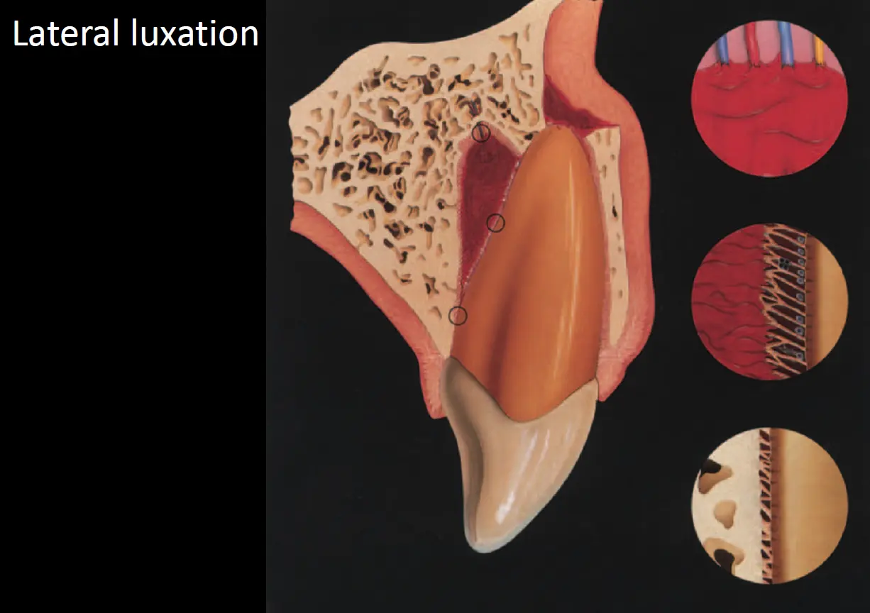
Clinical and radiographic findings
- crowns displaced lingually associated with vestibular fractures of socket wall
- increased apical PDL space on an occlusal or eccentric exposure
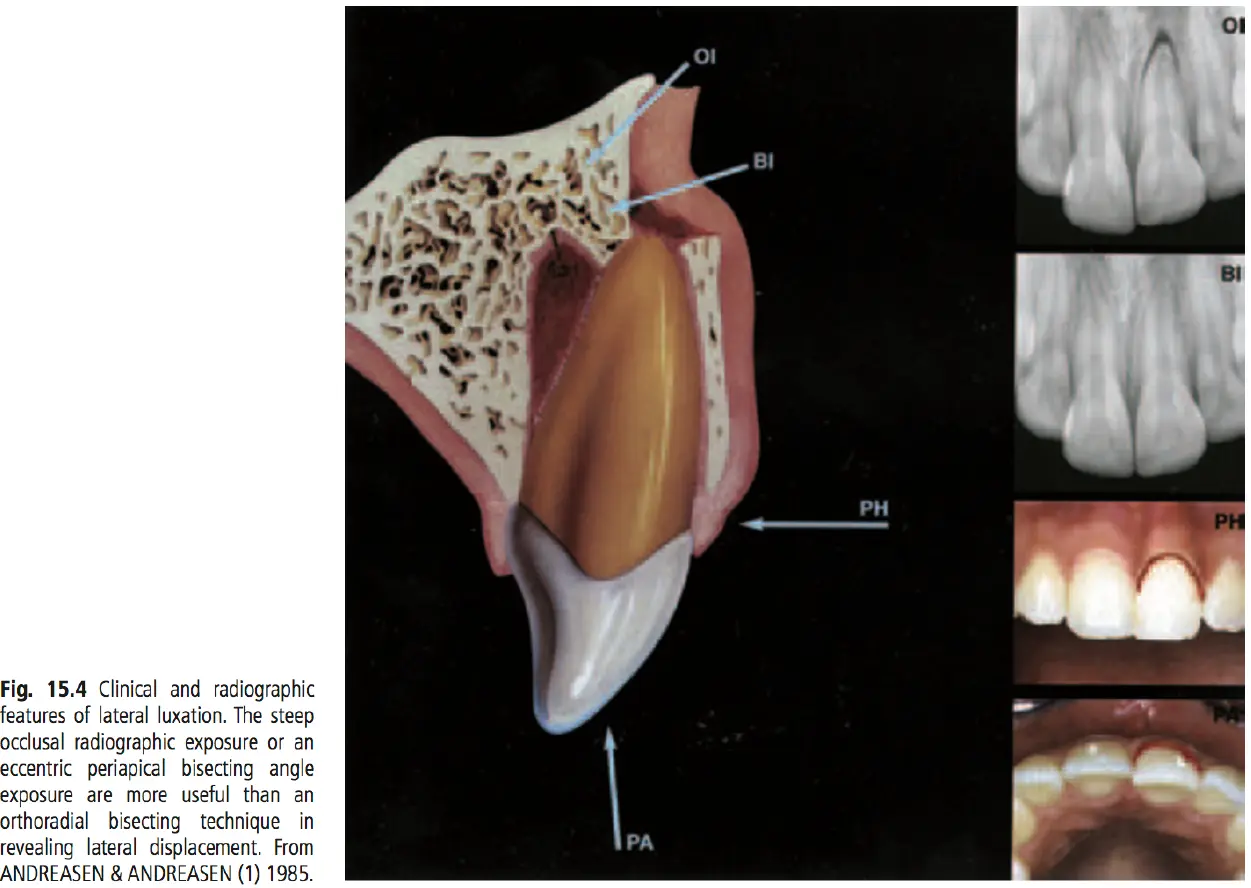
Management
- gently reposition using finger pressure or forceps
- splint tooth 4 weeks
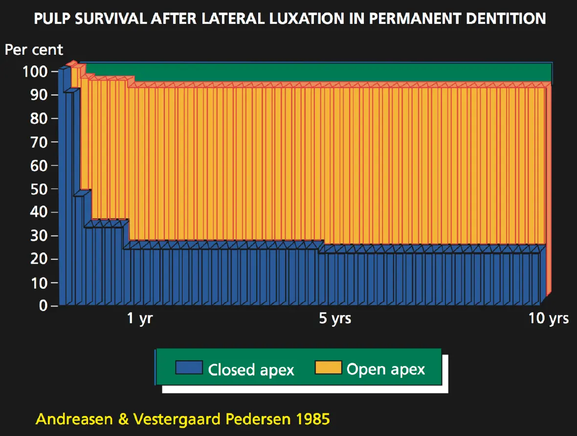
- monitor pulpal condition
- if pulp becomes necrotic, RCT indicated for prevention of root resorption
Intrusive luxation
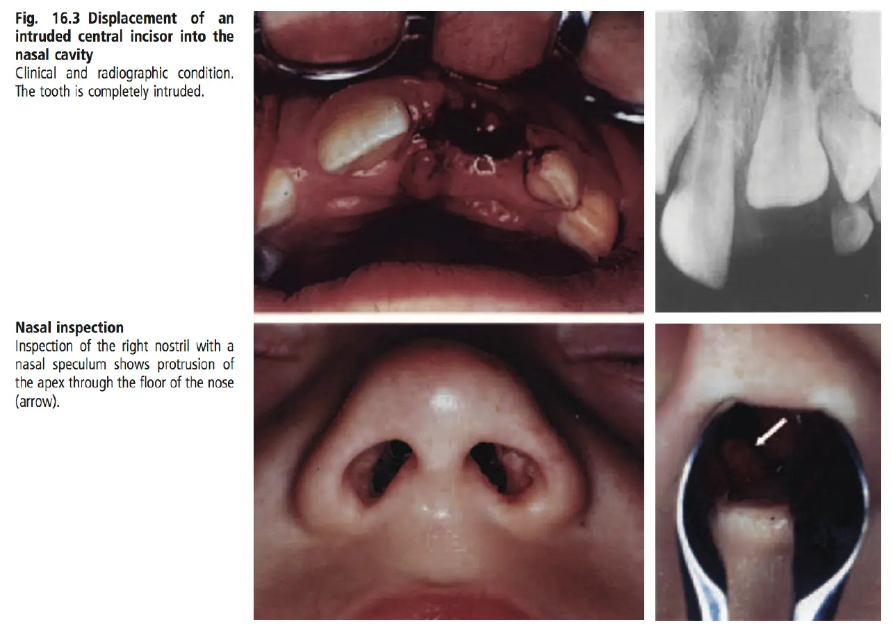
Clinical and radiographic findings
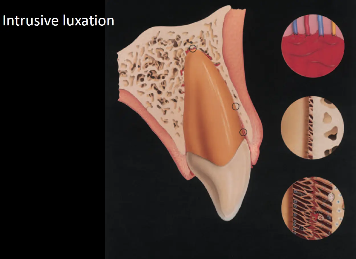
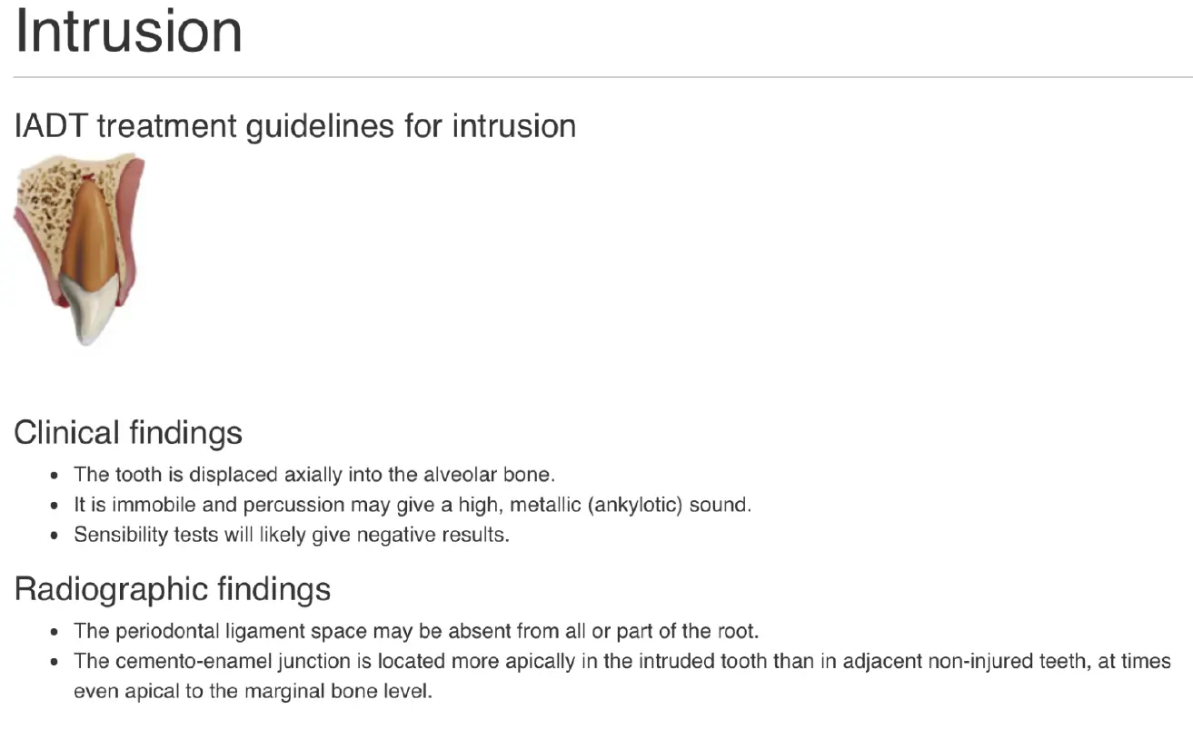
- displaced axially into alveolar bone
- no mobility, high pitched (metallic sound) to percussion like ankylosis
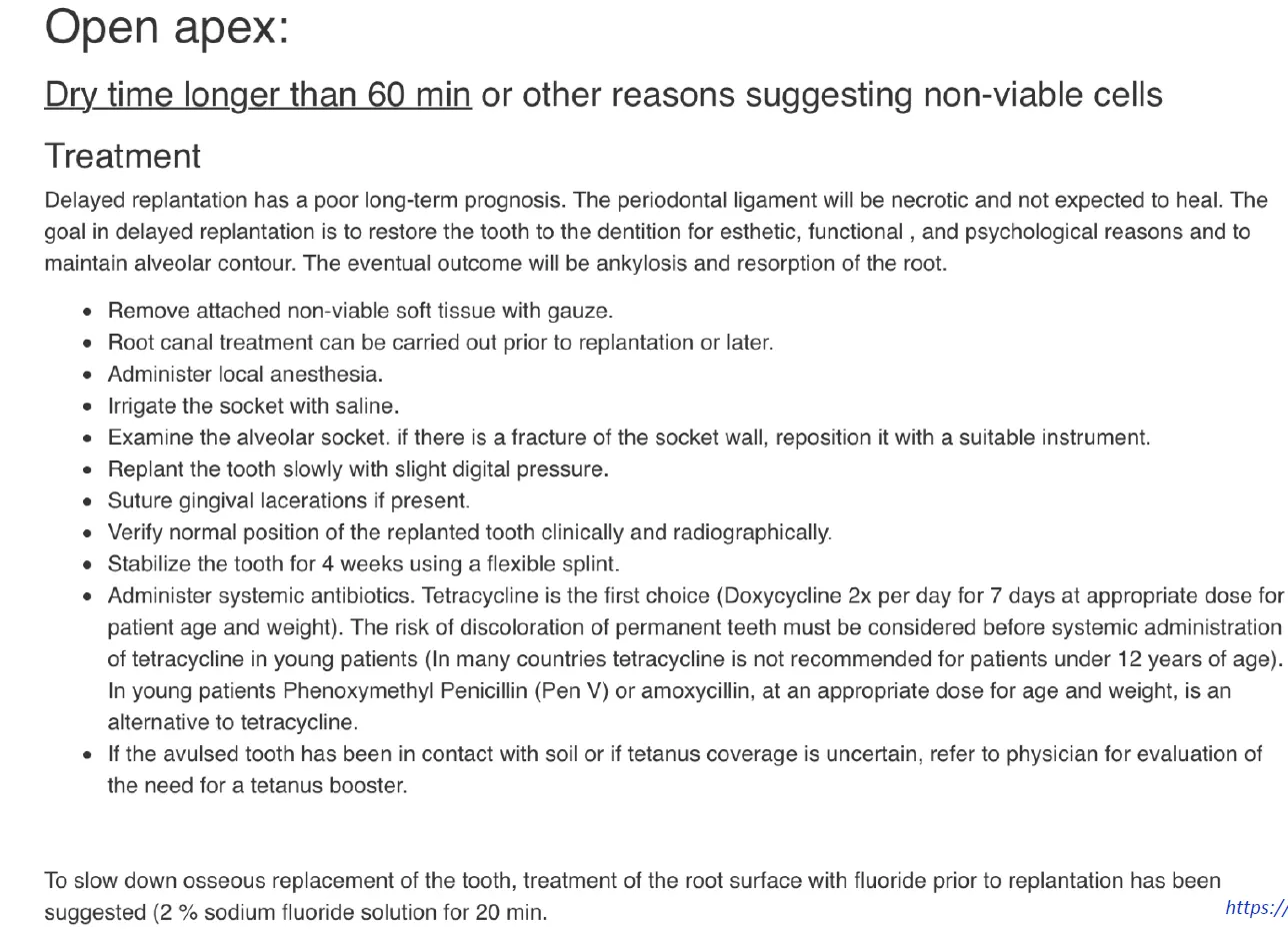
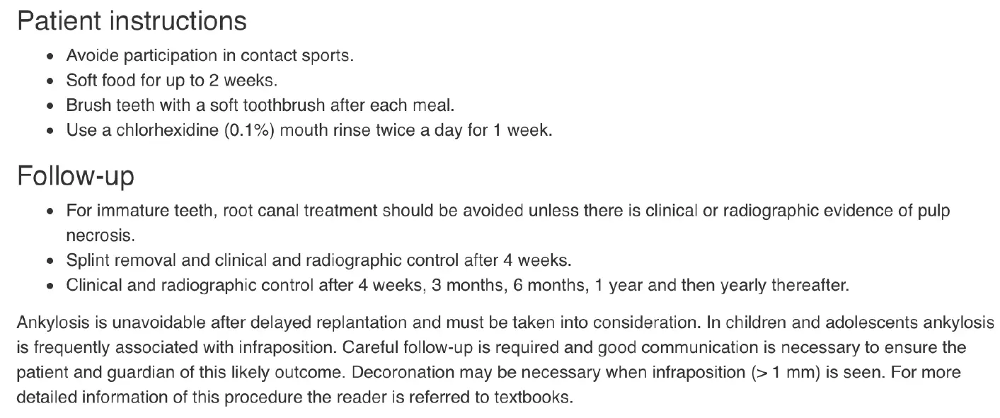
- may be no PDL space on periapical film
- crowns displaced lingually associated with vestibular fractures of socket wall
- CEJ located more apically than non affected teeth
Treatment
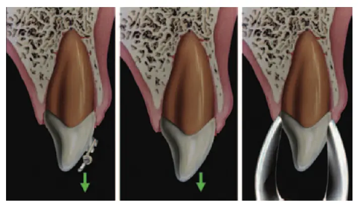
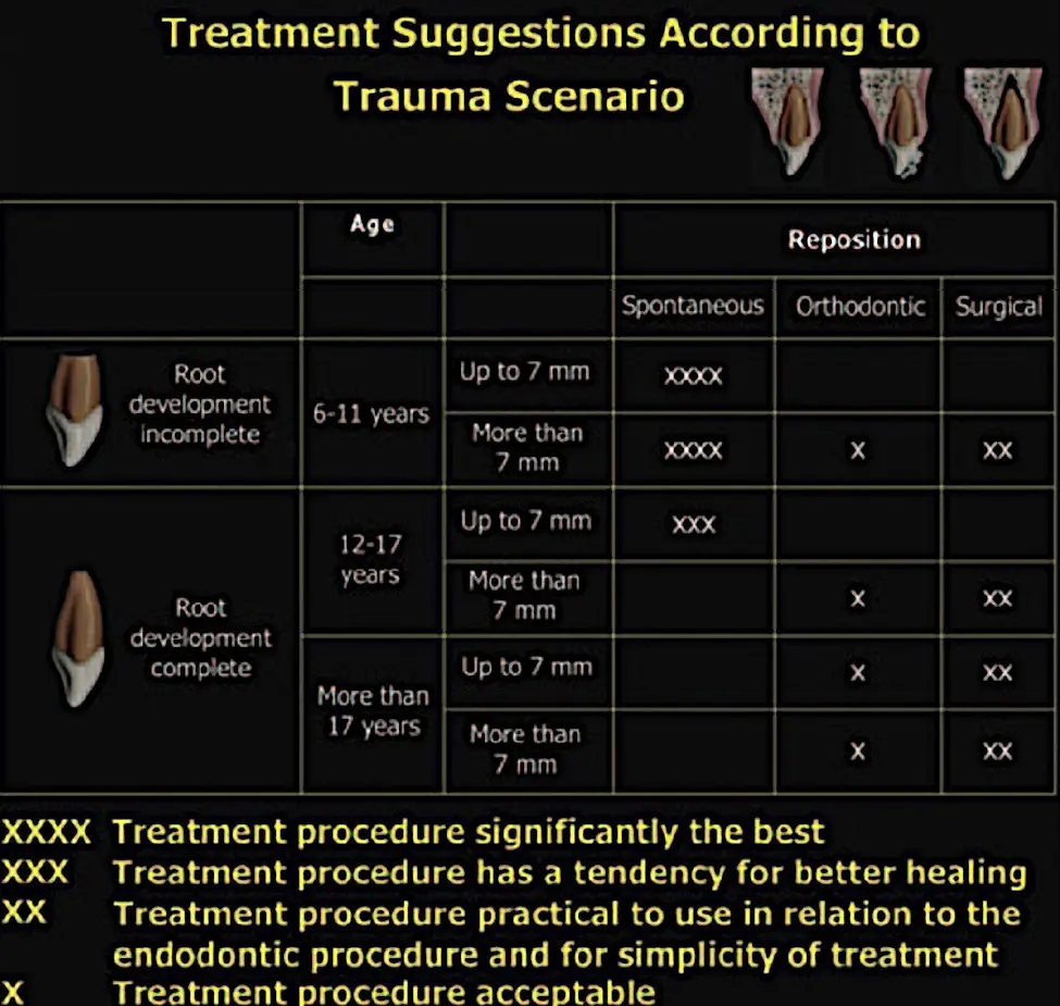
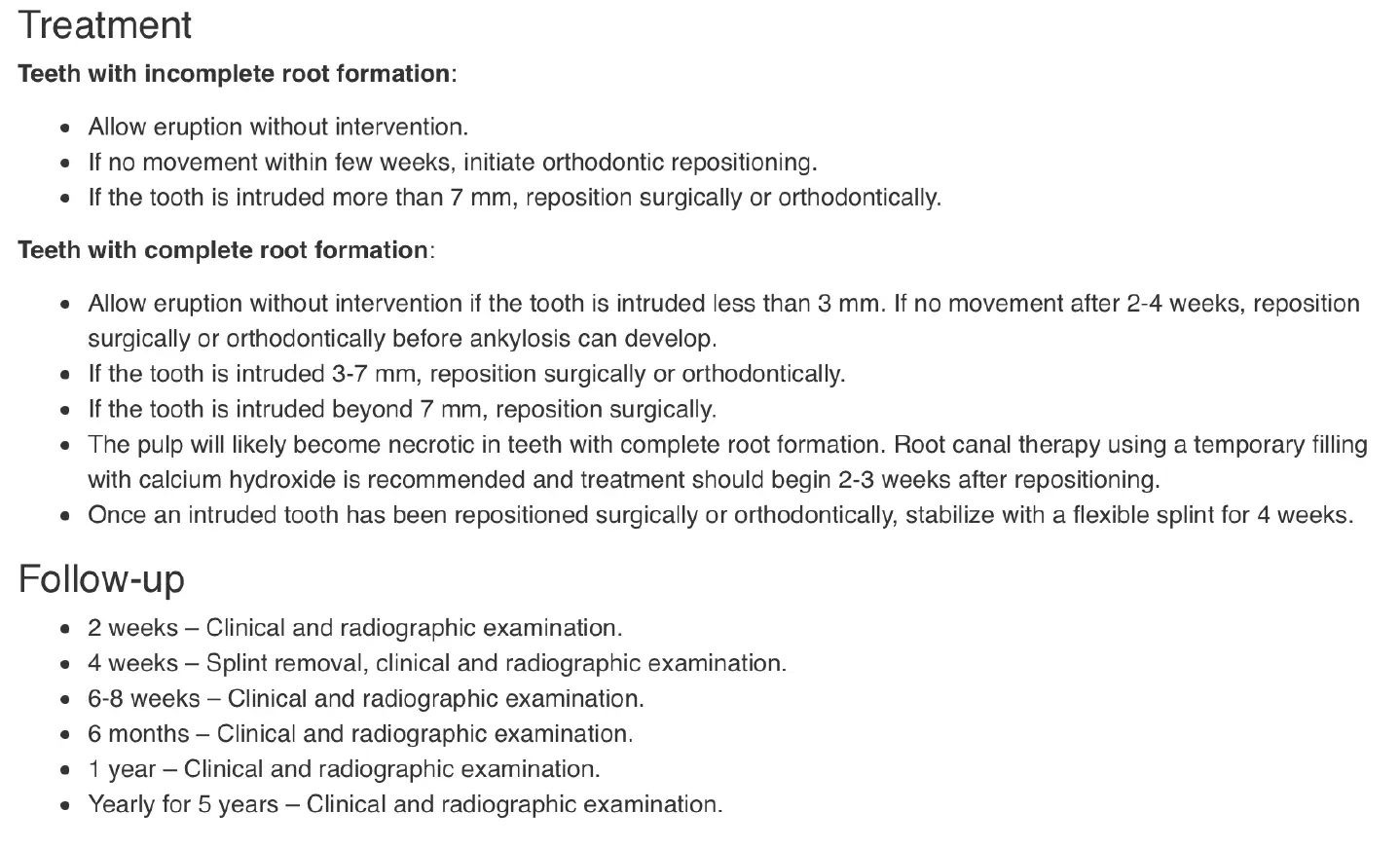
- repositioning
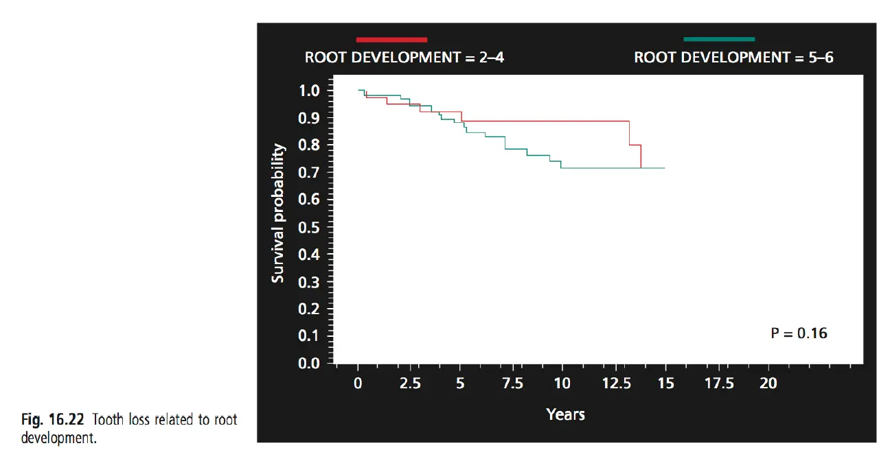
- spontaenous re eruption - treatment of choice for immature teeth (takes about 6 months for full eruption)
- orthodontic eruption (Cases with completed root development)
- surgical repositioning - where tooth is intruded more than 6mm or multiple intrusions
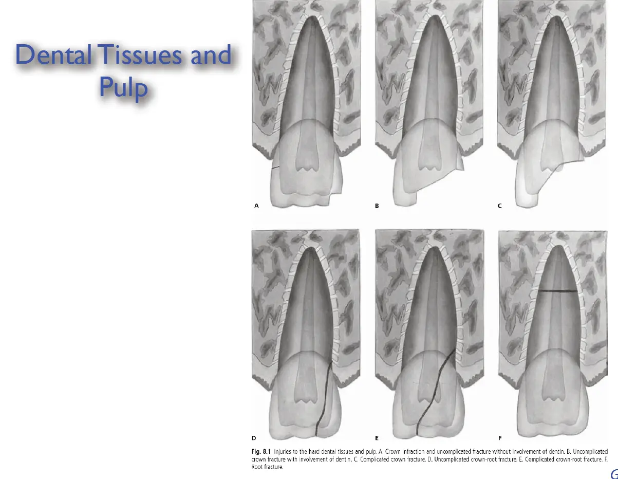
Crown Fractures
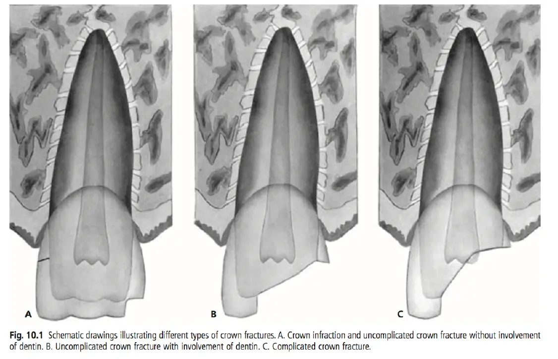
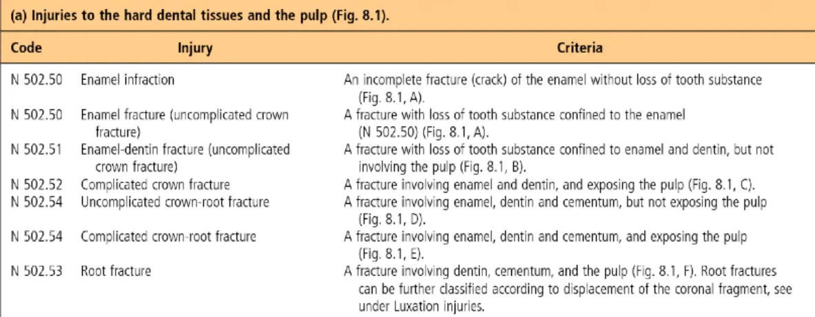
Enamel
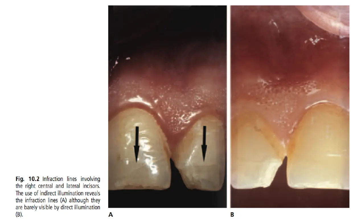
Enamel Dentine
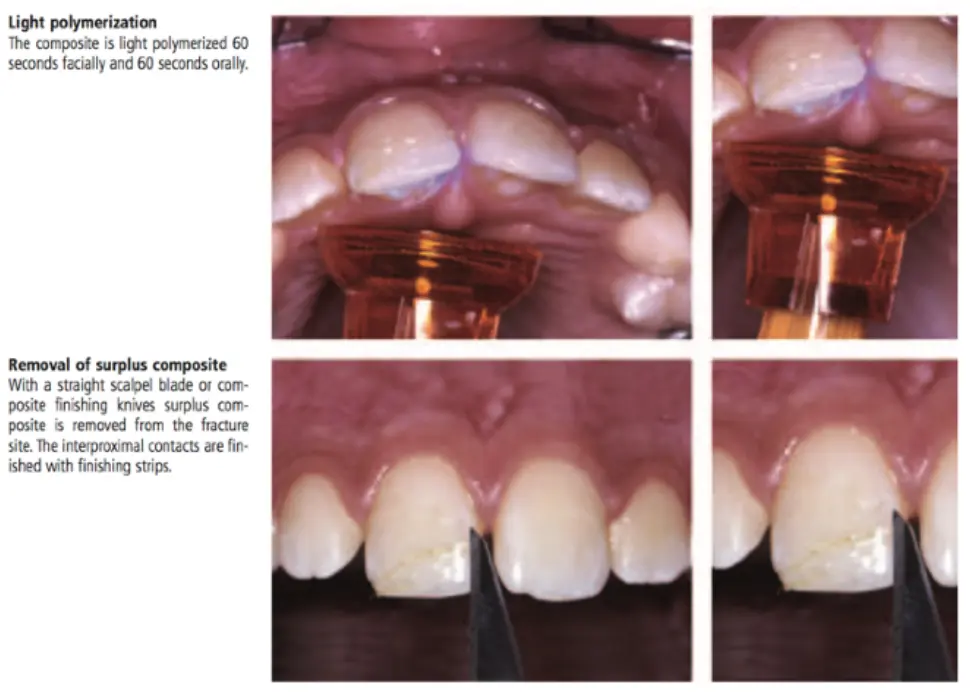
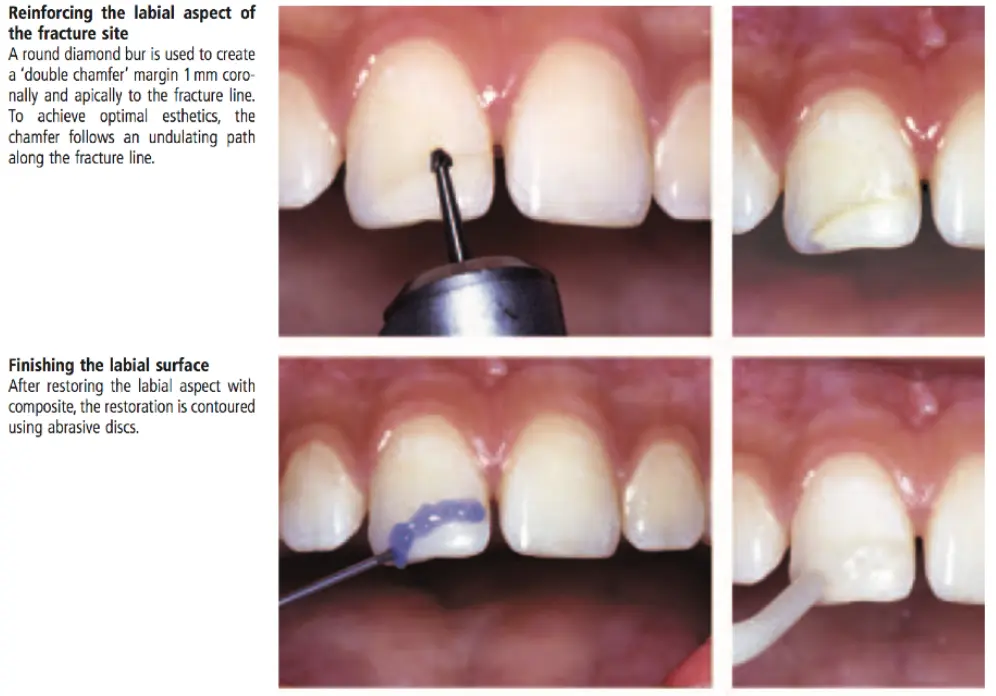
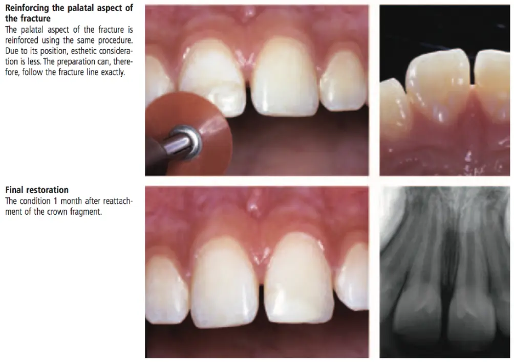
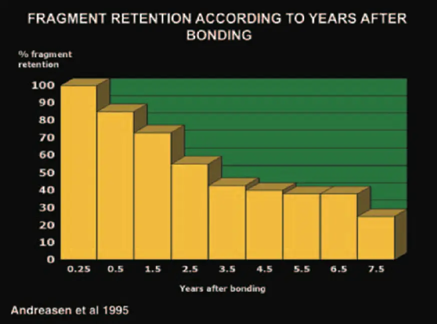
Rebonding crown fragment
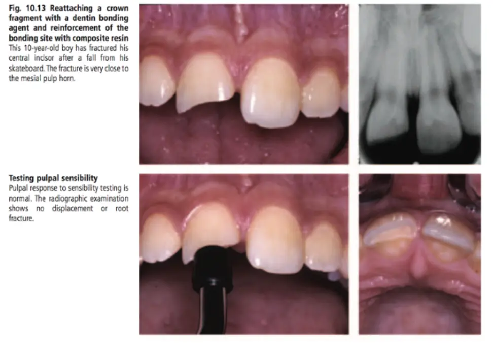
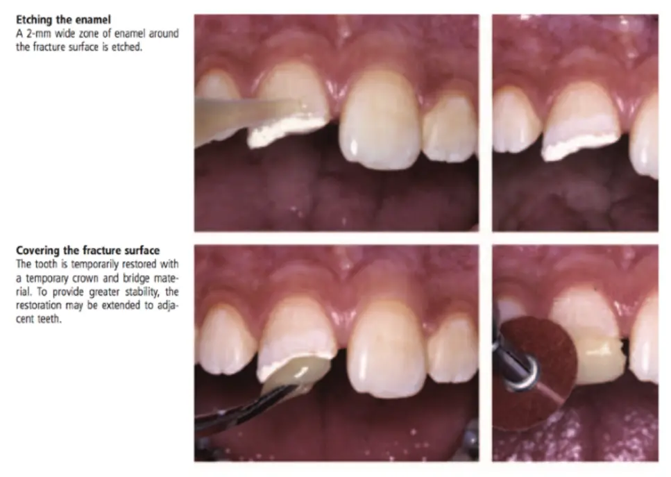
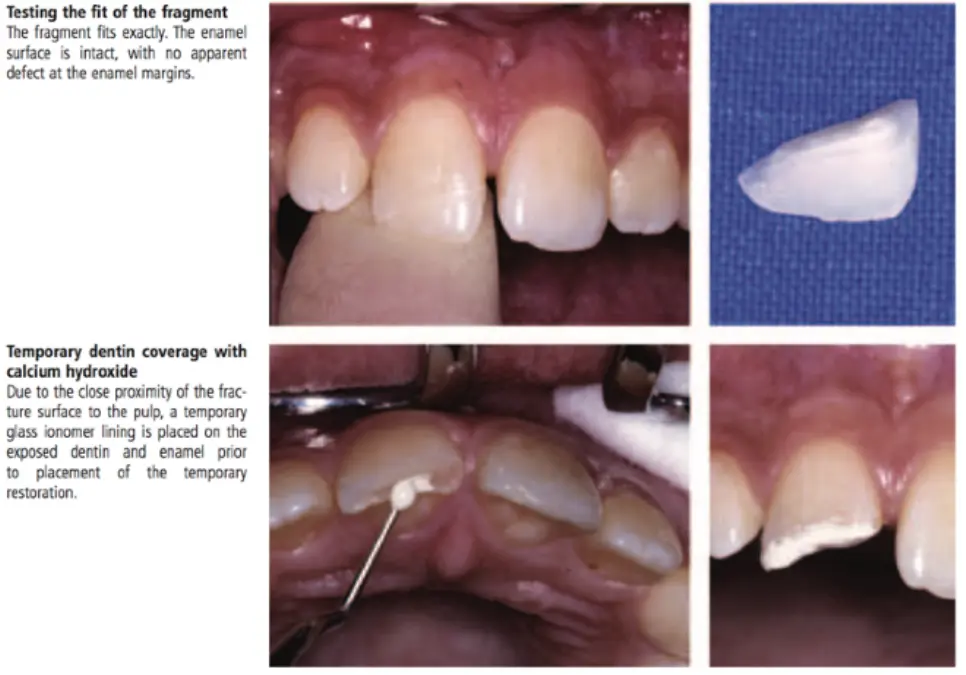
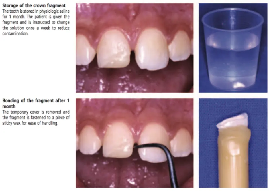
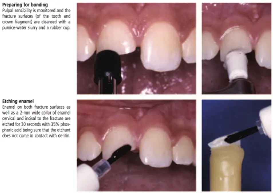
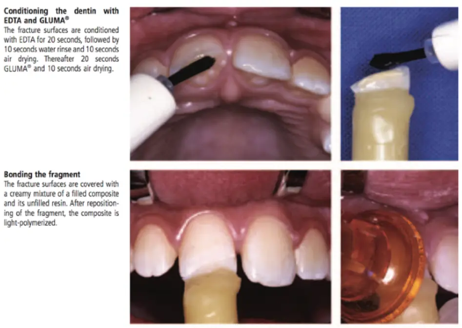
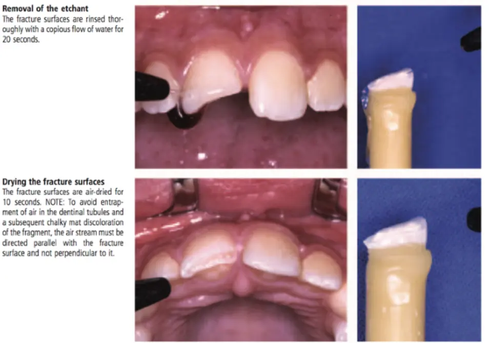
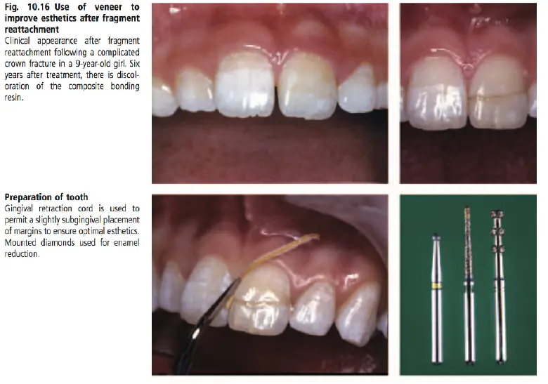
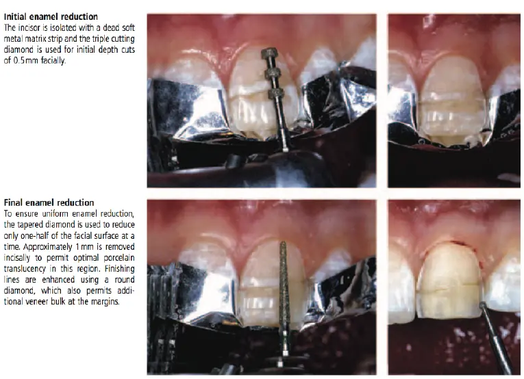
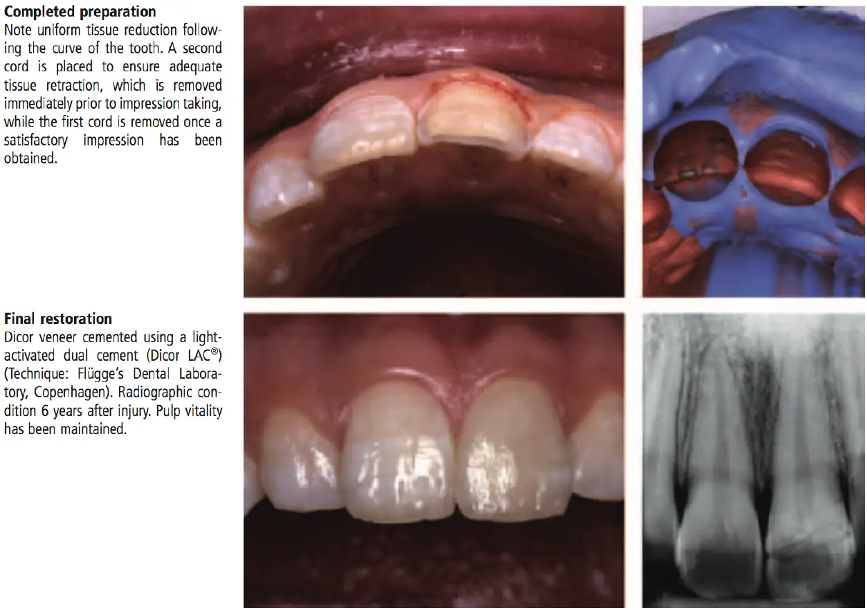
Veneer to improve aesthetics after fragment reattachment
Complicated crown fractures
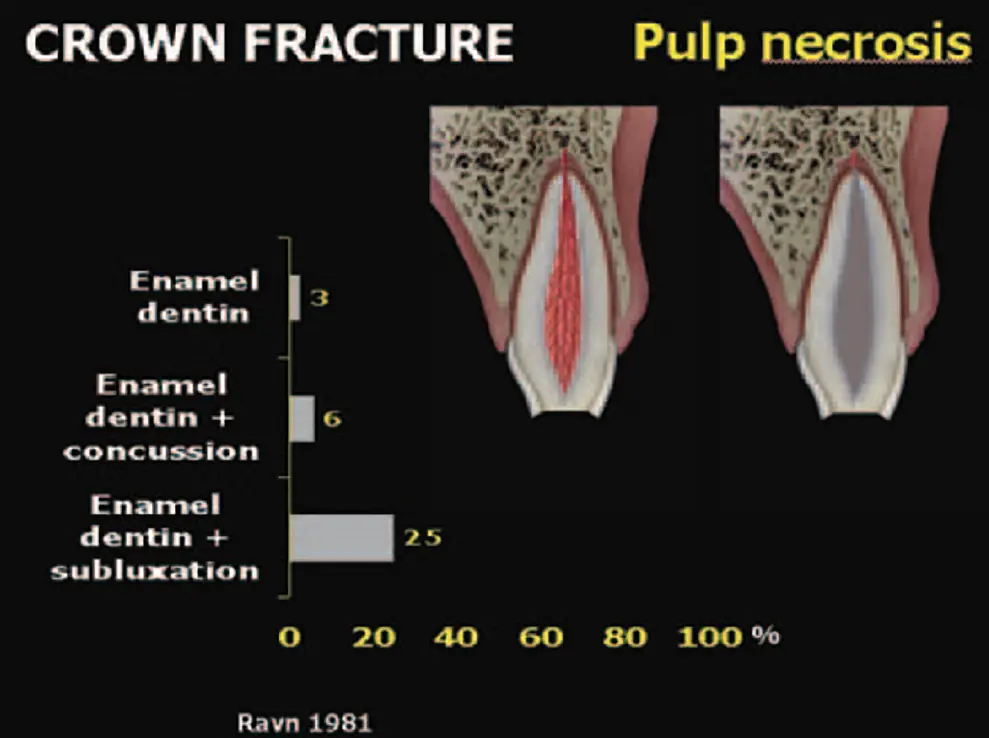
- immature teeth with pulpal involvement
- pulp status
- vital or necrotic
- vital - assess exposure size
- pulp cap
- apexogenesis (Cvek pulpotomy)
- necrotic
- apexification
- regeneration
- pulp status
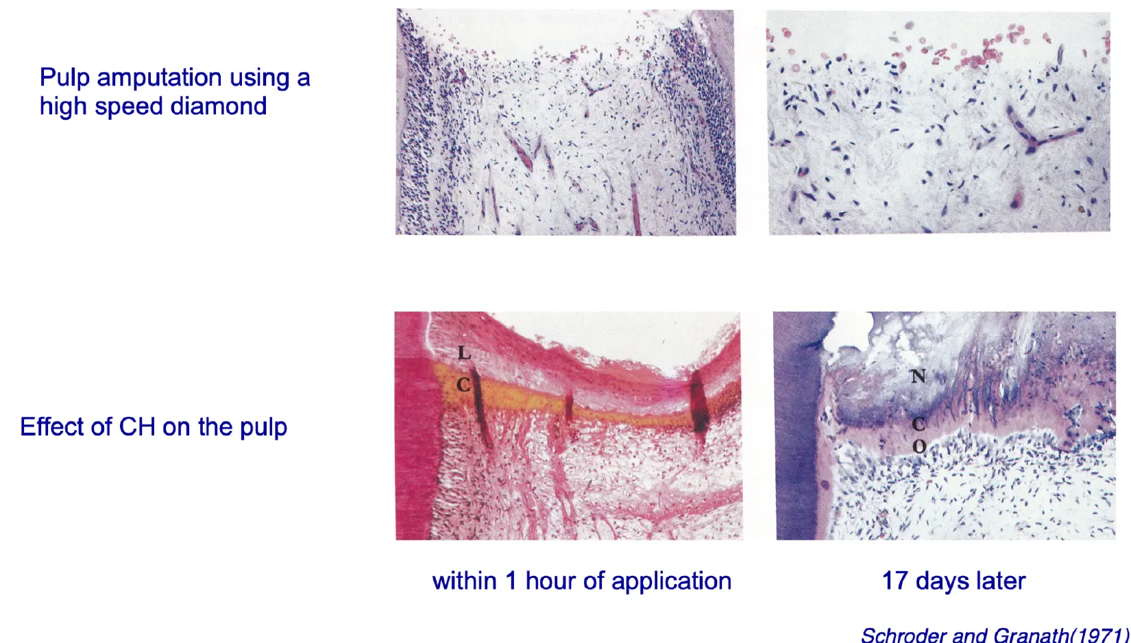
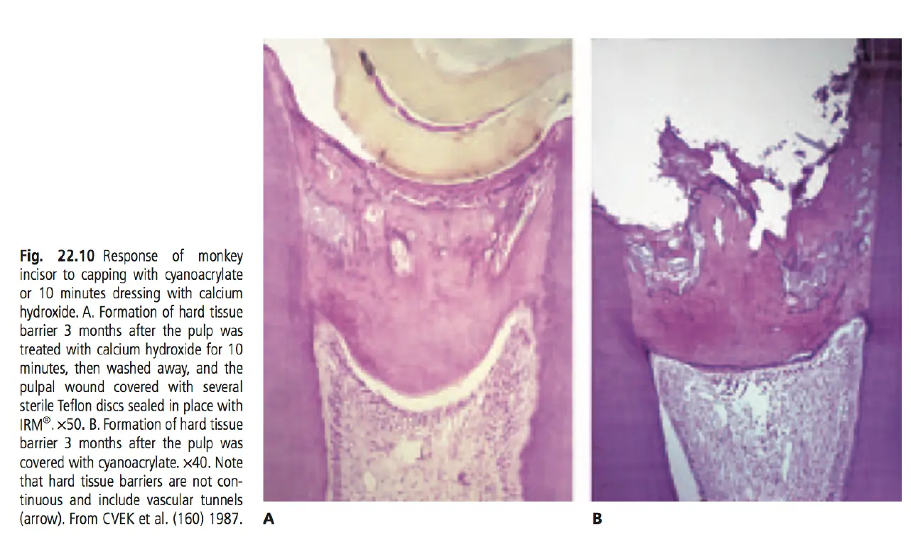
cvek pulpotomy
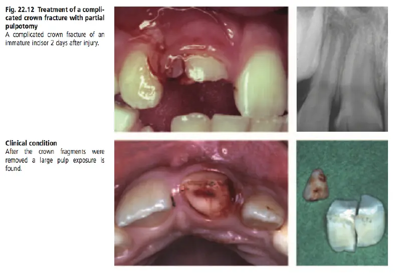
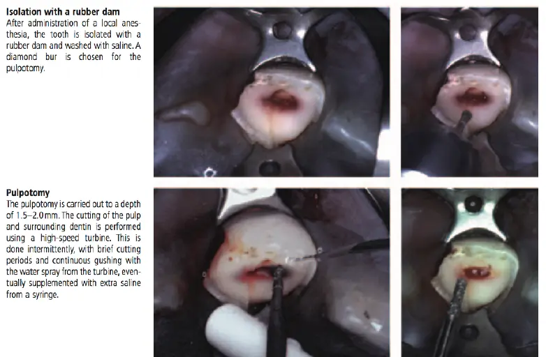
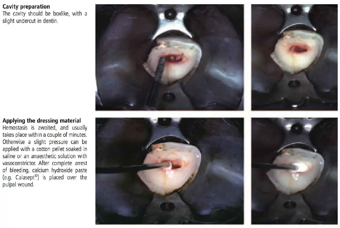
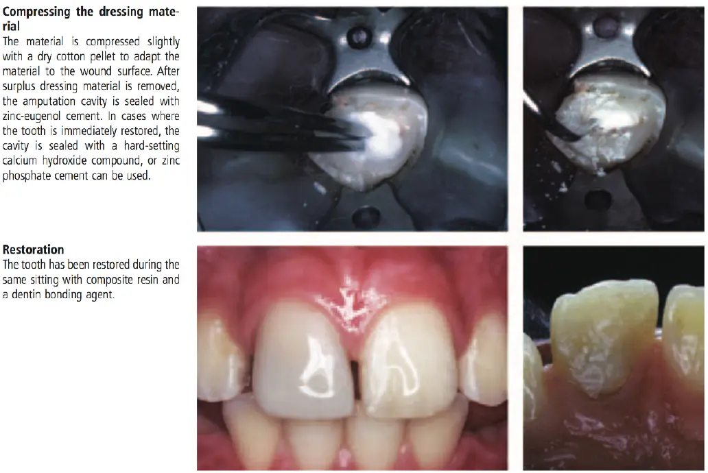
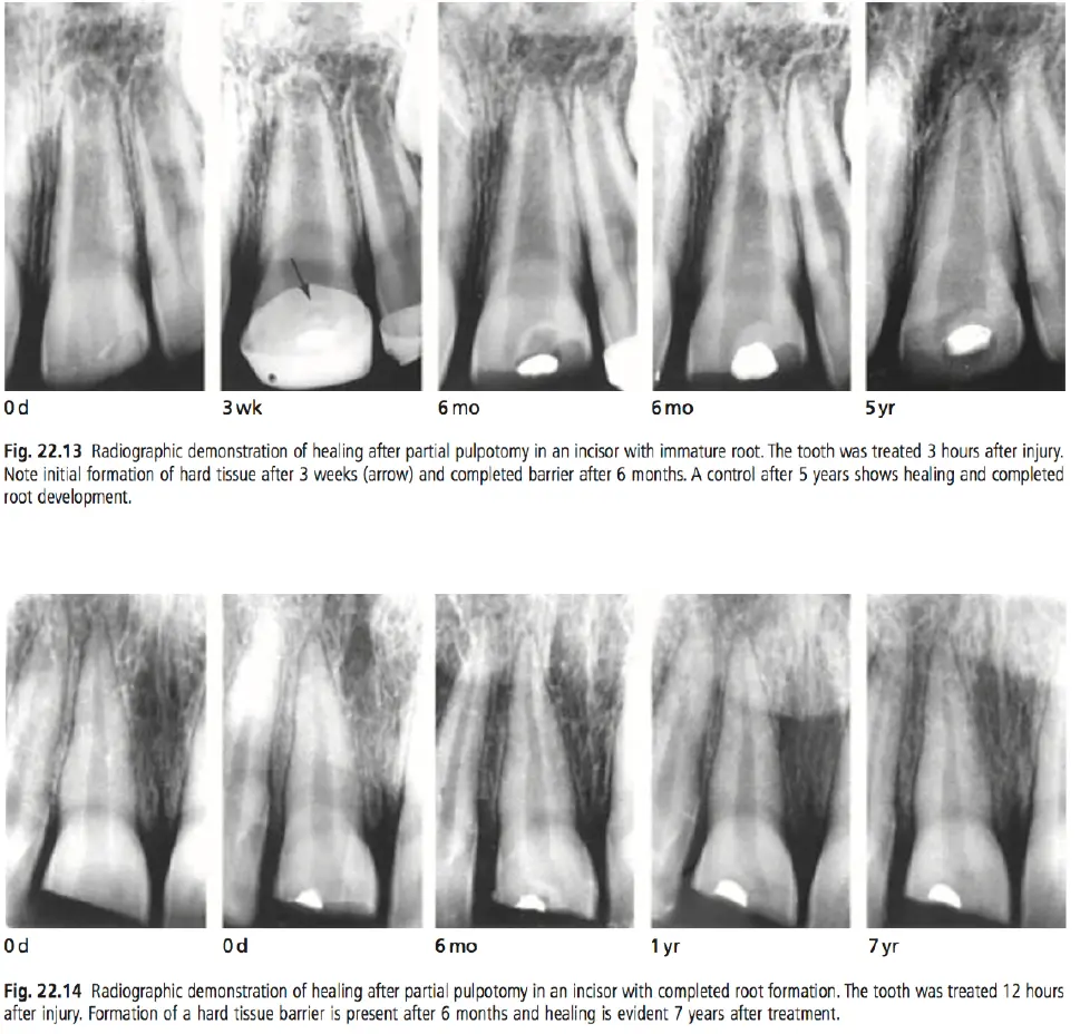
- 96% success rate from 31mo follow up, pulpotomy completed ranging 1hour-90 days
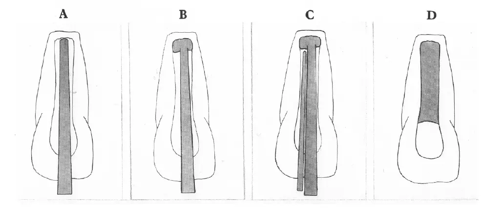
Apexification
- must be resporable
- imaturature teeth with pulpal necrosis
- surgery not alternative
- **technique
- isolation
- access
- pulp extirpation
- working length
- instrumentation
- dry canal
- calcium hydroxide
- coronal seal
- recall
- hard tissue barrier
- Obturation
- modified lateral condensation
- master GP cone -chemically softened
- Thermoplasticised GP
- Follow-up
- apical barrier
- location
- composition
- bone
- cementum
- connective tissue
- CaOH2
- Hertwigs root sheath
- apical barrier formation
- remnants in PDL
- role of calcium hydroxide
- stimulate barrier formation
- provide suitable environment
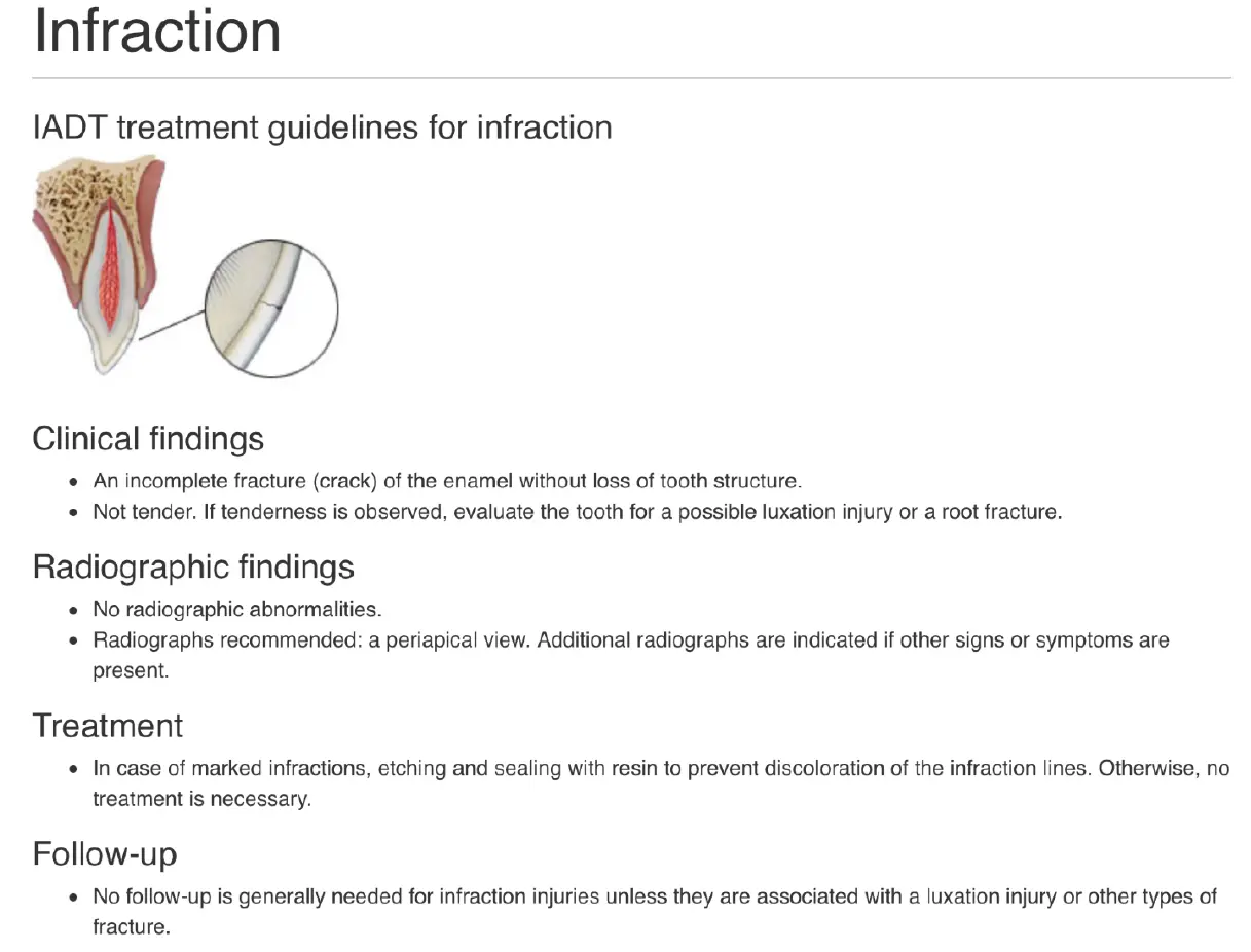
Infraction
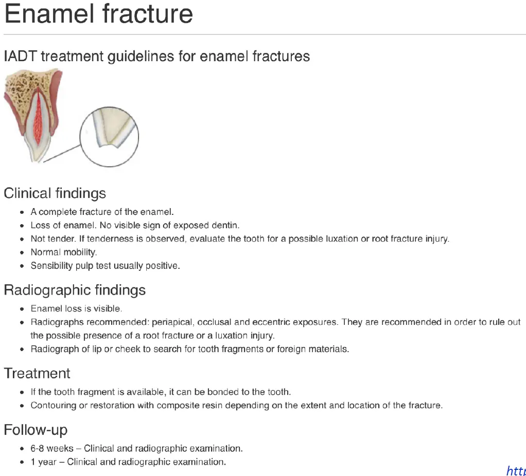
Enamel Fracture
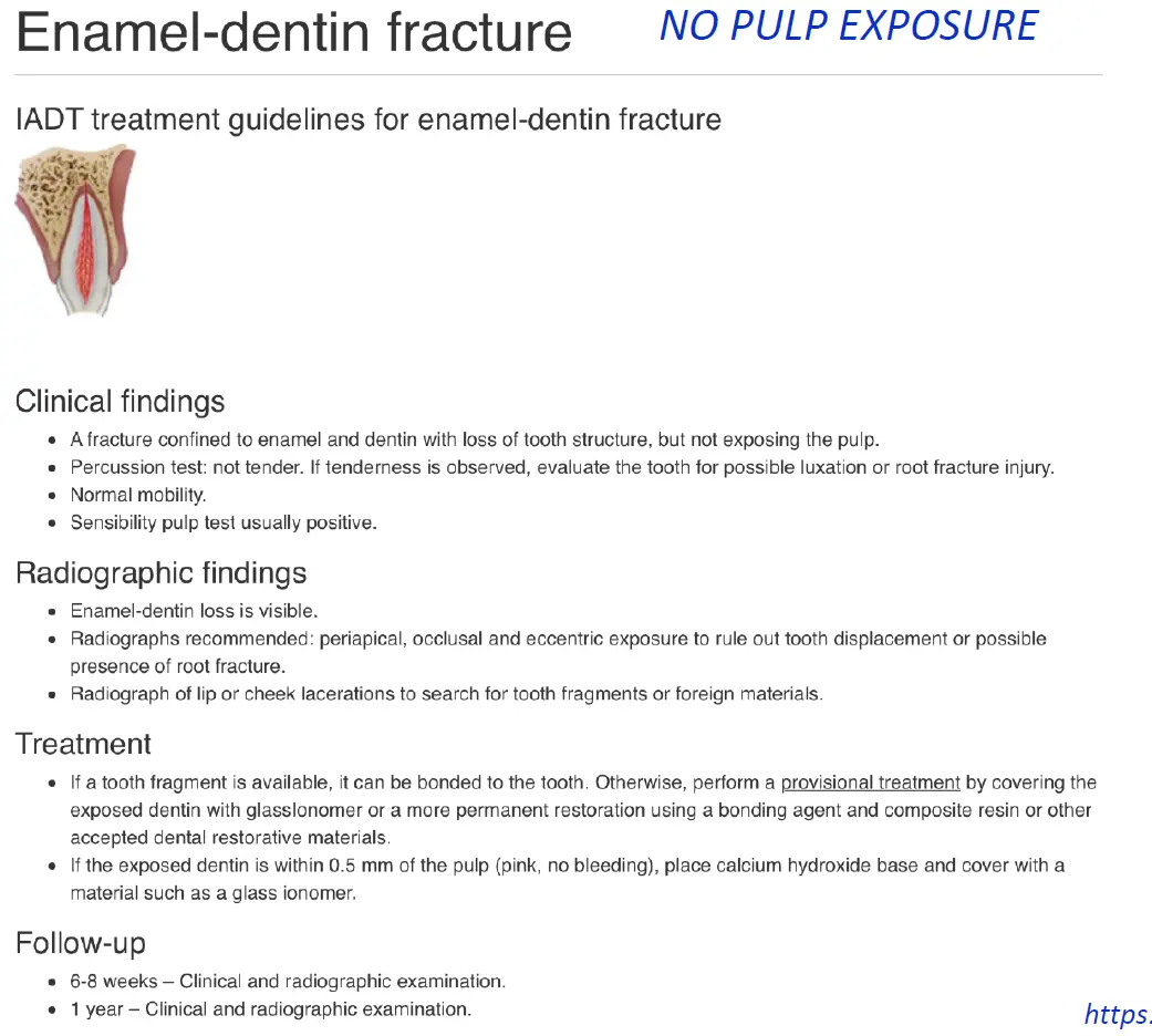
Enamel Dentine Fracture
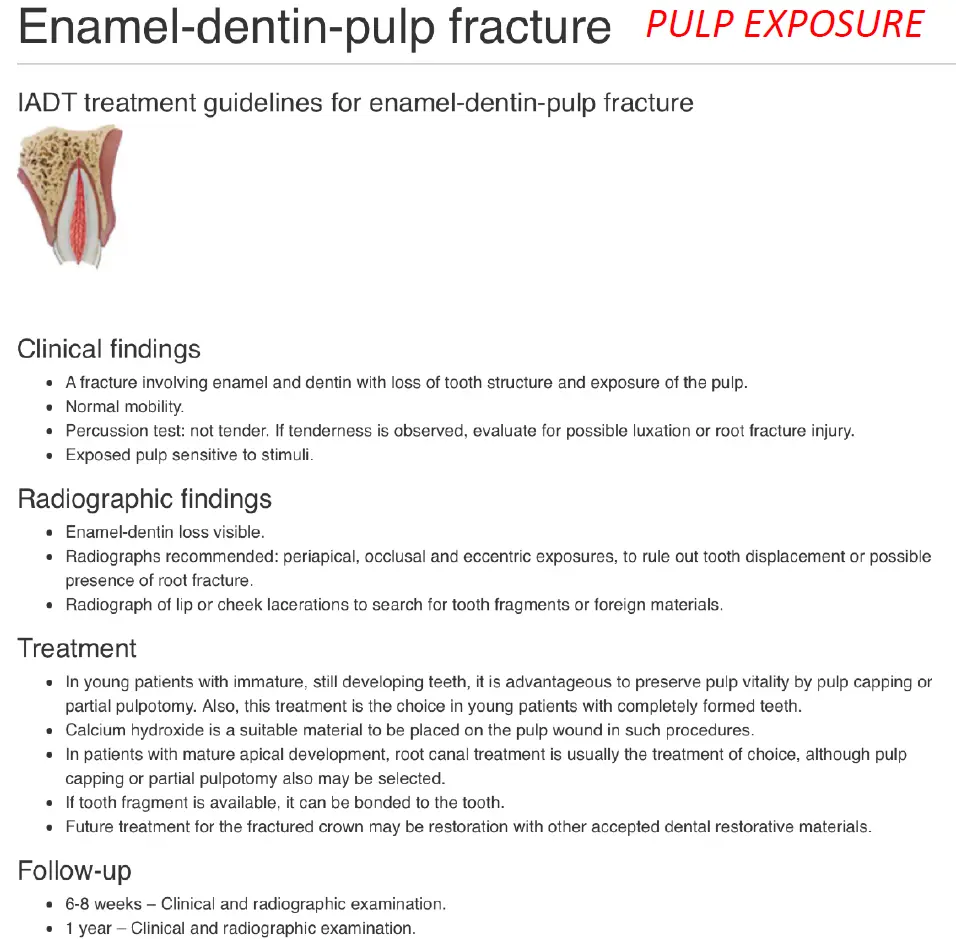
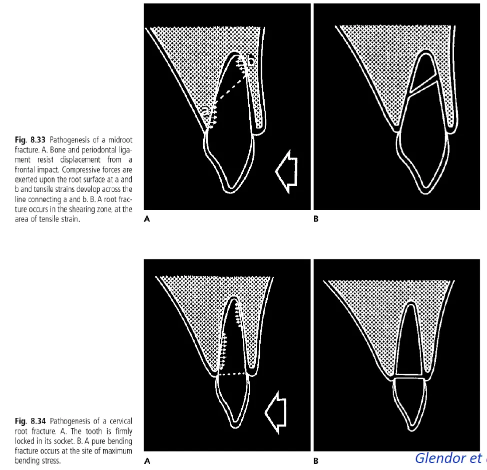
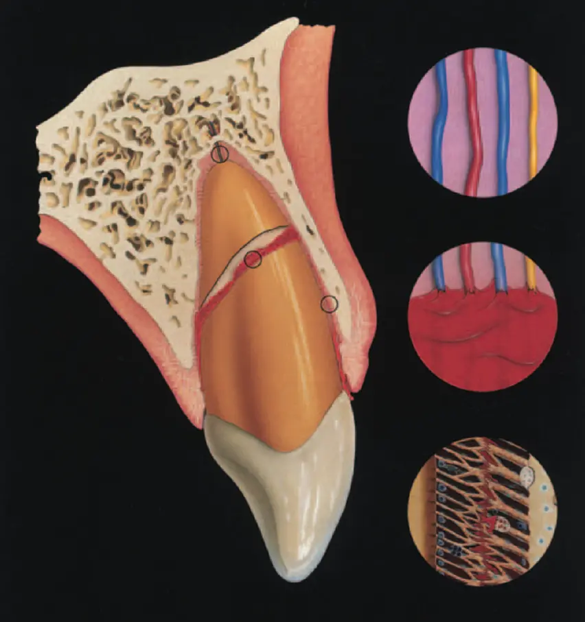
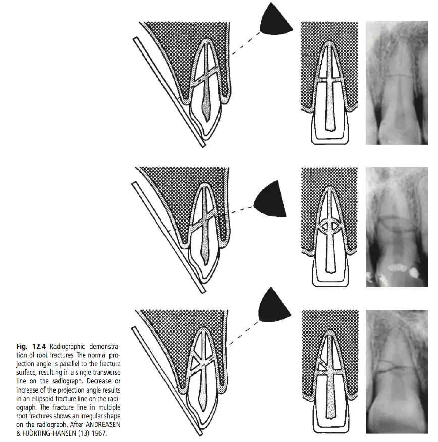
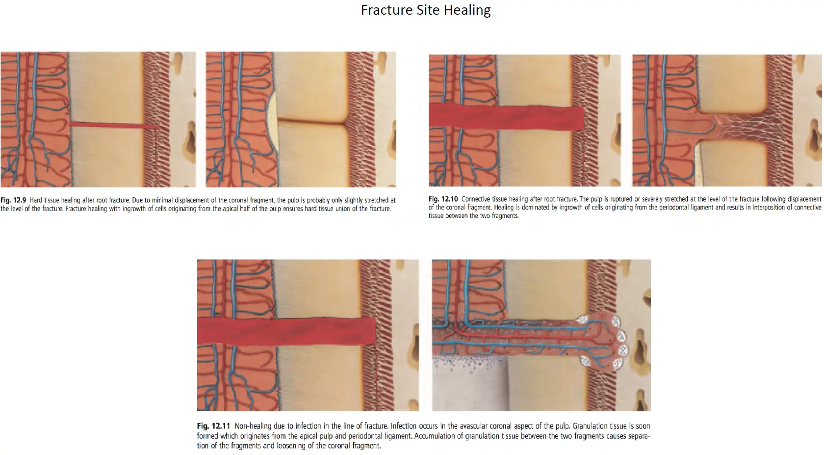
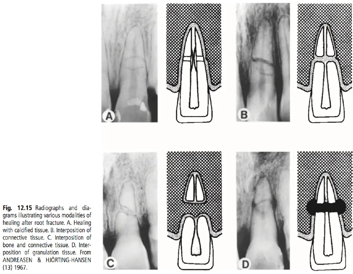
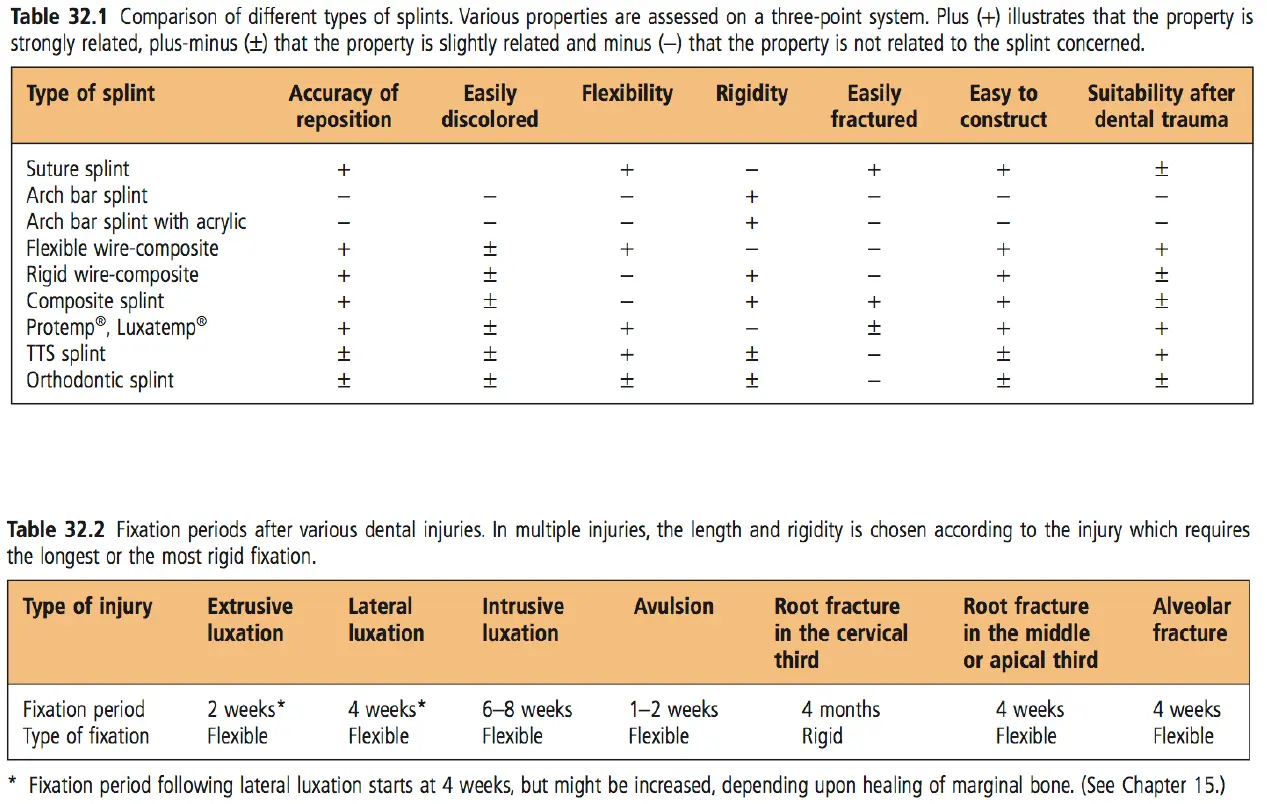
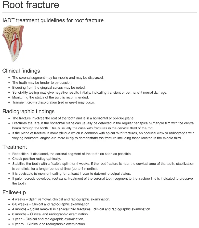
Root fractures
Treatment
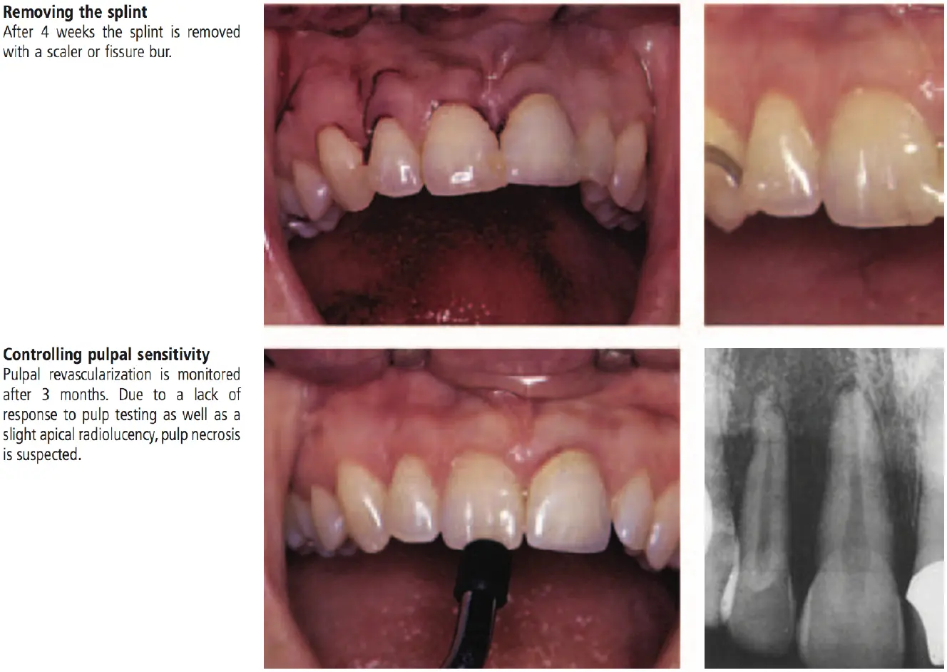
- reduction of displaced coronal fragment
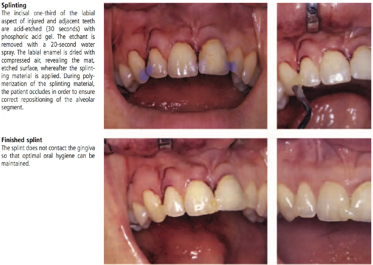
- immobilisation
- semi rigid fixation - acid etch / resin splint
- fixation for about one month
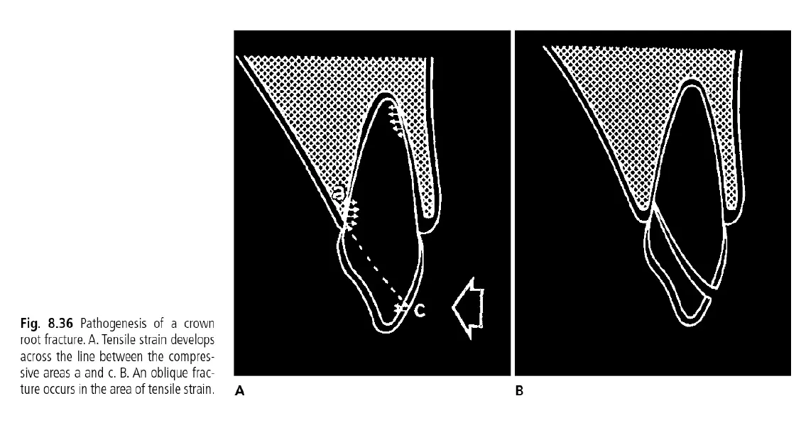
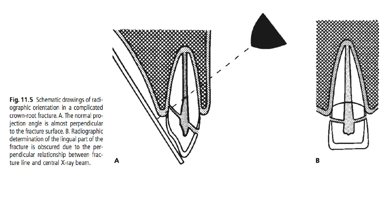
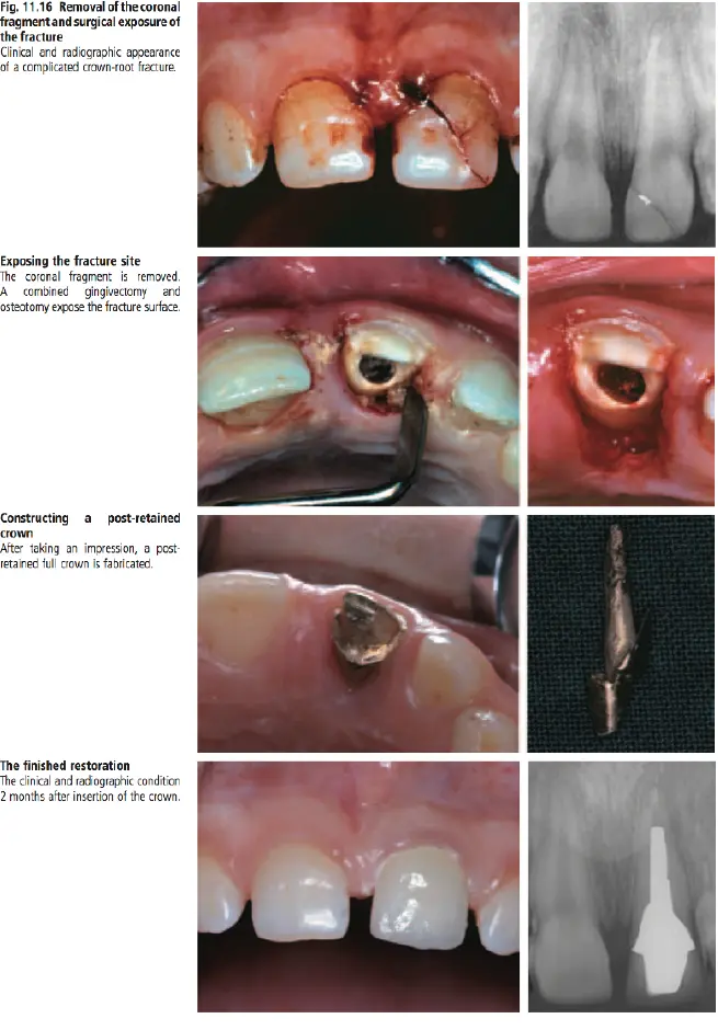
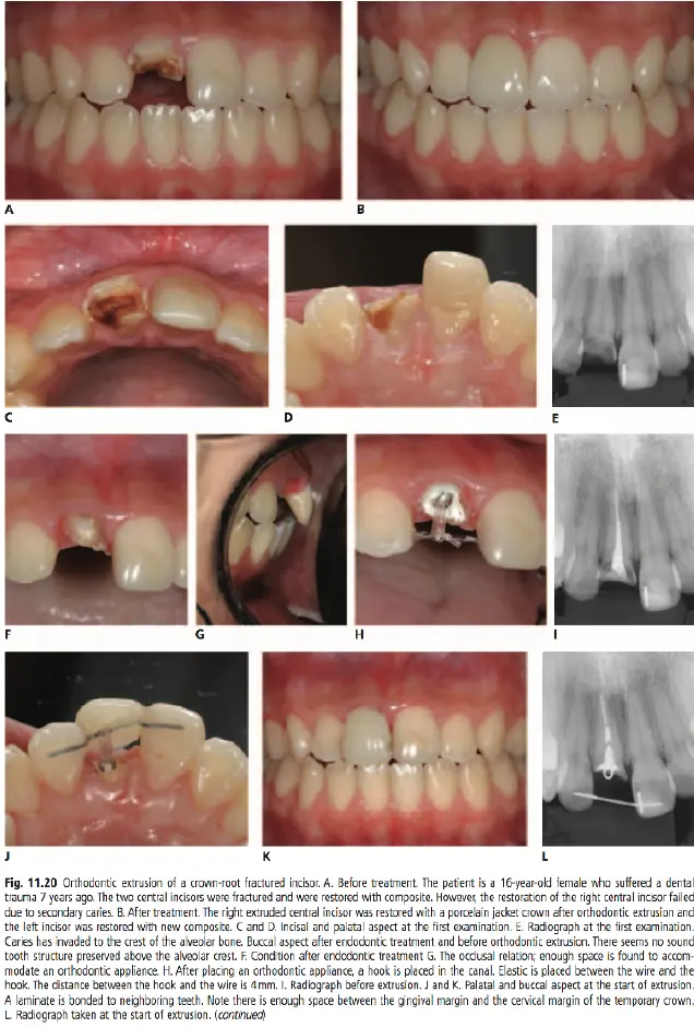
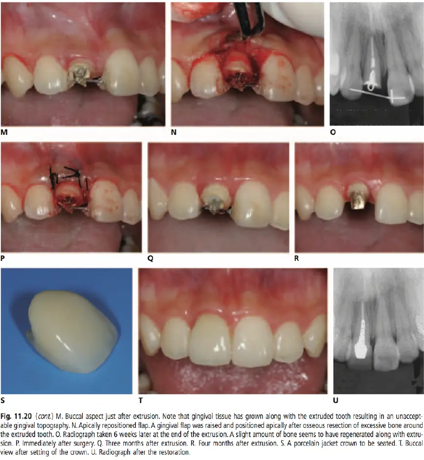
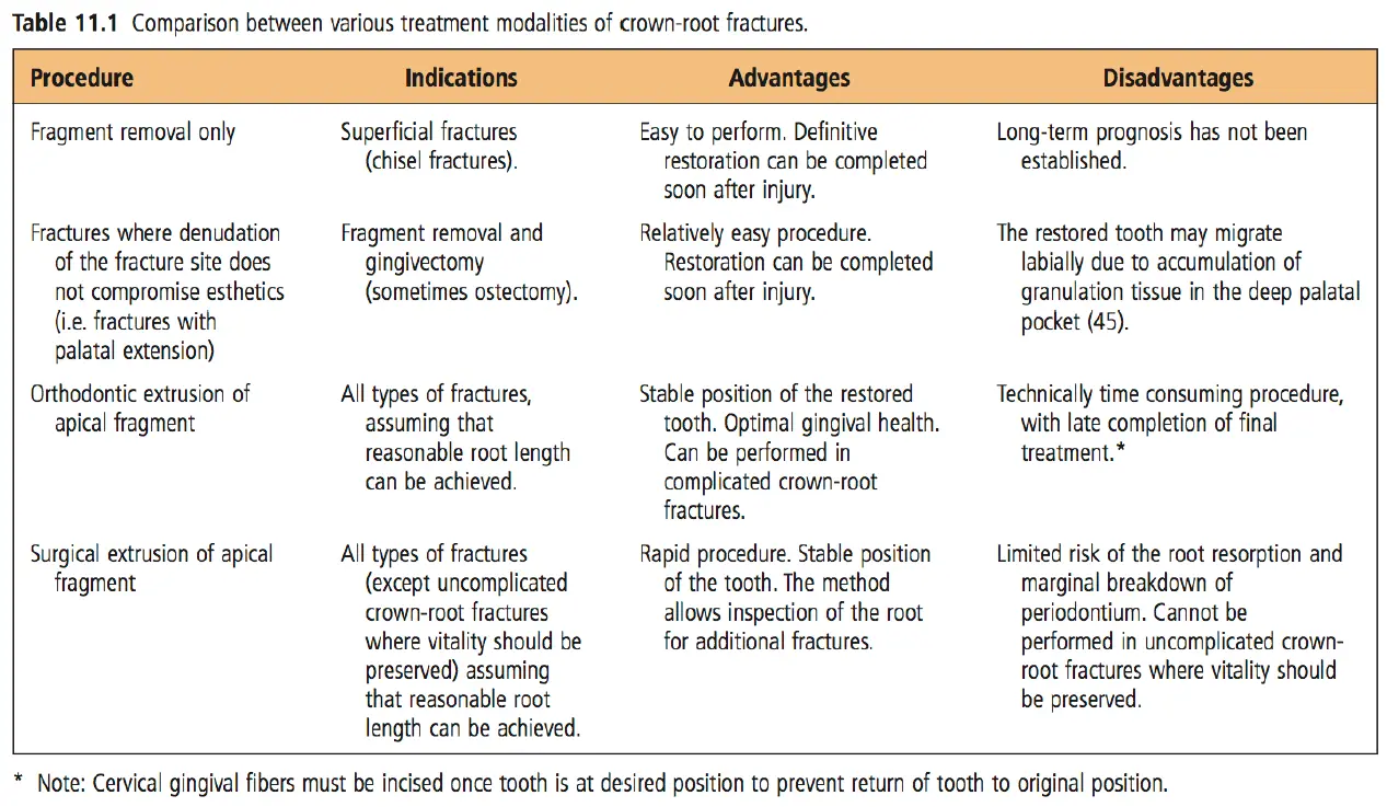
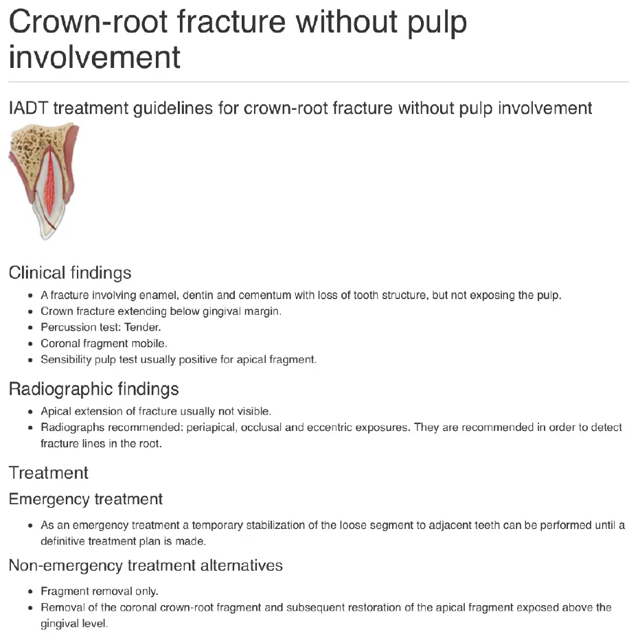
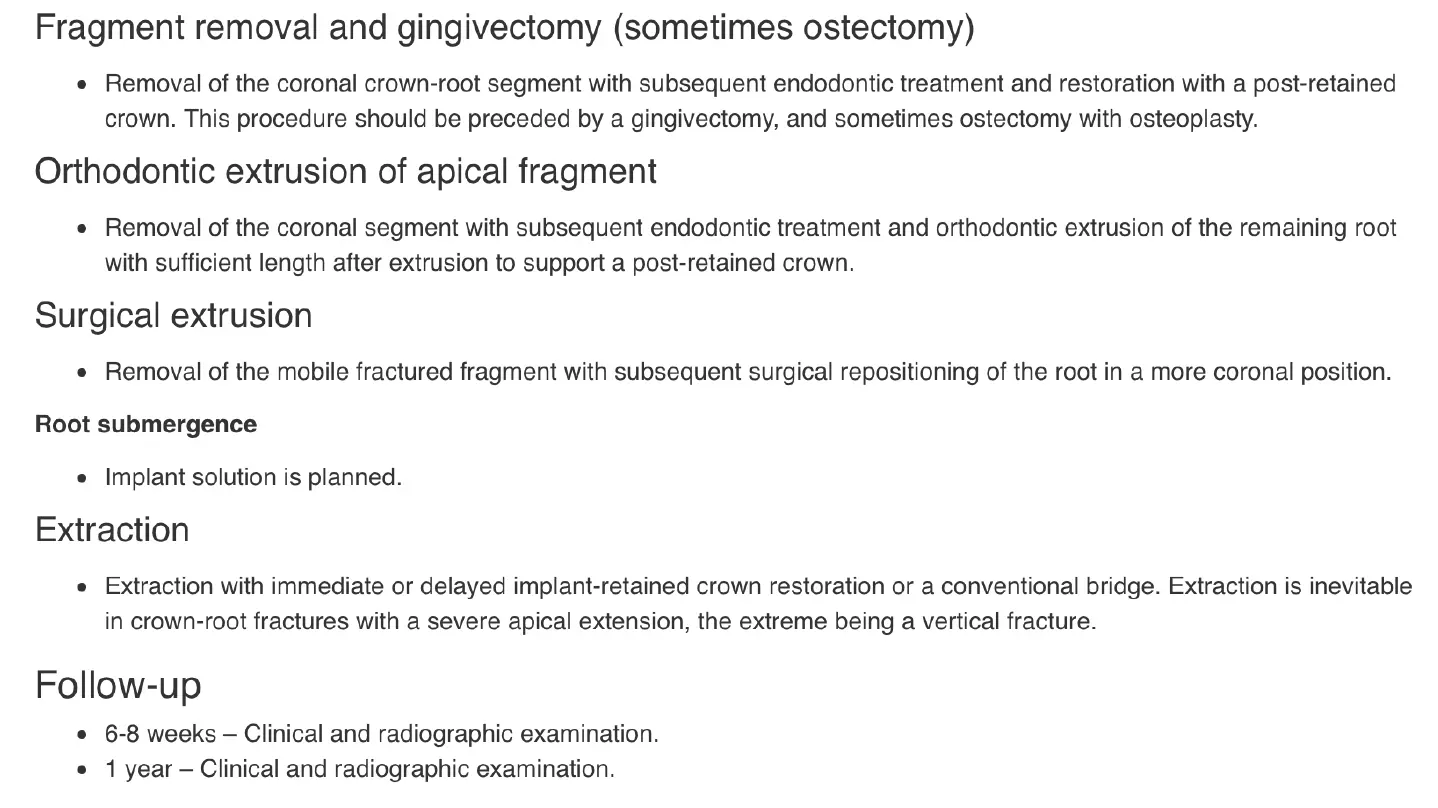
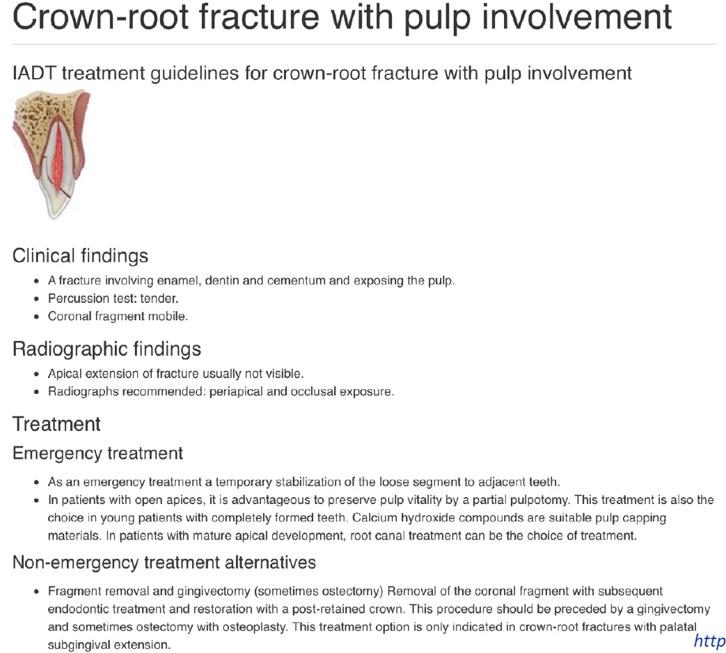
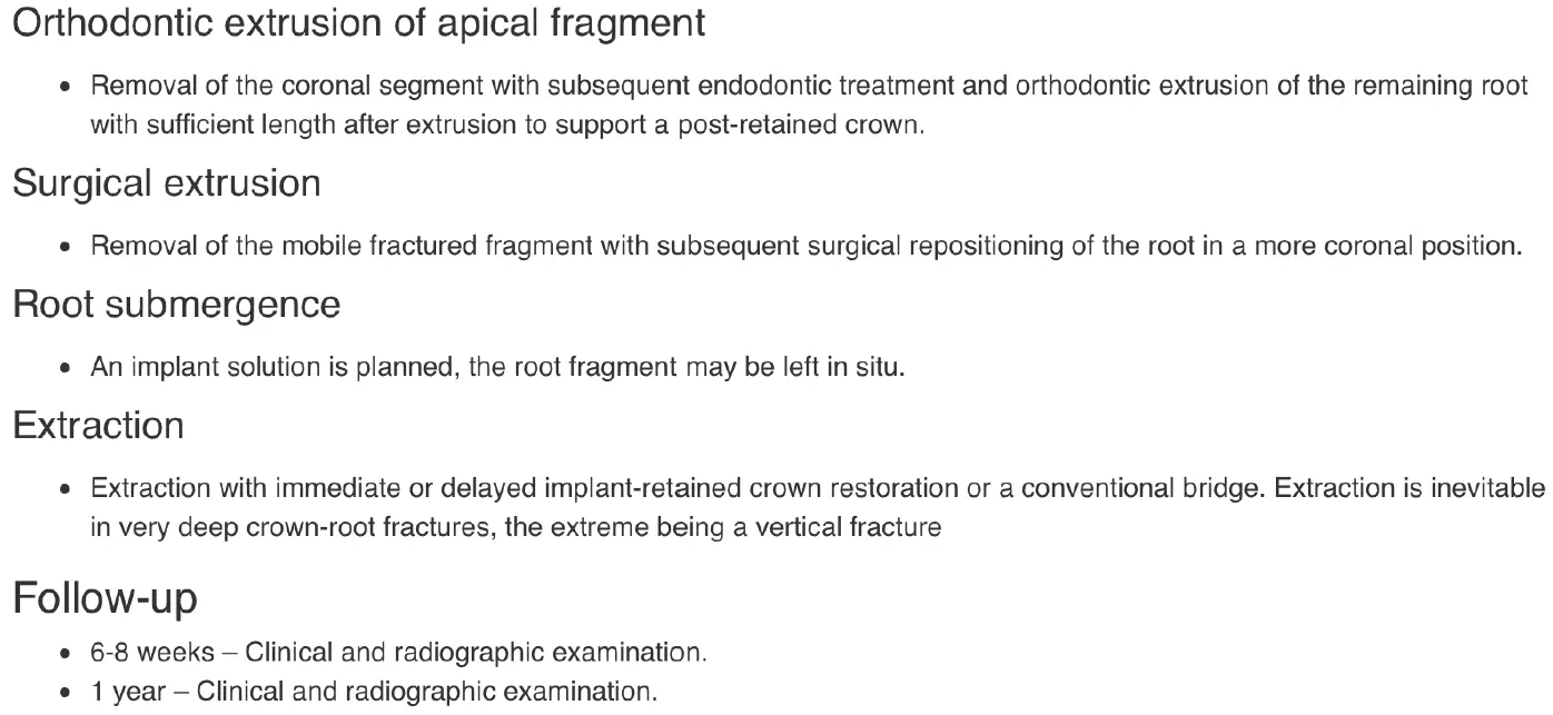
Crown root fracture
- fracture involving enamel, dentine, cementum
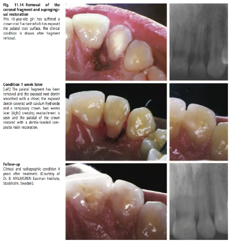
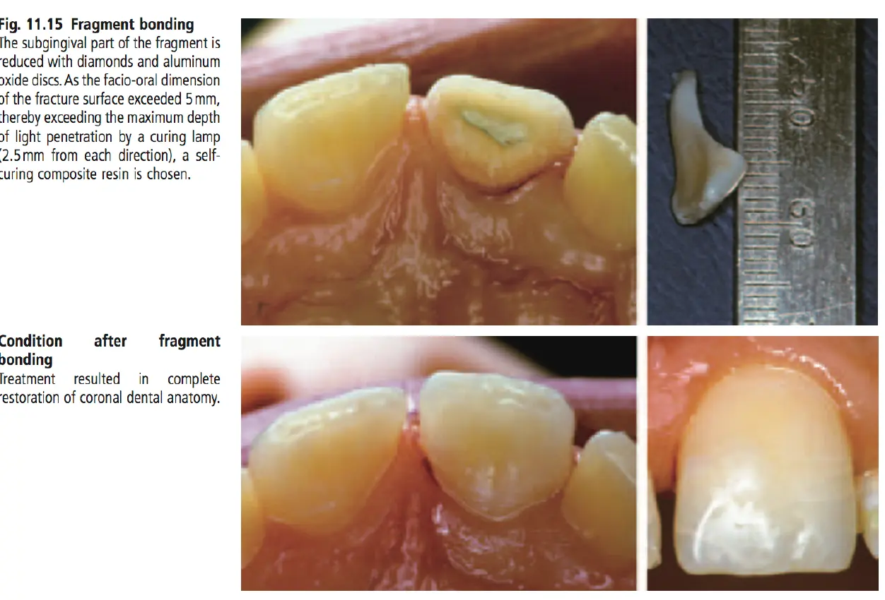
perio surgery
ortho extrusion
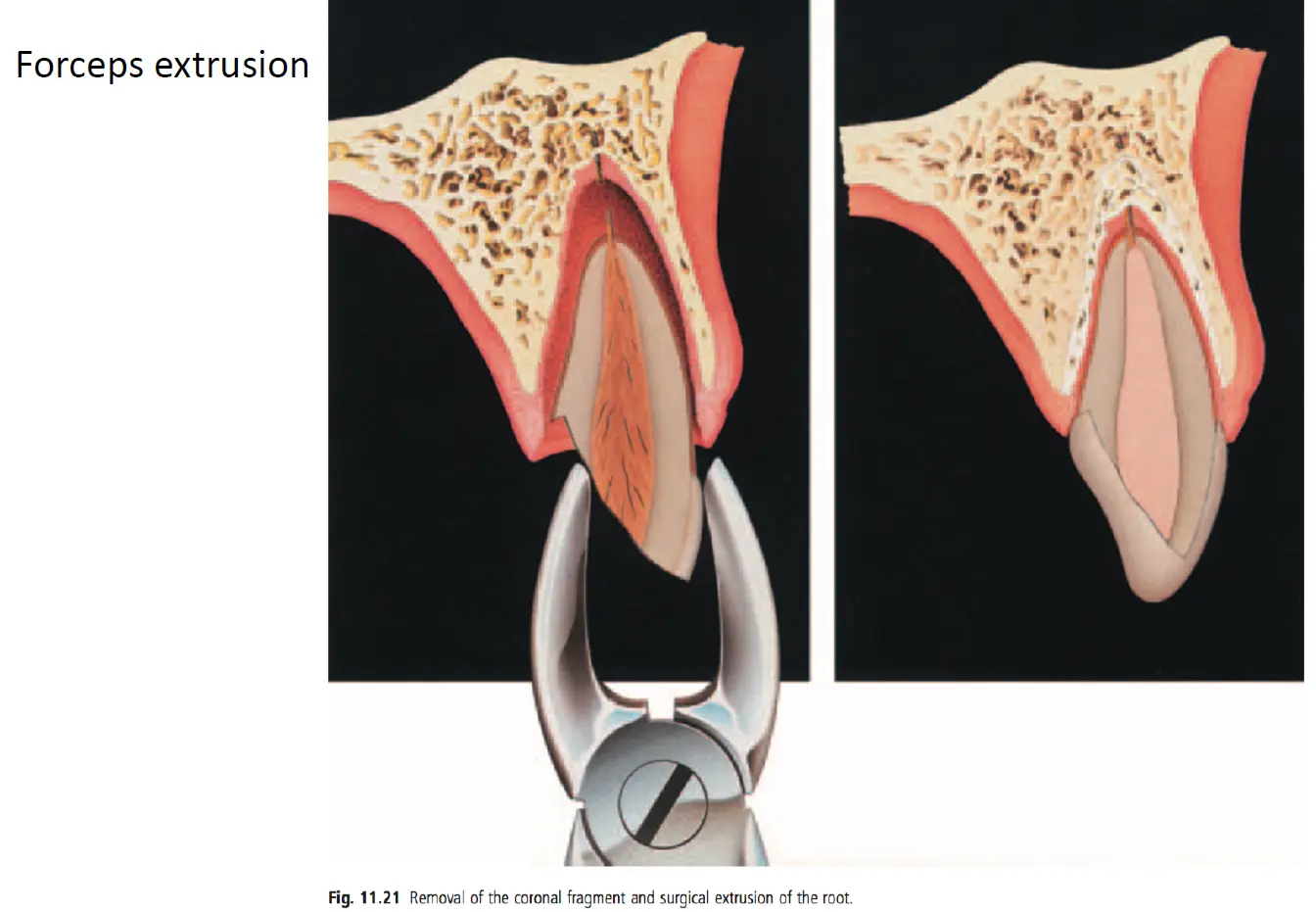
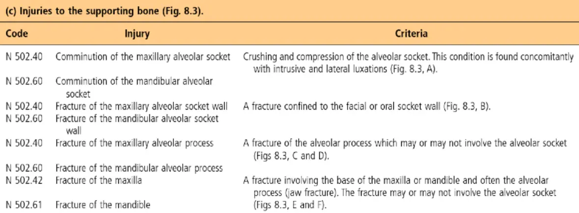
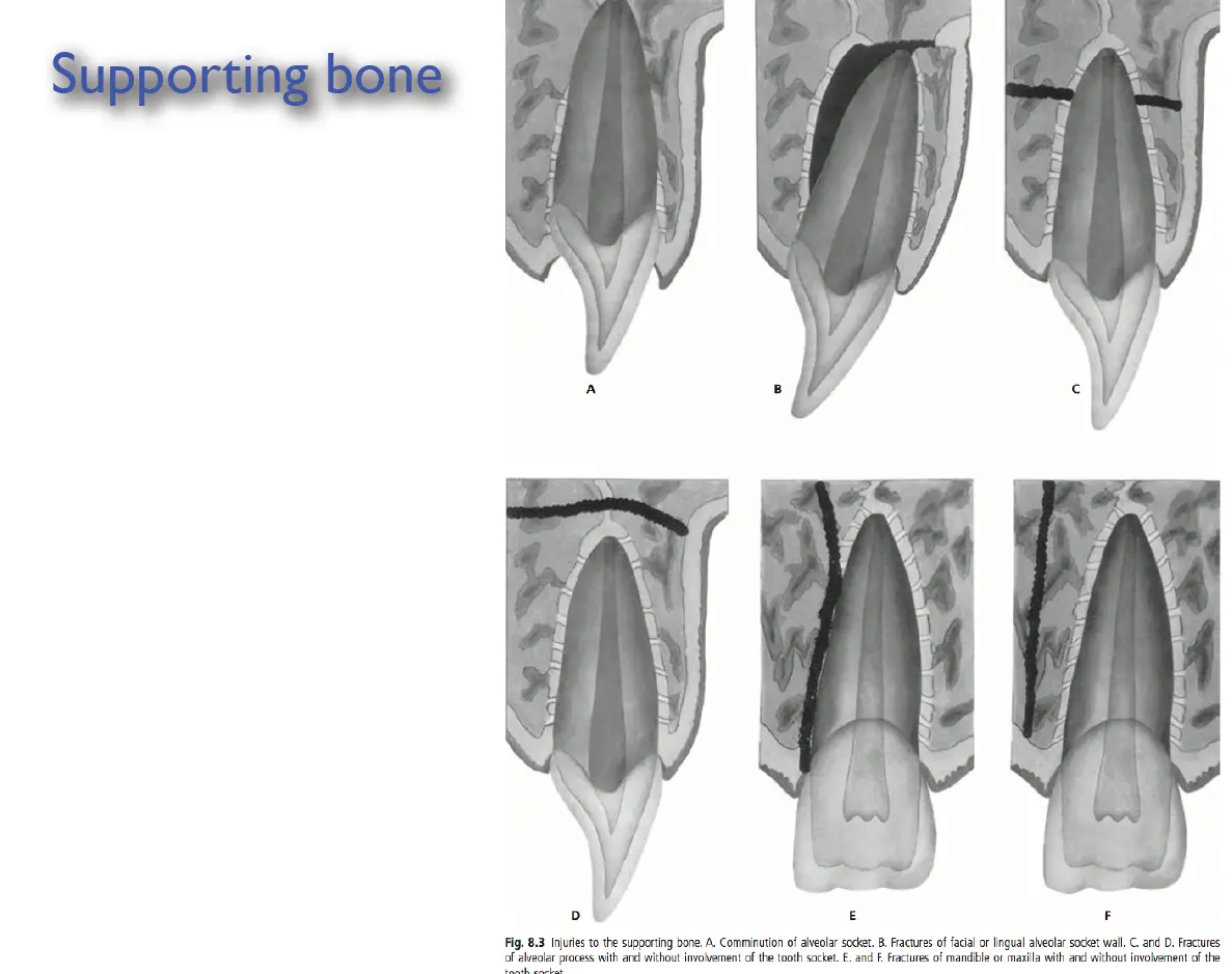
Injuries to supporting bone
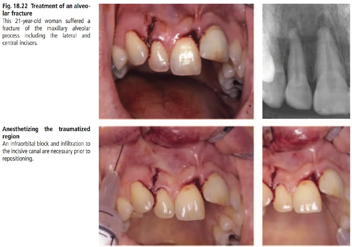
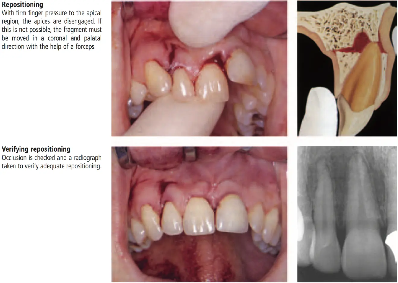
- comminution of alveolar socket
- fracture of socket wall
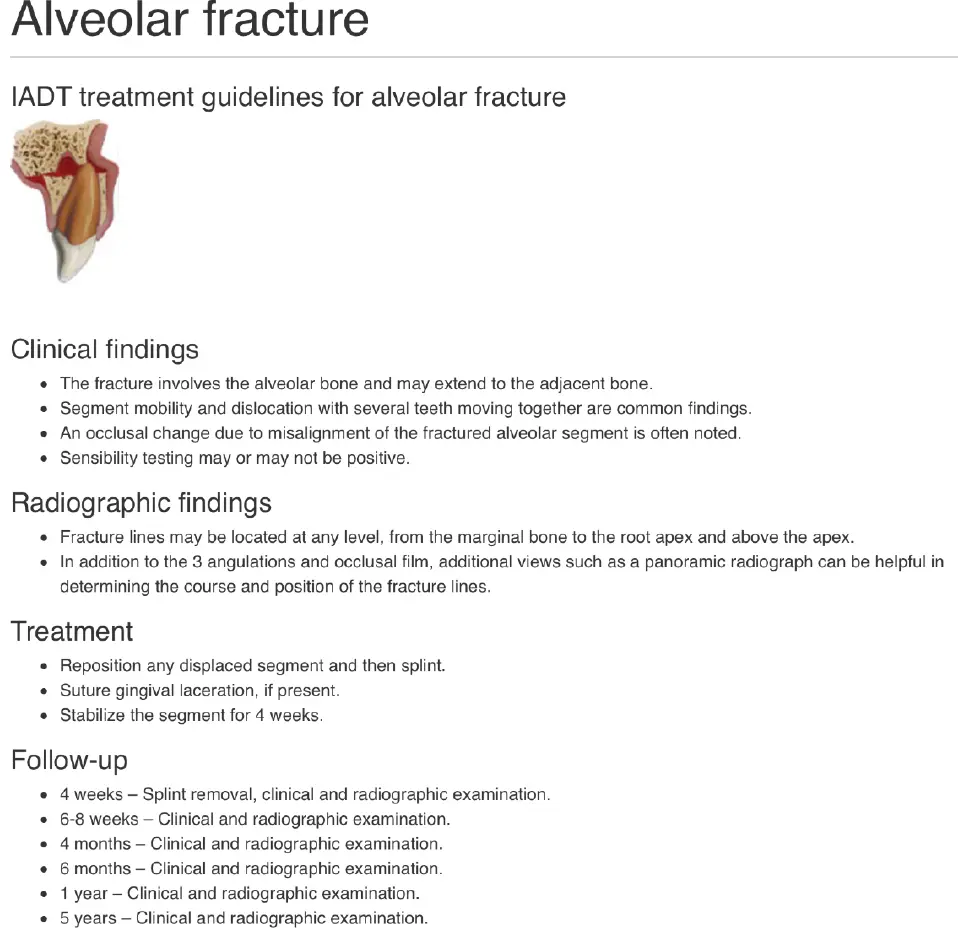
- fracture of alveolar process
- fracture of maxilla or mandible
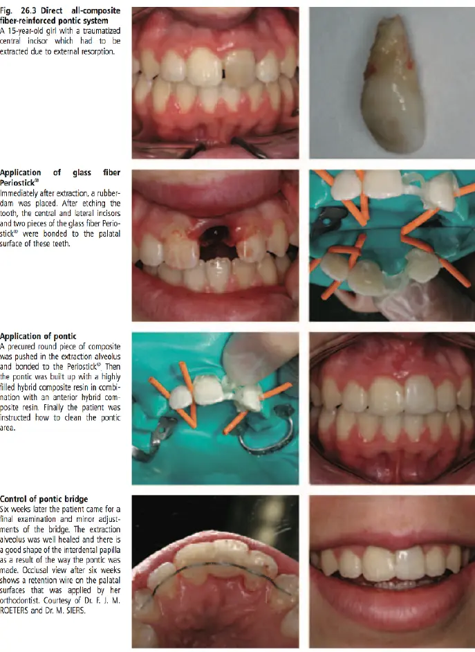
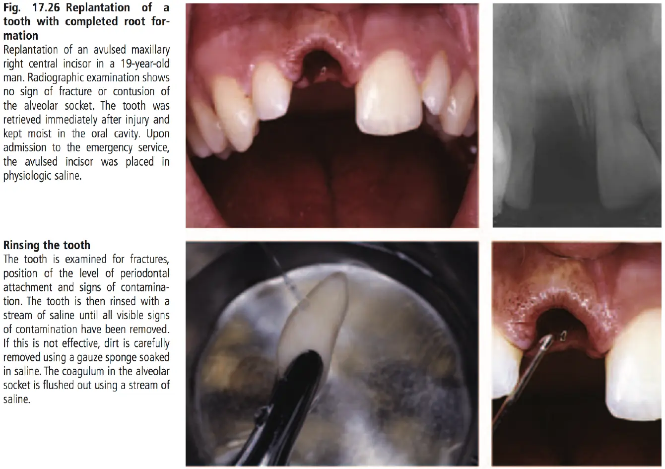
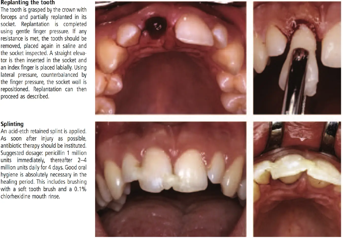
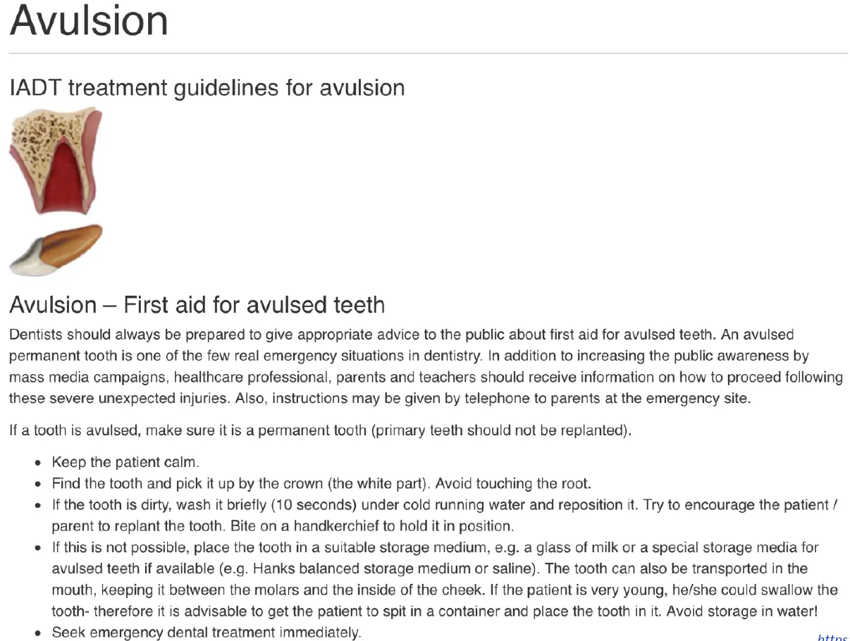
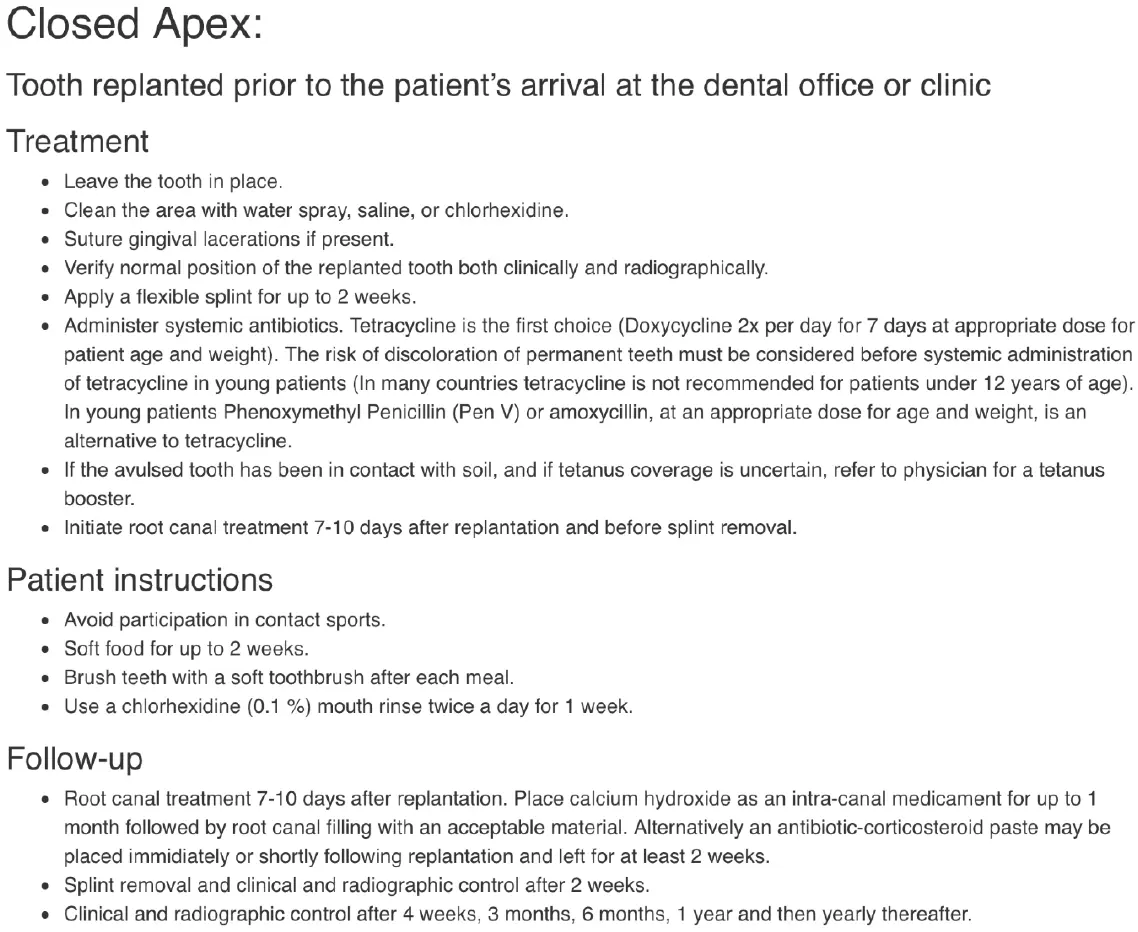
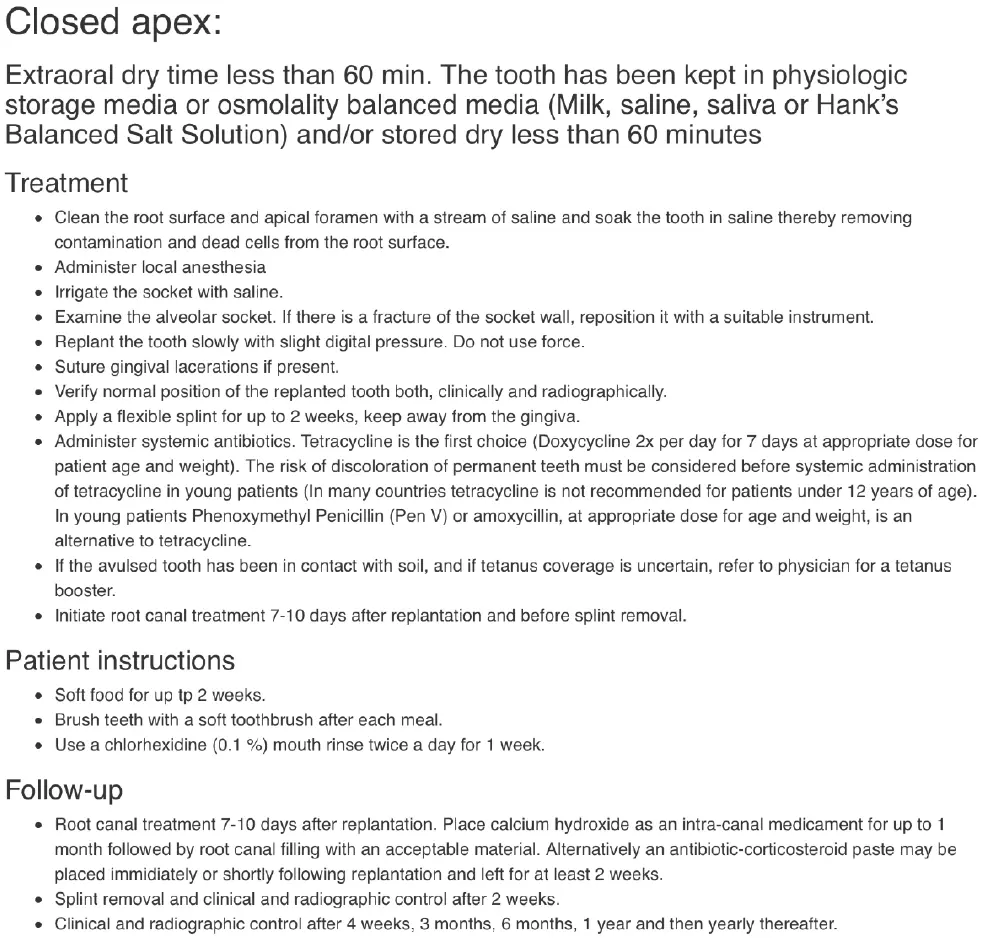
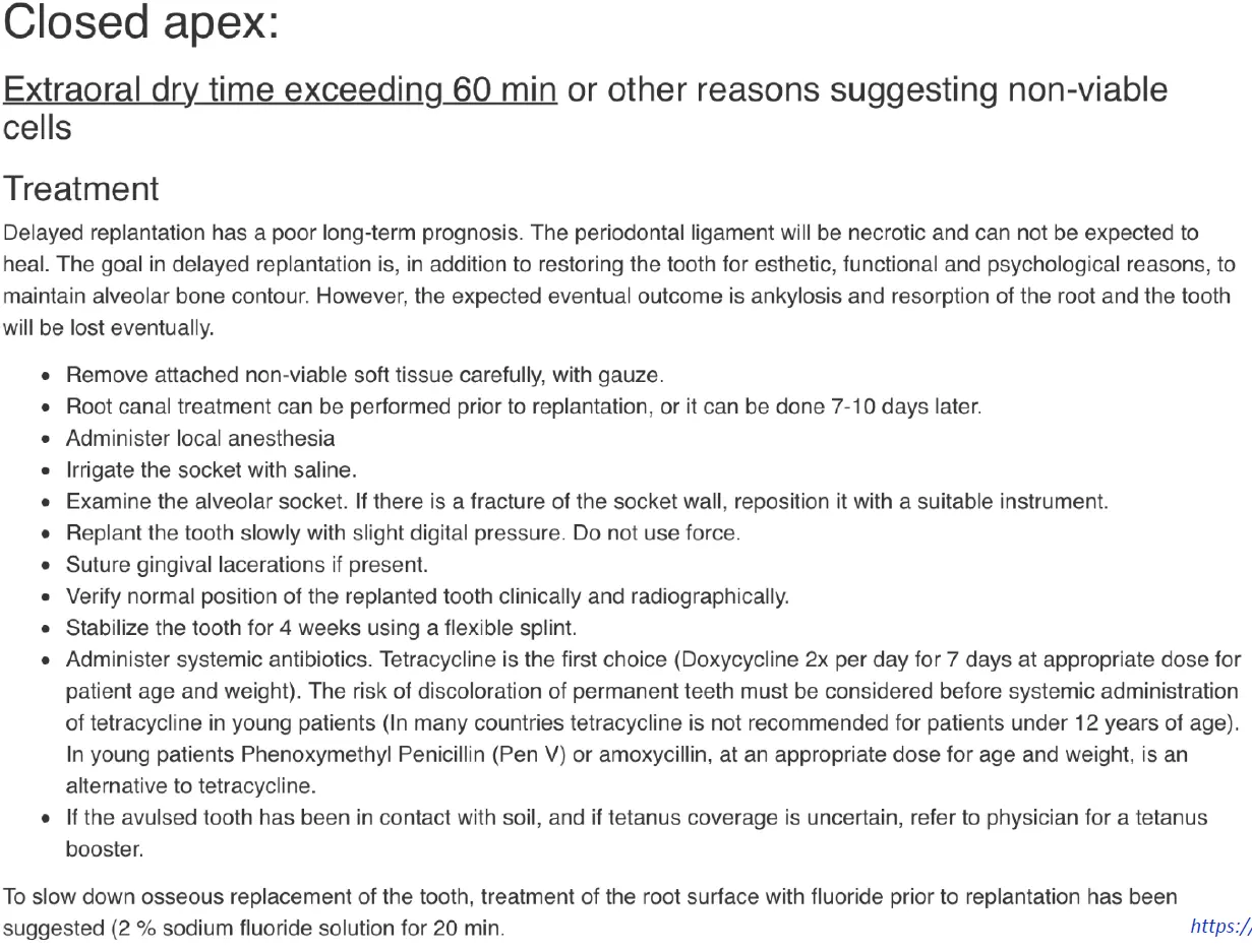
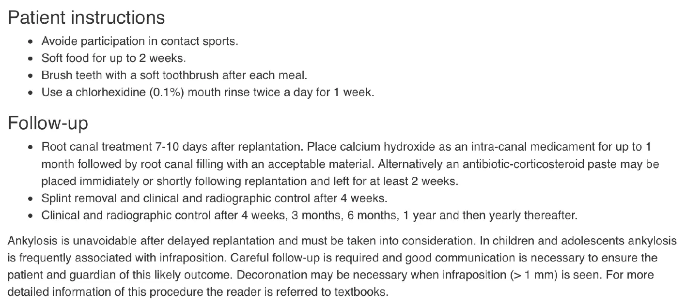
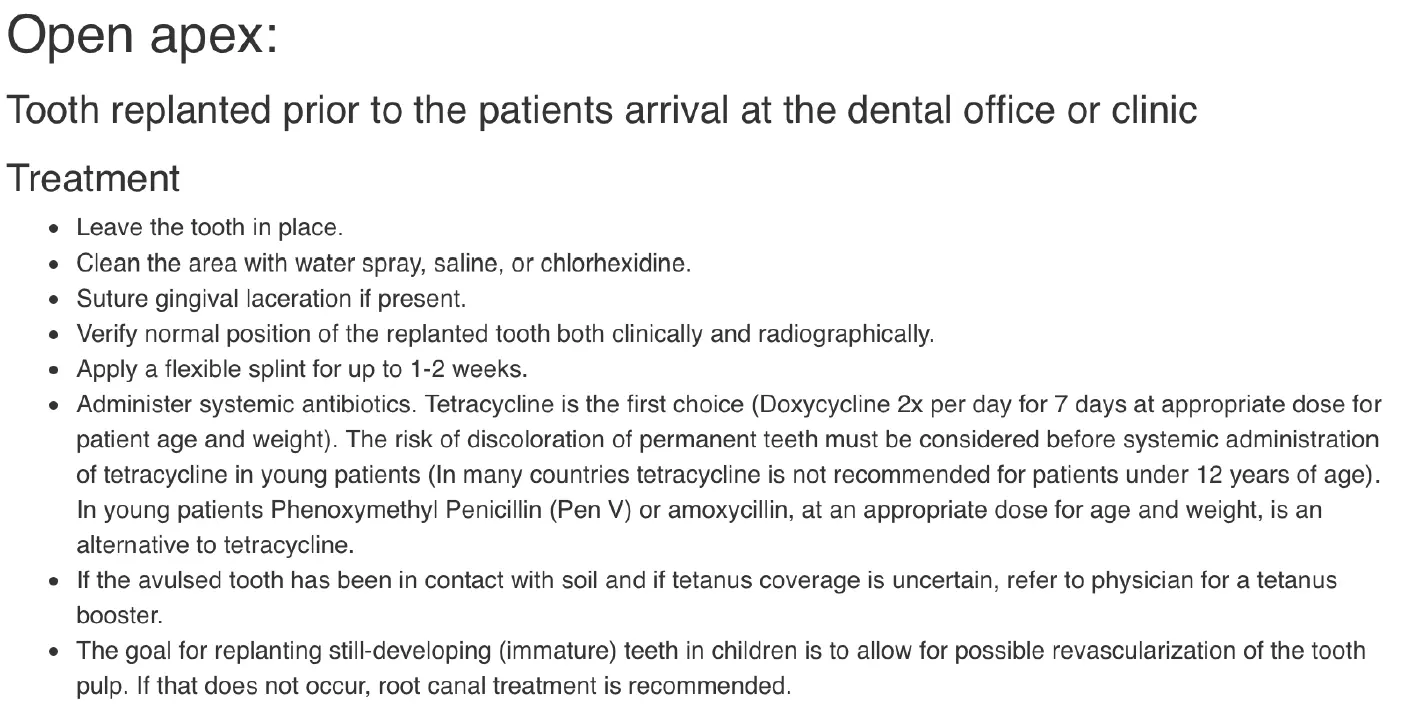
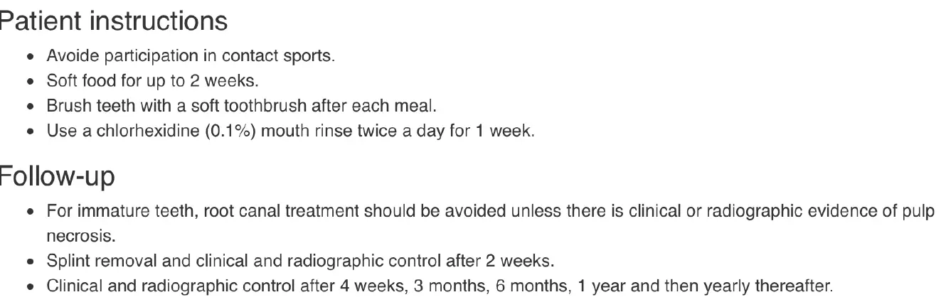
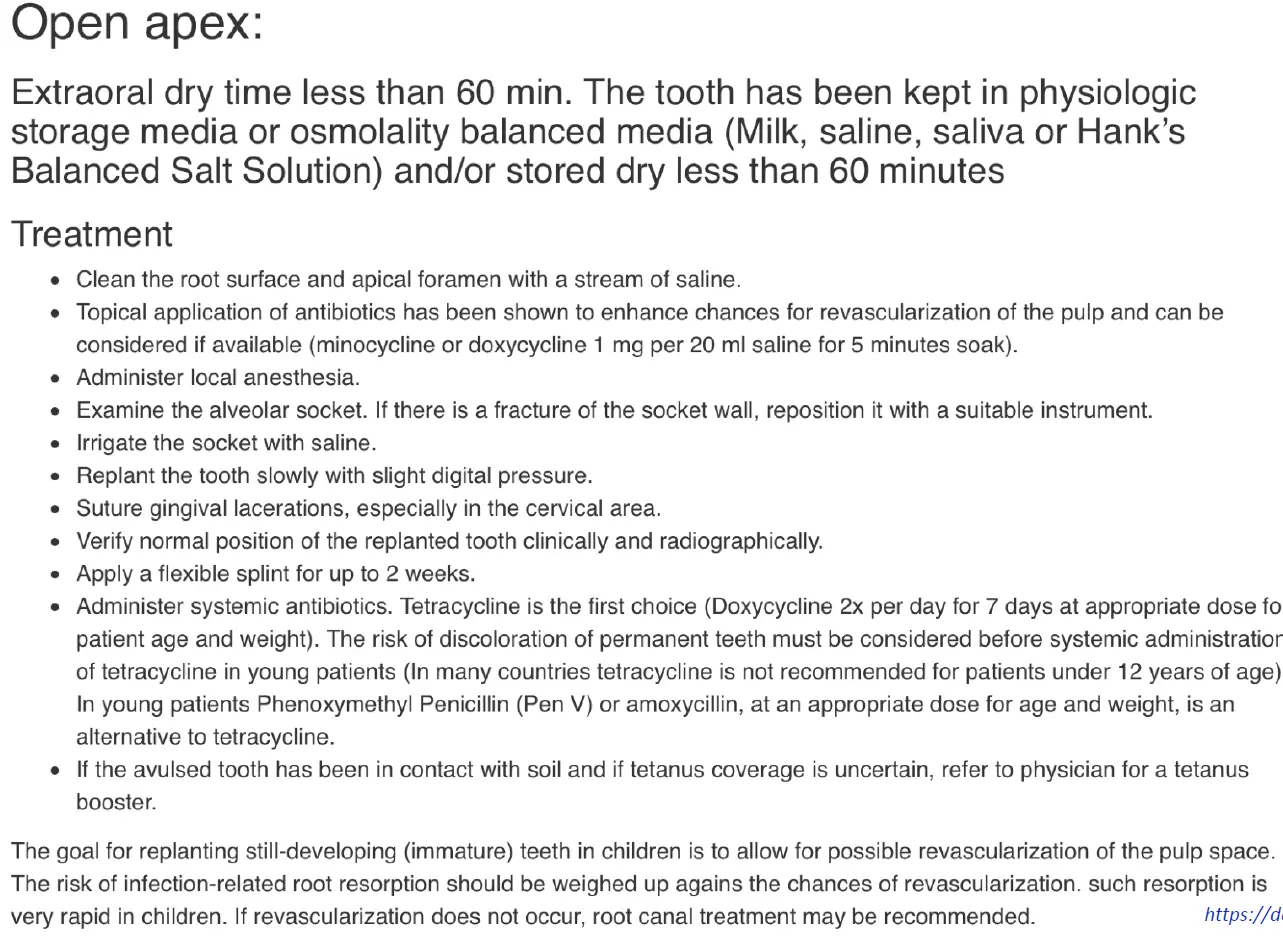
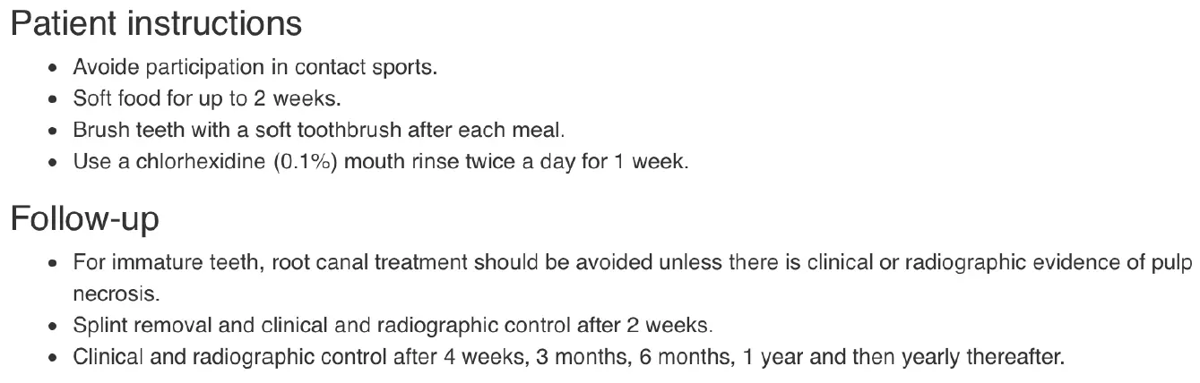
Avulsion
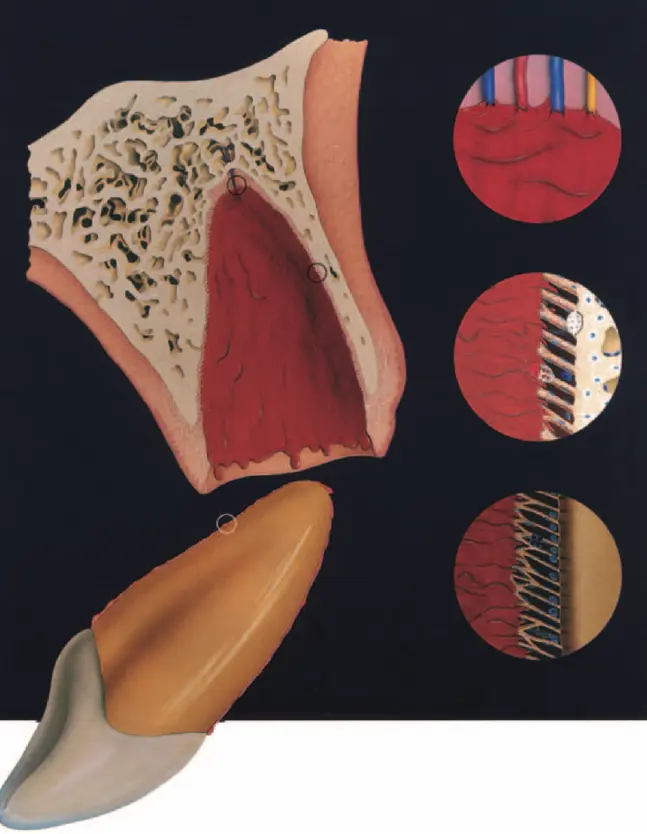
Periodontal healing reactions
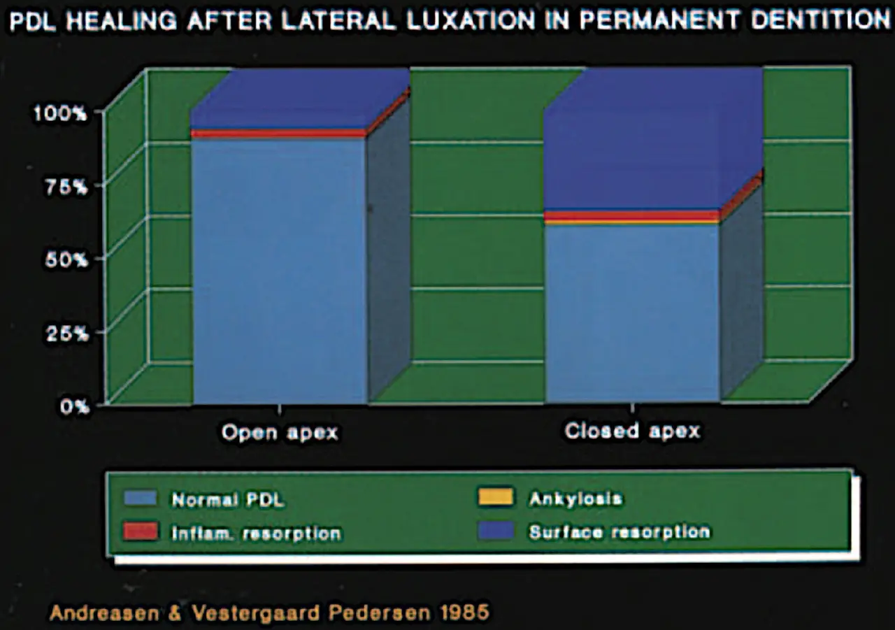
- healing with normal PDL
- healing wit hsurface resorption
- healing with replacement resorption (ankylosis)
- healing with inflammatory root resorption
PUlpal healing reactions
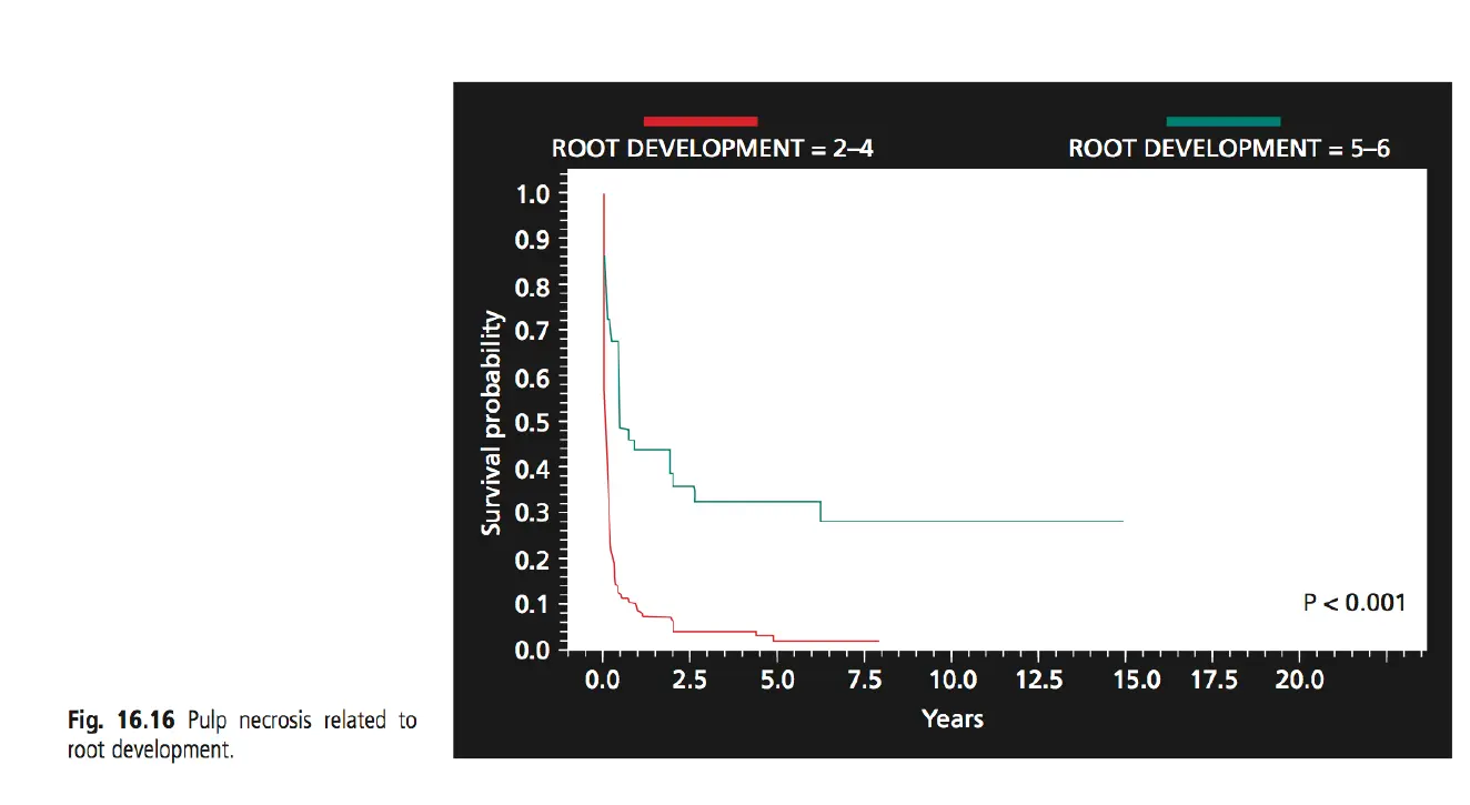
- revascularisaiton or necrosis
- necrosis only diagnosed after 2-4 weeks
- predictors for revascularisation
- width and length of RC
- mature apices (<1mm apical foramen) only if immediate replantation otherwise necrosis
- open apices - greater contact area for revascularisation
- storage period and storage medio
- dry storage / non physiologic storage
- physiologic media - saline / milk - weak relationship with time
- width and length of RC
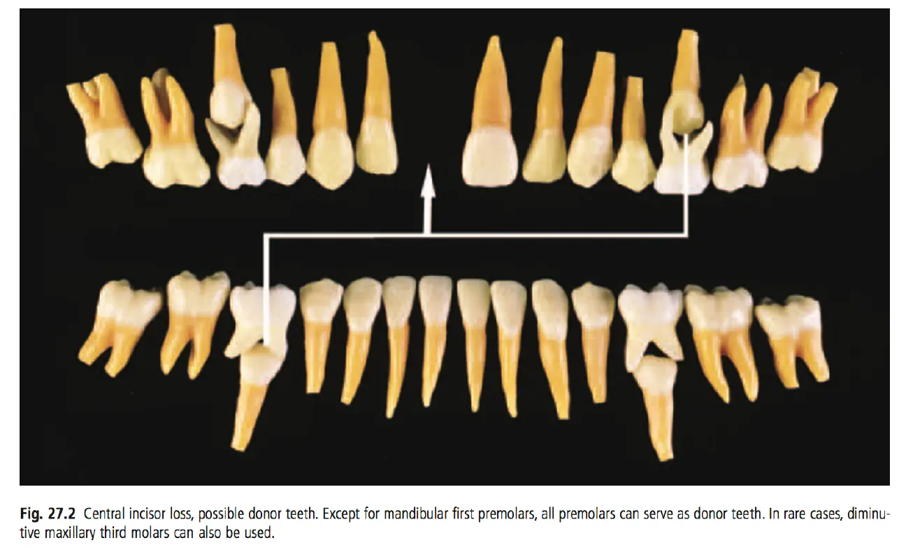
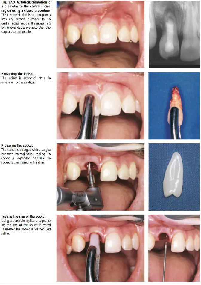
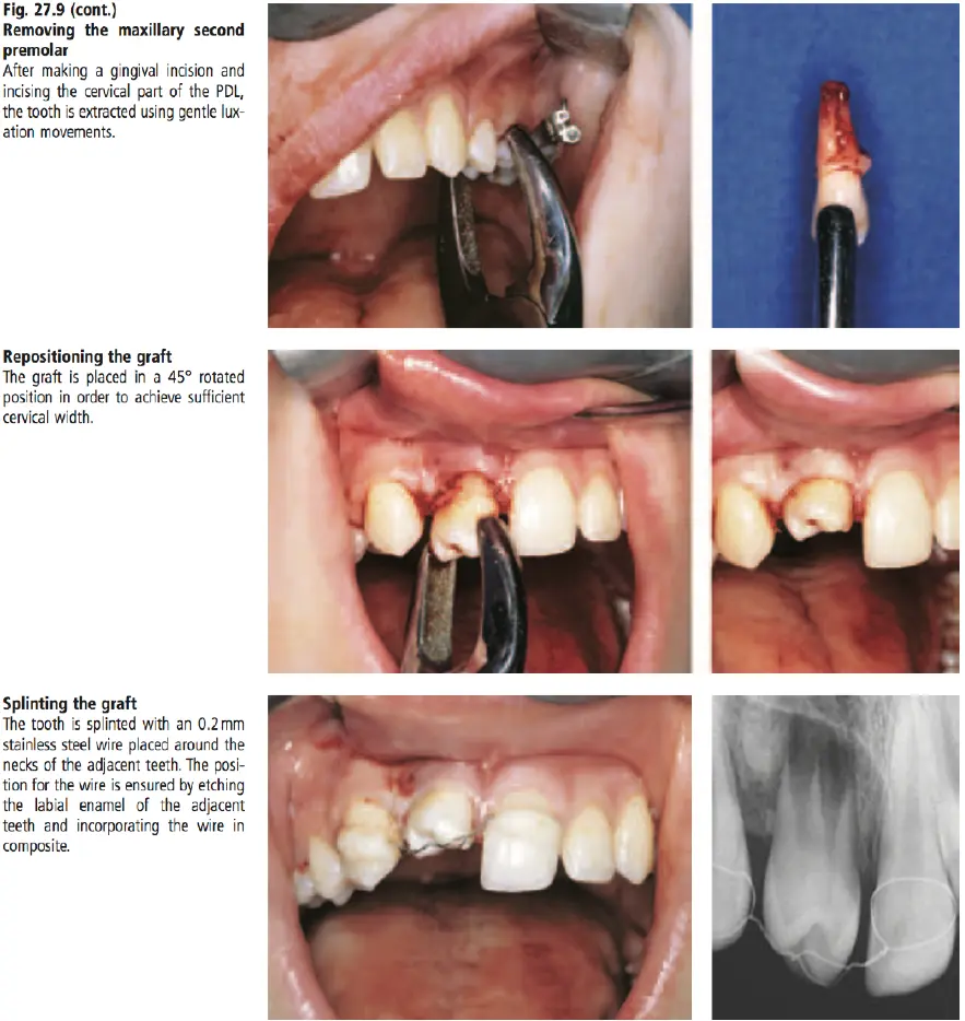
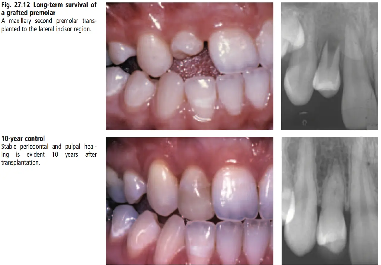
Autotransplantation