Module 8
#DEN4002
#D4/S2/W8
#OralSurgery
Facial Trauma
Facial trauma
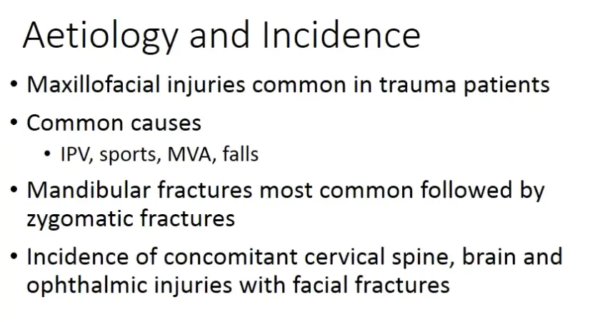
- epidemiology
- 5% paediatric, primary cause is falling
- primary dentition 11-30%
- permanent mixed dentition 5-20%
- M:F 2:1
- common causes
- children: contact sports, playground activities, child abuse
- adult: motor vehicle collisions, contact sports, assult/intentional violence, industrial accident
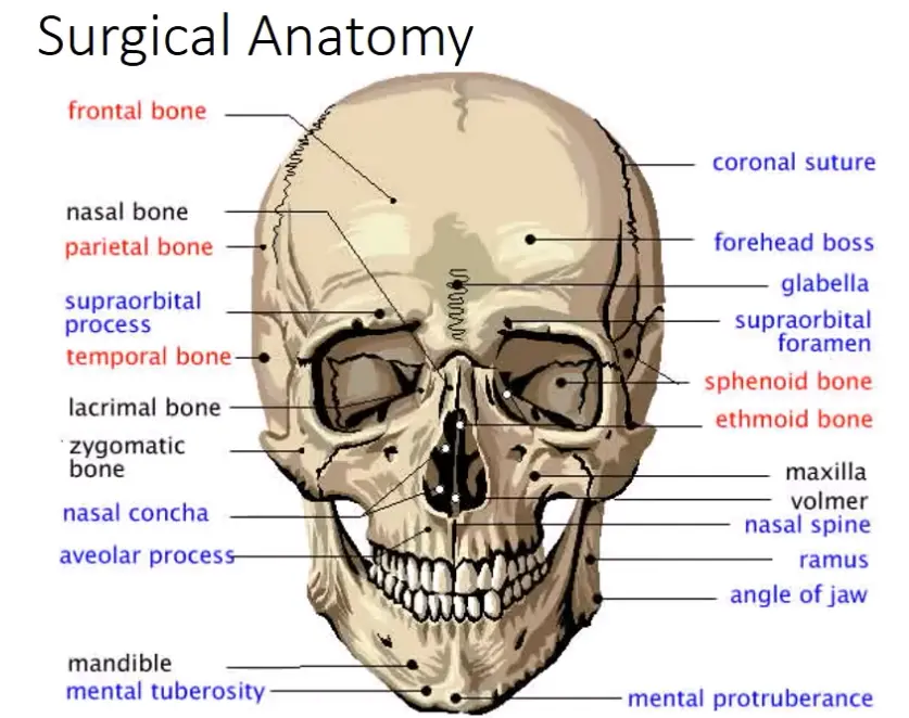
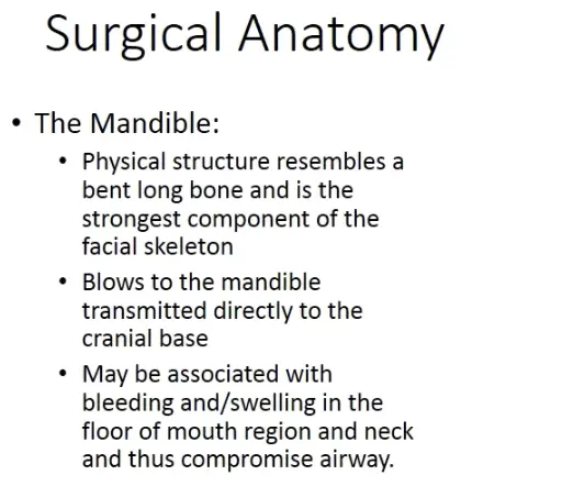
- skull is like a frame
- top area is frontal bone
- lateral struts are lateral orbital margins and ZMC
- lower platform is mandible
- when damaged, the frame is reconstructed first
Emergency Management
Emergency management
**Management **
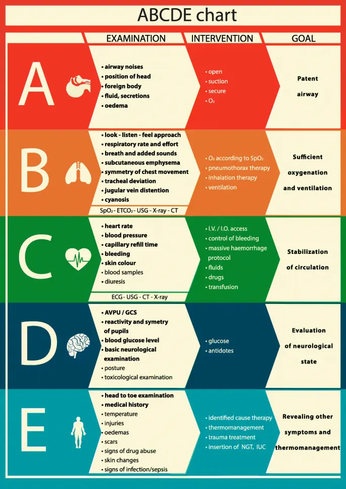
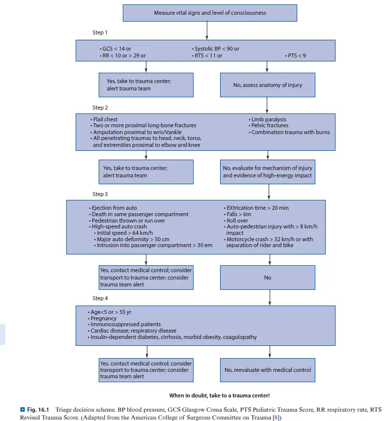
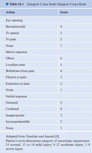
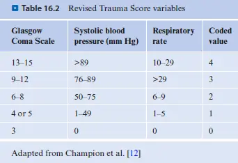
- ABCDE
- airway
- most urgent
- mandibular fracture?
- tongue trauma
- intubation/surgical airway
- cricothyroidotomy
- tracheostomy
- no nasotracheal intubation, tube can be displaced through middle cranial base into brain
- intubation/surgical airway
- airway control
- chin lift
- jaw thrust
- oropharyngeal suctioning
- manually move tongue forward
- maintain cervical immobilisation
- haemorrhage/circulation control
- rarely develop shock from facial bleeding alone
- direct pressure
- fracture reduction
- soft tissue closure
- nasal haemorrhage may require anterior and posterior packing
- anterior via adrenaline soaked gauze
- posterior via folley catheter
Physical Exam
Inpsection
- initial patient appearance
- foreign bodies
- swelling and ecchymosis
- facial elongation
- lefort fracture
- asymmetry
- deformities and cranial nerve injury (muscle palsy)
- inspect for malocclusion, bleeding, step off
Palpation
- tenderness
- step off (step in bone)
- facial stability
- crepitus
- subcutaneous air
- cutaneous anaesthesia, paraesthesia
- palpate the mandible for tenderness, swelling, step off
in order
periorbital
- check visual acuity
- how many fingers / eyechart
- check pupils for roundness and reactivity to light
- examine eye/eyelids for lacerations
- test extra ocular muscles
- full range of eye movement
- palpate around entire orbits
- change in sensation (supraorbital and supratrochlear)
- penetrating injuries
- occult globe penetration (retinal haemorrhage)
- eyelid laceration
- racoon eyes
- bilateral periorbital eccymosis
- likely skull base fracture concern
Nose
- septal cartilage, examining for septal haematoma
- surgical emergency to evacuate haematoma, can cause necrosis of septal cartilage and permanent nasal deformity
- cerebrospinal fluid leak (CSF rhinorrhea)
- clear fluid leak from nostril (skull base fracture)
- need CSF test to pathology
- referral to neurosurgeon
Ears
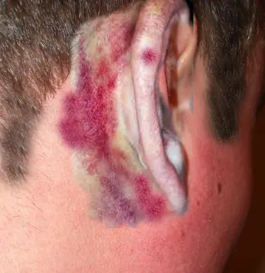
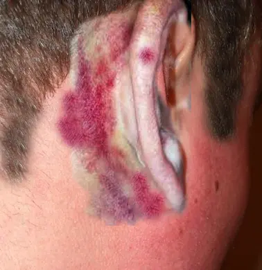
- battles sign
- ecchymosis behind ear in mastoid process
- likely basilar skull fracture
- can lead to permanent brain injury, meningitis, etc
Oral and mandibular exam
- under tongue/floor of mouth
- ecchymosis suggestive of fracture
- occlusion
- open bite/ AOB suggestive of mandibular fracture
- palpate alveolus
- tenderness with movement indicating fractured region
- break/breach in mucosa
- fracture into mucosa
- mandible deviation
- deviation to one side is contralateral condylar fracture
- paraesthesia
- if involving IAN, tongue lip teeth
- tongue blade test
- bite onto wooden spatula and pressure in biting should cause mandibular movement in fracture
Example cases
Mandible
Fracture pattern
- usually young male patient after blow to face
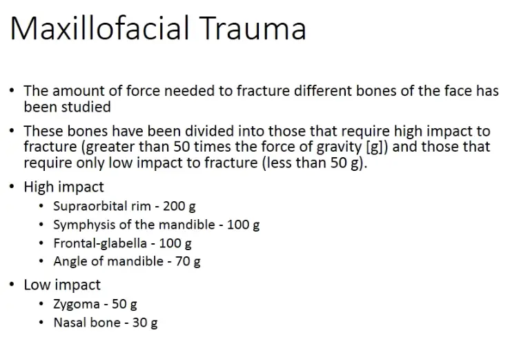
- angle of mandible 20-33%
- body of mandible 15-25%
- condlye of neck 15-36%
- parasymphaseal 14-15%
- ramus 5%
- coronoid process 1-3%
- alveolar ridge 2%
Clinical exam - can be associated with cervical spine fracture, extremely urgent
- can displace tongue
- step off in occlusion, likely mandibular fracture
Classification 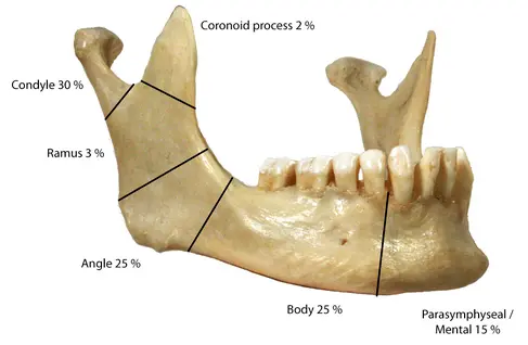
- displaced / non displaced
- open / closed (piercing skin vs not)
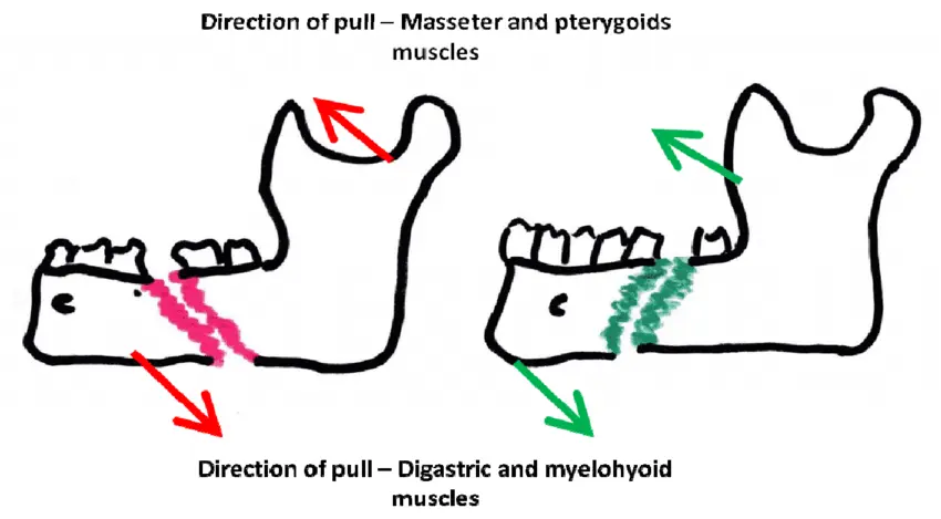
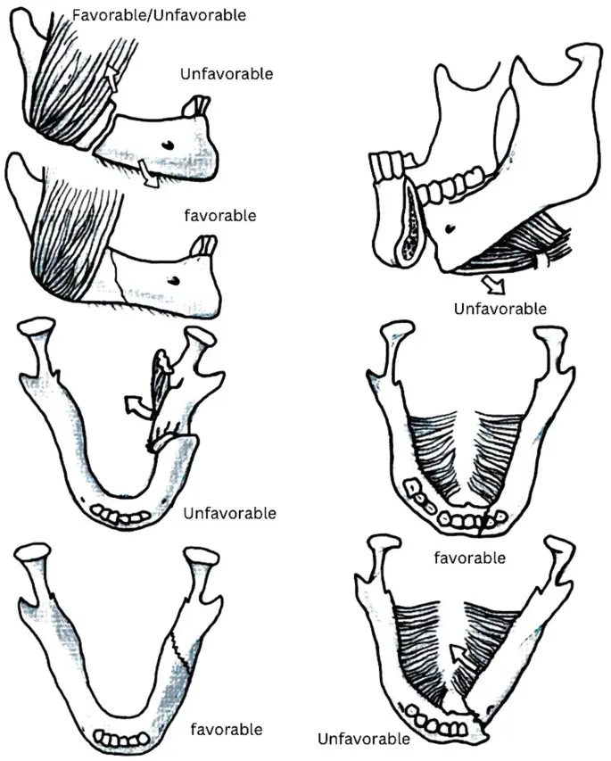
- favourable / unfavourable (based on muscle pull direction)
Radiographic interpretation- ,
Treatment options - .
Followup - .
Orbit
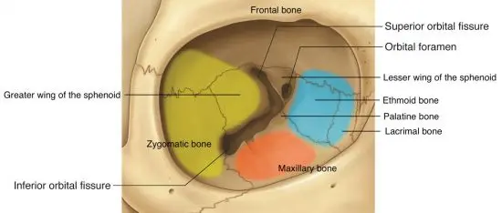
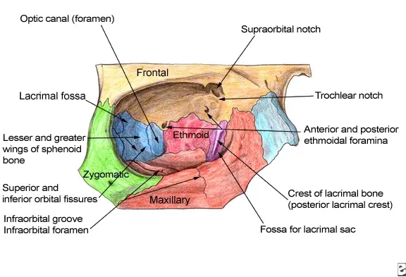
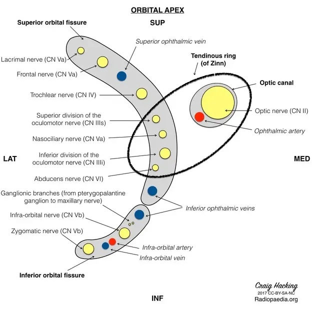
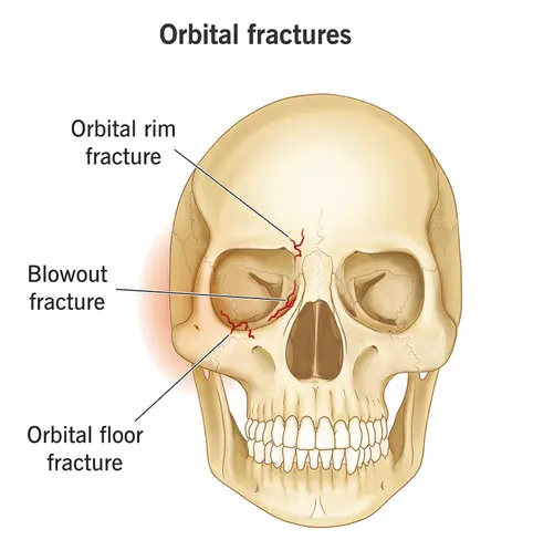
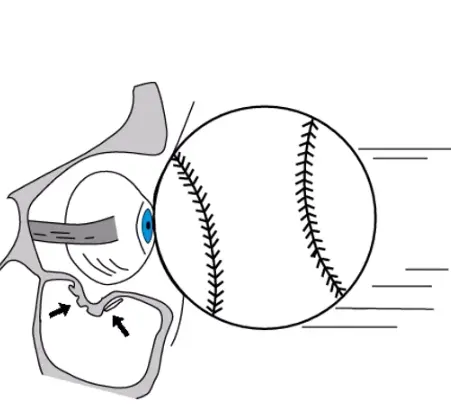
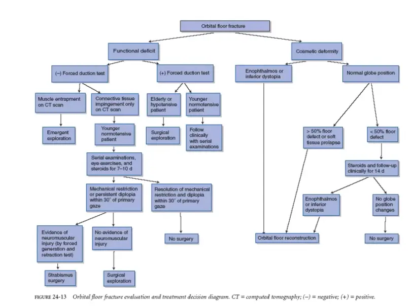
Fracture pattern
- when projectile or fits hits the eye
- orbital rim fracture
- bony outer edges of eye socket
- typically car accident, takes a lot of force to break
- optic nerve function to be checked
- blowout fracture
- break in floor or inner wall of orbit/socket
- can trap/pinch muscles/nerves around the eye
- ball or fist
- orbital floor
- trauma/blow causes orbital rim to push bones back, and bones of socket floor buckle downward
Clinical exam
- trauma/blow causes orbital rim to push bones back, and bones of socket floor buckle downward
- urgent if (immediate surgical intervention)
- associated with muscle entrapment (inferior rectus)
- globe injury
- retrobulbal haemorrhage
- usually through floor or medial wall
- hydrolytic theory where great sudden expansion forces are created from direct blunt trauma to orbit, which cause buckling of adjacent walls (thinnest parts are floor/medial)
- presents with
- enophthalmos (sunken eye)
- anaesthesia infraorbitally (infraorbital nerve)
- diplopia (uncoordinated eye muscles)
- infraorbital stepoff deformity
- subcutaneous
- check for
- muscle motility (all rectus muscles)
- visual acuity (rule out damage to optic nerve)
- check infraorbital nerve supply
- gross inspection of area
Radiographic interpretation
- ,
Treatment options - surgery
- consult ophthalm and macsfacs
- prophylactic antibiotics (likely contamination of maxillary sinus bacteria), nasal congestants, avoid valsalva and nose blowing
Followup - .
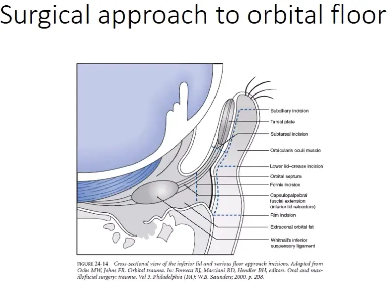
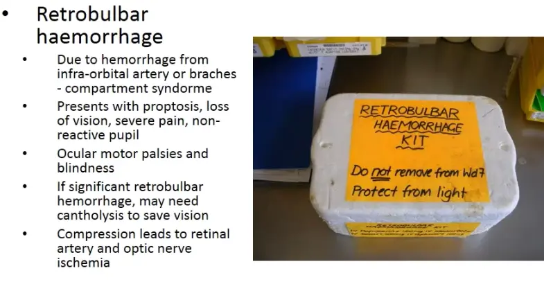
Riskf of orbital floor surgery
- retrobulbar haemorrhage
Zygomaticomaxillary complex
Fracture pattern
- tripod fracture
- zygo arch, infraorbital rim and lateral orbital wall
Clinical exam
- zygo arch, infraorbital rim and lateral orbital wall
- .
Radiographic interpretation - ,
Treatment options - .
Followup - .
Maxilla
Fracture pattern
- leforte 1
- floor of nose, hizontal fracture separating maxilla from pterygoid plates
- leforte 2
- pyramidal, involving nasal bridge, maxilla, orbital floor
- blow to lower maxilla/midmaxilla
- extend from nasal bridge/nasofrontal suture
- through frontal processes of maxilla
- inferolaterally through lacrimal bones and inferior orbital floor and rim (near inferior orbital foramen),
- inferiorly through anterior wall of maxillary sinus
- under zygoma and across pterygomaxillary fissure through pterygoid plates
- leforte 3
- transverse, entire midface separated from skull base
- start at nasofrontal and frontomaxillary sutures
- extend posteriorly along medial wall of orbit through nasolacrimal groove and ethmoid bones
- continue along floor of orbit along inferior orbital fissure, continuing superolaterally through lateral orbital wall
- through zygomaticofrontal junction and zygomatic arch
- intranasally extending through base of perpendicular plate of ethmoid, through vomer, through interface of pterygoid plates to base of sphenoid
Clinical exam
- can produce massive haemorrhage
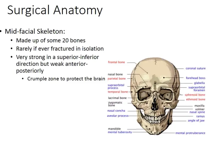
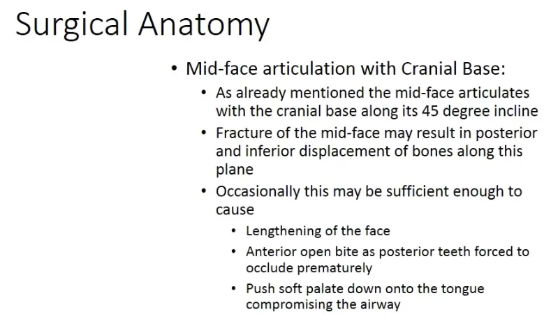
- can displace soft palate and cause airway compromise
- facial elongation and AOB
- midface crush/lefort
- urgent if
- airway compromise
Radiographic interpretation
- ,
Treatment options - .
Followup - .
Frontal
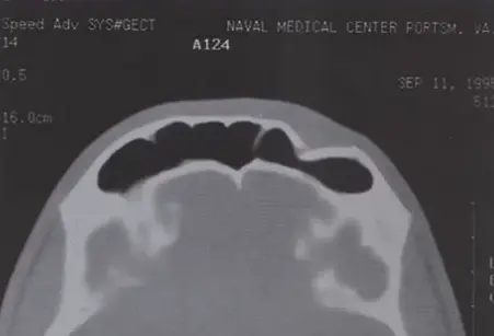
- outer table frontal sinus fracture penetrating into frontal sinus
- triangular fracture shape with intact inner table
- frontal sinus inner table communicates with cranial space
- frontal sinus drains through frontonasal duct into ethmoid sinus
Fracture pattern
- usually blunt trauma to front of face from motor vehicle accident or assault
- treatment depending on severity and displacement
- small fracture may be managed with minimally invasive incision
- large fracture hair line incision will full forehead retraction
- very thick bone with great force required to fracture
- classification
- isolated anterior table/wall
- combined anterior and posterior table/wall
- with/without CSF leak
Clinical exam
- consequence of fracture
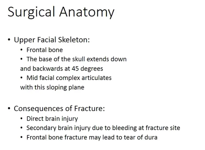
- direct brain injury
- secondary brain injury due to bleeding at fracture site
- frontal bone fracture may lead to tear of dura
- forehead depression
- inner table fracture with dura tear
- intracranial injury
- CSF leak
- risk of meningitis
Radiographic interpretation
- ,
Treatment options - neurosurgeon referral if concern of intracranial injury
- where frontal sinus remains functional (sinus lining intact and drainage patent), no sinus treatment required
- sinus affected
- surgery indicated
Follow up
- surgery indicated
- .
Nasoethmoid complex
Fracture pattern
- high impact force anterior to nose and transmitted posteriorly through ethmoid
- very complex area with many small bones, very difficult to treat and diagnose
Clinical exam - flattened nasal bridge or saddle shaped deformity of the nose
- widening of nasal bridge (telecanthus)
- damage to inner canthal tendon of lacrimal bone
- concerns of CSF rhinorrhea or epistaxis
- from fracture to cranial base
- tenderness, crepitus and mobility of nasal complex
- intranasal palpation reveals movement of medial canthus
Radiographic interpretation - ,
Treatment options - .
Followup - .