Vital Pulp Therapy
Paradigm shift with contemporary treatment
- why is it necessary to retain or recreate a vital pulp
- diagnostic considerations and what is pulpal disease
- current concepts on progression of pulp disease
- new concepts of progression and arrest of caries
- how do we rationalise different treatment modalities to arrive at a treatment plan
- how can we treat teeth with wide open apices
Why is it necessary?
Dilemma of deep caries
- felt that traditional non-selective management may be destructive of tooth substance, there is a push to find more sonservative methods of treatment
- is it possible to avoid classical RCT and exo?
- find balance of being too conservative allow disease to progress vs invasive intervention requiring extensive treatment
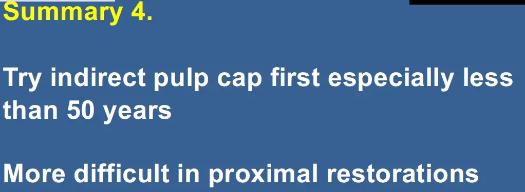
Indirect pulp cap
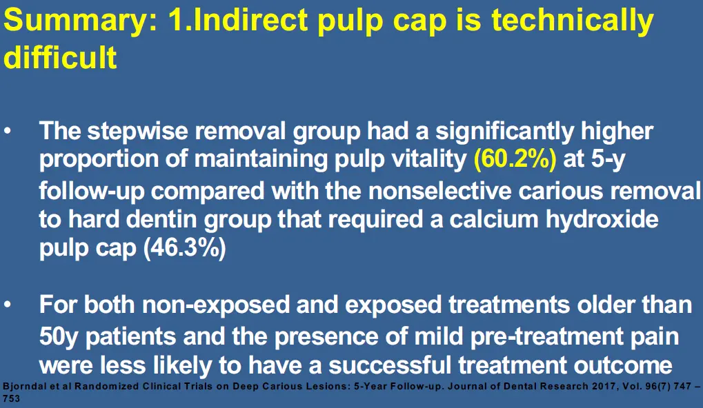
- technically difficult
What is vital pulp therapy
- maintenance or recreation of integrity of pulp - dentine - PDL complex
- purpose is to maintain or create functional and vascularised pulp to enhance dentine maturation and dentine development following damage to dentine and or pulp through pulps inflammatory response or creation of a new pulp
- use molecular and cellular mechanisms of the dentine and pulp to aid in the repair and promote development of the pulp
- Apexogenesis
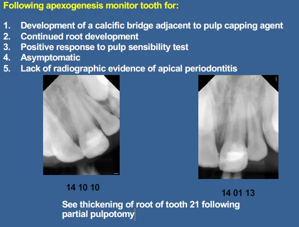
- if pulp is vital we want to allow contiuned maintenance of vascular status of pulp to ensure the continued maturation of underdeveloped root
- Revitalisation / Regenerative endodontic procedure
- if pulp is devitalised and necrotic we use regenerative endodontics to provide bvioligic based procedure to arrest necrotic disease pulp process, prevent its recurrence while favouring repair or replacement of damaged structures in pulp dentine complex
- Apexification
- under developed root treated by creating biological apical barrier in devitalised/necrotic pulp (historical?)
WHy do vital pulpectomoy with vital caries pulp exposure in past?
- Traditionally rational for pulpectomy is if caries was close enough to the pulp to allow apulpal exposure, most likely micro-organisms in carious dentine also extend into pulp
- creating a pulpal infection and inflammation that has probably reached a level where itselimination is not possible without removal of the entire pulp.
Why are we now saying preferable to retain vital pulp?
- teeth with RCT have lower survival than teeth with vital pulp
- teeth with vital pulps undergo continuing changes in mineralisatoin of the dentine, changes allow dentien to adapt to forces and age changes that are placed on the tooth, evident in immature and mature teeth
Different terms for immature root in vital pulp therapy
- apexogenesis
- vital pulp procedure performed to encourage continued physiological development and formation of the root end in a tooth with a still vital and functioning pulp
- apexification
- method to induce an apical barrier in a root with an open apex or continued apical development of an incomplete root in teeth with necrotic pulp
Diagnostic considerations
Diagnosis
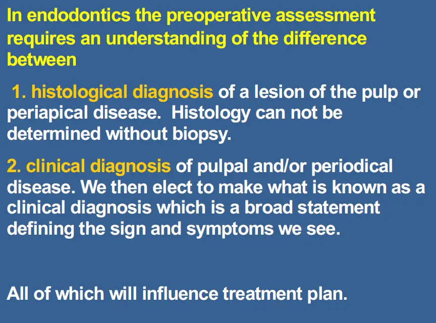
- not a fact but a status (opinion) based on certain relative values of
- patient perceived pain
- pulp sinsibility
- pulp sensitivity
- radiographic interpretation
- other special test
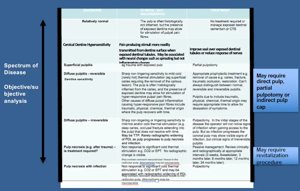
- diagnosis is part of a spectrum
- when patients present with symptoms of pulpitis, consistency of type of inflammation found in pulp
- but, inflammation may be confined to certain areas of the pulp and not uniform throughout
- clinical irreversible pulpitis usualyl assocaited with pulpal infection, but may be localised
- zimmet preference
- mainly treat aymptomatic cases with VPT as we are not yet able to distinguish cases that have vital but infected pulps
- diagnosis still very rudimentary
Current concepts on progression of pulp disease
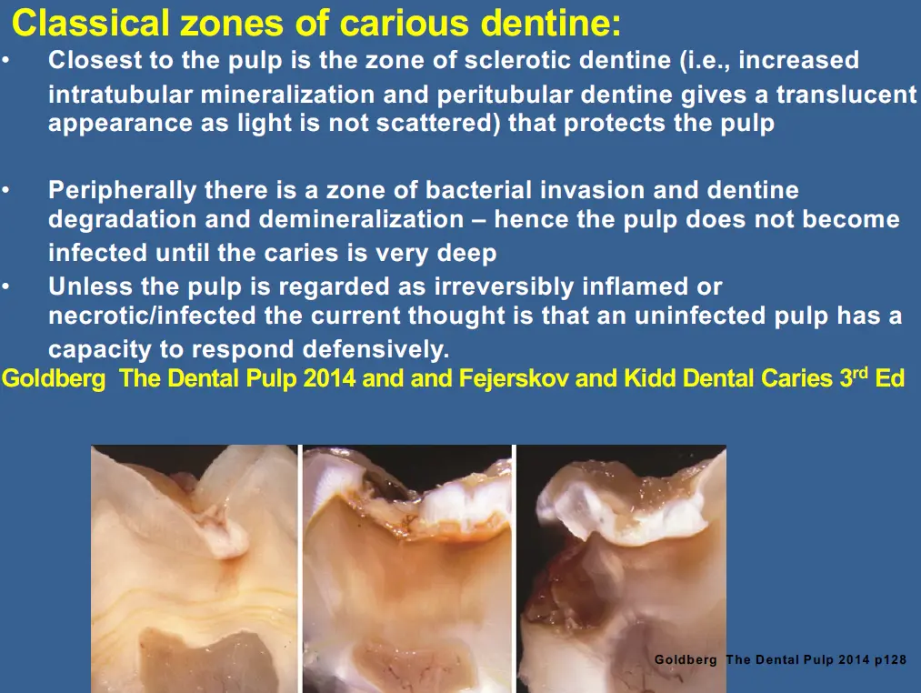
**How does dentine respond to caries,
- **what are odontoblasts
- peripheral area of pulp where odontoblasts reside - odontogenic zone
- cells are arrange in palisading patterns cells on tall, columnar forming layer of 3-5 cells in depth
- shape may vary
- coronal pulp columnar
- midportional cuboidal
- apical region flattened
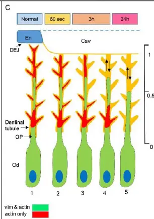
- Odontoblastic process
- how far processes extend into dentine is controversial
- degree of extension of OP is contentious, number of groups suggesting full extension to DEJ beneath intact dentine surface
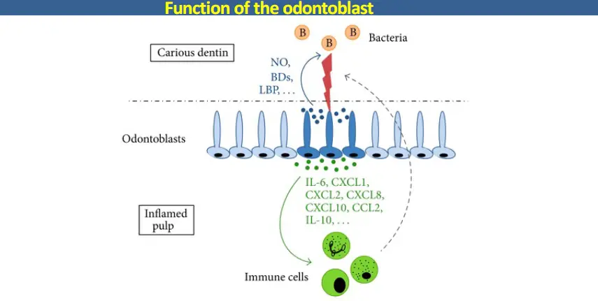
- Functional of odontoblast
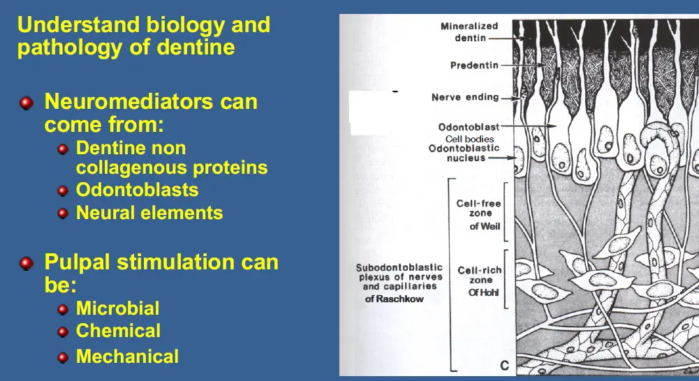
- initially due to complex interaction between release of bacterial enzymes, host enzymes with pattern of recognition that stimulate odontoblasts to initiate a neurogenic inflammatory response by release of neurotransmitters
- creates sensitisation of nerves creating allodynia
- odontoblast process directly contributes to dentine mineralisation, occurs along entire odontoblast process increasing peritubular and intertubular mineralisation
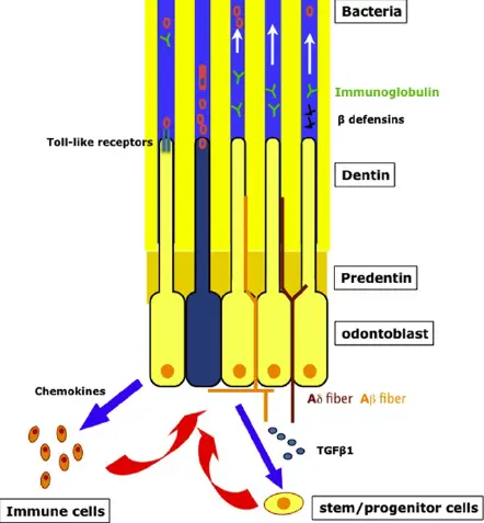
- odontoblast defence
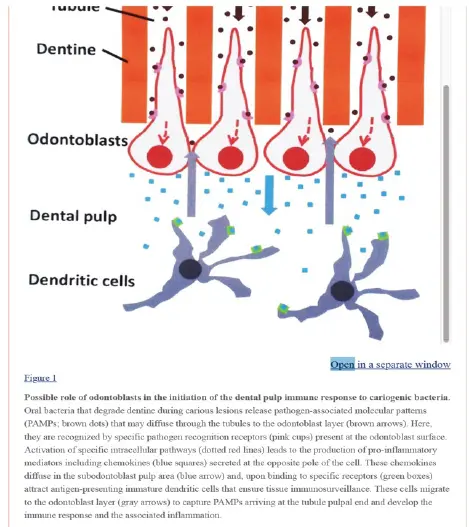
- bacteria contain Pathogen Associated molecular patterns (PAMPS) that stimular Pattern recognition receptors (PRR) that activate odontoblast adjacent to lesion
- production of antibacterial molecules which diffuse through dentine tubule in attempt to destroy invading microorganism
- proinflammatory and immunomodulatory mediators secreted into pulp area to mobilise immune cells towards pulp dentine interface to combat bacteria and coordinate immune defence response
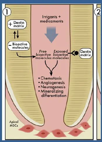
- dentine also release signal bioactive molecules (non collagenous matrix proteins) that can act as bio mediators that are associated with defence and repair
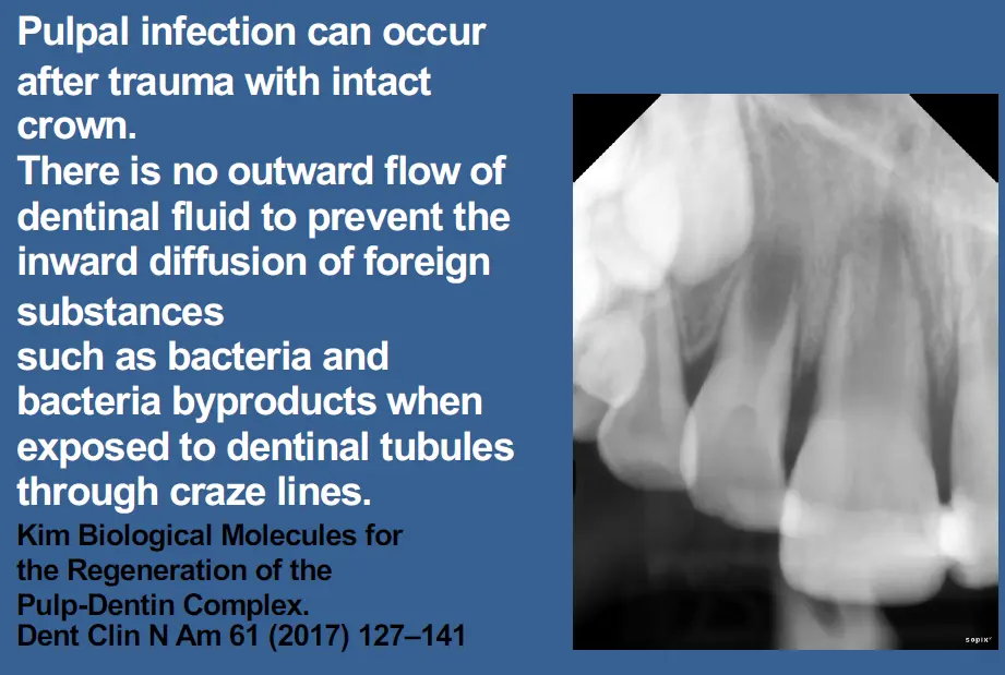
Creation of tertiary dentine
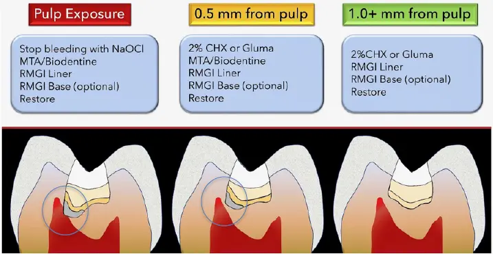
- indirect pulp cap relies on release of biomediators from odontoblasts or non collagenous proteins from demineralised dentine as loss of odontoblasts = reparative dentine
- direct pulp cap relies on pulp capping agent to provide biomediators as loss of odontoblasts = reparative dentine
- further awar from pulp odontoblasts remain intact = reactionary dentine
New concepts of progression and arrest of caries
3 Eras of carious diagnosis and management
- Black: remove caries
- Fusyama: Remove affectd caries
- modern:
- caries is a biofilm bases disease due to imbalance between mineralisation and demineralisation, infected dentine can remineralise
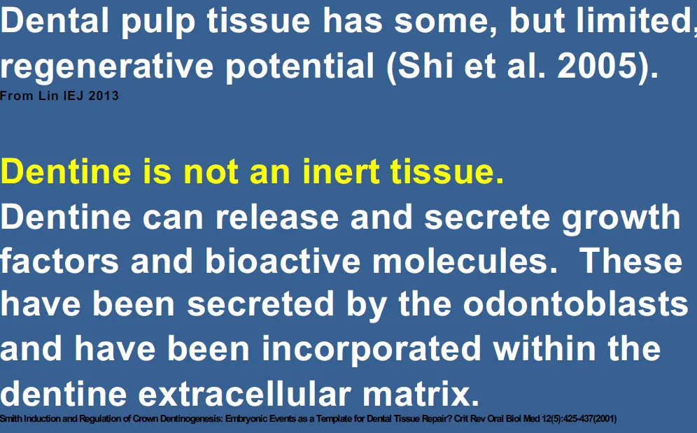
- caries is a biofilm bases disease due to imbalance between mineralisation and demineralisation, infected dentine can remineralise
Newer concepts on defensive/reparative potential of the pulp
- pulp has remarkable reparative abilities
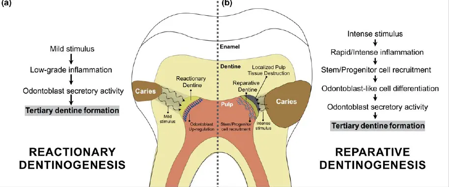
- responds to irritation by producing tertiary (reparative and reactionary dentine) and mineralising any affected dentinal tubules
- dentine contains biomediators that were secreted by odontoblasts during dentine formation to induce tertiary dentine
- mild to moderate irritation results in continued peritubular dentine formation and intertubular calcification
- tertiary dentine types
- reactionary
- reparative
- formation depends on speed of caries progression and depth of penetration
- reactionary dentinogenesis
- occur following moderate dentine injury if primary odontoblasts are present to secrete dentine, occurs in shallow and slowly progressing carious lesion
- reparatie dentinogenesis
- occurs if odontoblasts have been damaged or destroyed and if formation of tertiary dentine is performed after stem/progenitor cell recruitment by odontoblast like cells. occurs in rapidly progressing very deep carious lesion
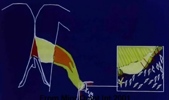
The dentine bridge
- causes death of pre-existing odontoblasts leads to reparative dentinogenesis involving the recruitment and proliferation of progenitor and or stem cells to site of injury and their differentiation into a second generation of odontoblasts of odontoblast like cells
New paradigm, when biofilm over caries is removed and restoration placed over residual
- level of bacterial growth in carious lesion in dentine is reduced when overlying biofilm is removed and lesion sealed
- complexity of bacterial microflora was reduced
- reduction of microorganism correlated with clinically detecteable change of retained dentine
- if pulp is irreversibly inflamed there is no choice but to perform RCT
- removal of overlying biofilm may temporarily stop sequence of events of caries progression leading to further unwanted inflammatory response in pulp as long as it is not yet irreversibly inflamed
- if there are any remaining bacteria in dentine after sueprficial caries removal with biofilm removal followed by placement of restoration with a good seal, caries progression should arrest by isolating bacteria from substrate and decrease production of acid
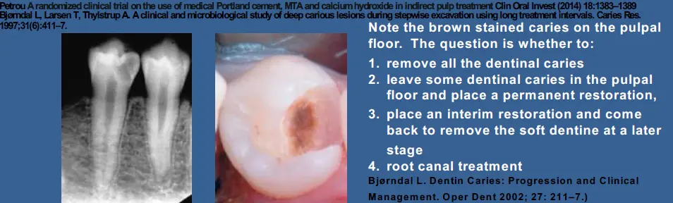
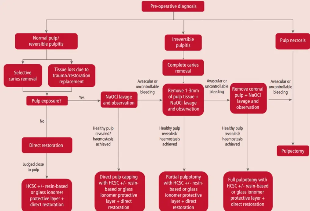
Management of caries in inner third of crown, where vital pulp therapy based on level of pulp preservation where caries close to pulp
- ART vs convensional restorative treatment
- Silver modified ART
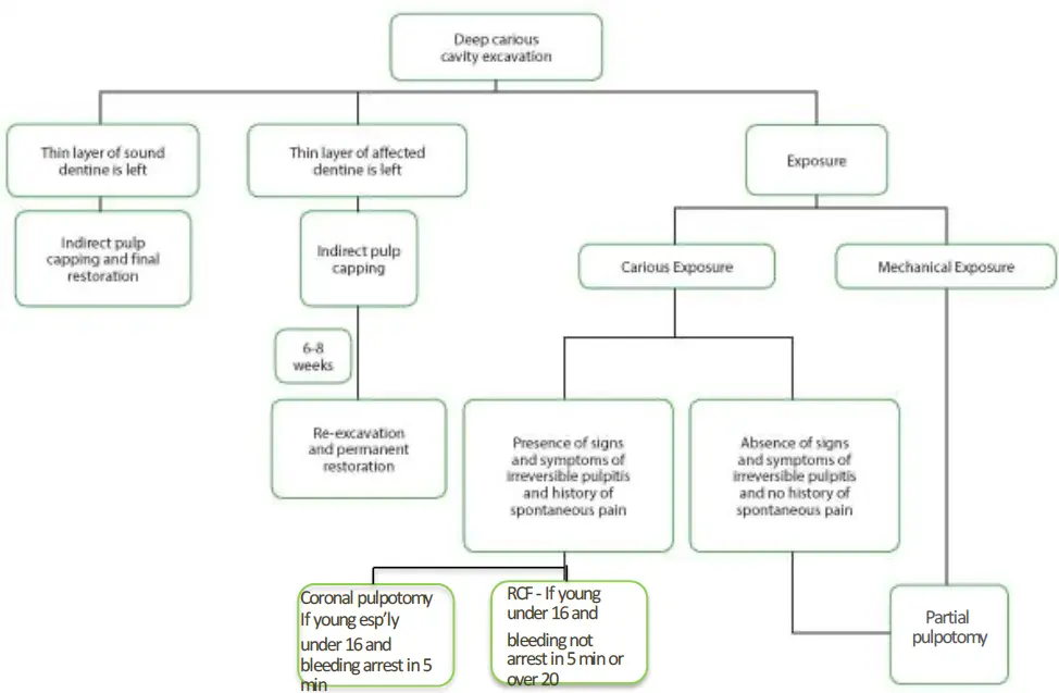
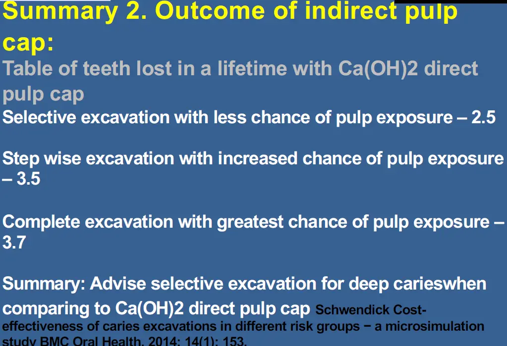
- selective removal to soft dentine that results in indirect pulp capping
- stepwise selective removal of soft dentine in 2 stages eneventually reaching firm dentine
- non selective caries removal that may result in pulp exposure requiring direct pulpal coverage
- DPC
- miniature pulpotomy
- partia/cvek pulpotomy
- coronal/complete pulpotomy
For deep carious lesion in teeth with sensible pulp
- ART, SMART
- non selective removal to hard dentine (now regarding as over treatment)
- stepwise removal, soft dentine left in first step (proximal to pulp), seal and and revise in 6-12months until firm dentine remains
- selective removal to soft dentine, carious tissue removal to hard dentine peripherally
- sealing using crowns
Atraumatic restorative treatment
- usually for under-served areas of world
- minimally invasive, hand instruments online
- adhesive restoration
Silver modified ART
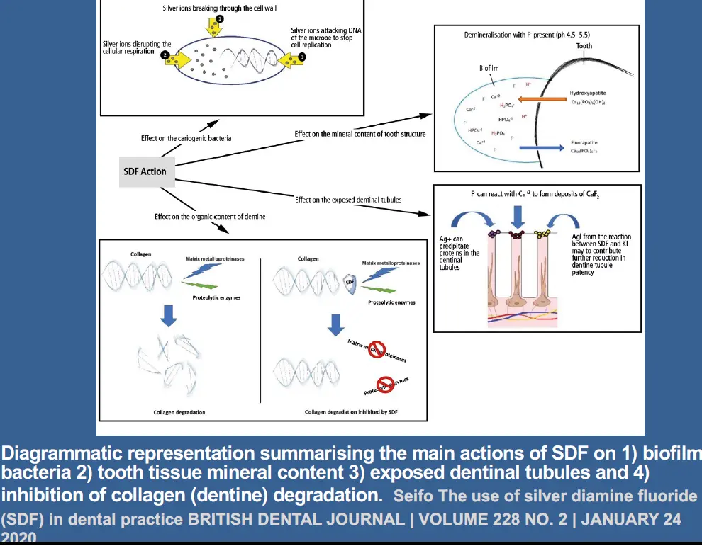
- silver diamine fluoride enhance remineralisation of demineralised soft dentine
- SDF better than ART alone
- discolouration may be reduced with application of potassium iodide after SDF
- may apply GIC over cavity with SDF to reduce further plaque accumulation
Silver Diamine Fluoride
- 38% weight per total volume (w/v) Ag(NH3)2F, 30% weight per total weight)
- colourless topical medicament comprising 25% to 29% (w/v) silver and 5% to 6% fluoride
- pH 10
- silver acts as antimicrobial
- fluoride promotes remineralisation
- ammonia stabilise high concentration in solution
- dry, etch, and apply, potassium iodide after
- when applied every 6 months, arrests more than 90% of caries
- 1 SDF on active lesion better than fluoride varnish 4x a year all surfaces
Selective removal to soft dentine with indirect pulp cap
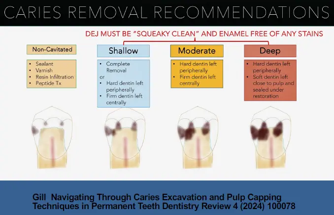
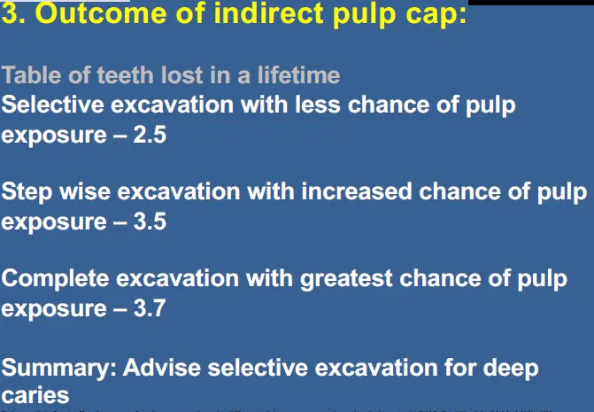
- pulpally remove carious tissue until soft dentine reached
- enough tissue removed to place durable restoration avoiding pulp exposure
- periphery of cavity clean to hard dentine
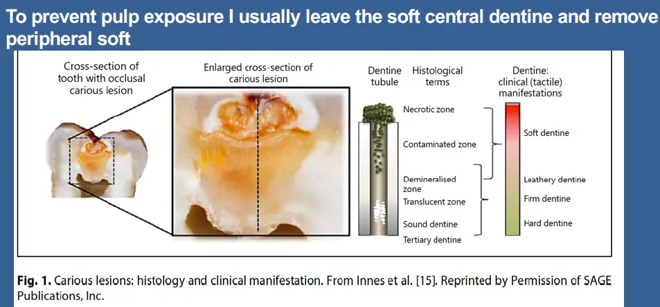
- indirect pulp cap evidence show not to use for irreversible pulpitis as degree of pulpal infection cannot be predicted
- no consensus on best material for pulp cap
Stepwise excavation
- residual caries remains and seal caries affected dentine with interim restoration
- expected that reactionary dentine is created with potential dentine remineralisation over next 16 weeks
- dentine is re entered and remnant soft caries is removed
- greater risk of pulp exposure than compared to selective caries removal as any remaining soft dentine is removed that may lead to pulp exposure
- no study or evidence for speed and remin amount
- tendency not to re enter as a 2 step procedure
Non selective caries removal that may result in pulp exposure require pulp cap
- compete caries removal pulpally and cavity periphery
- greater risk of pulp exposure
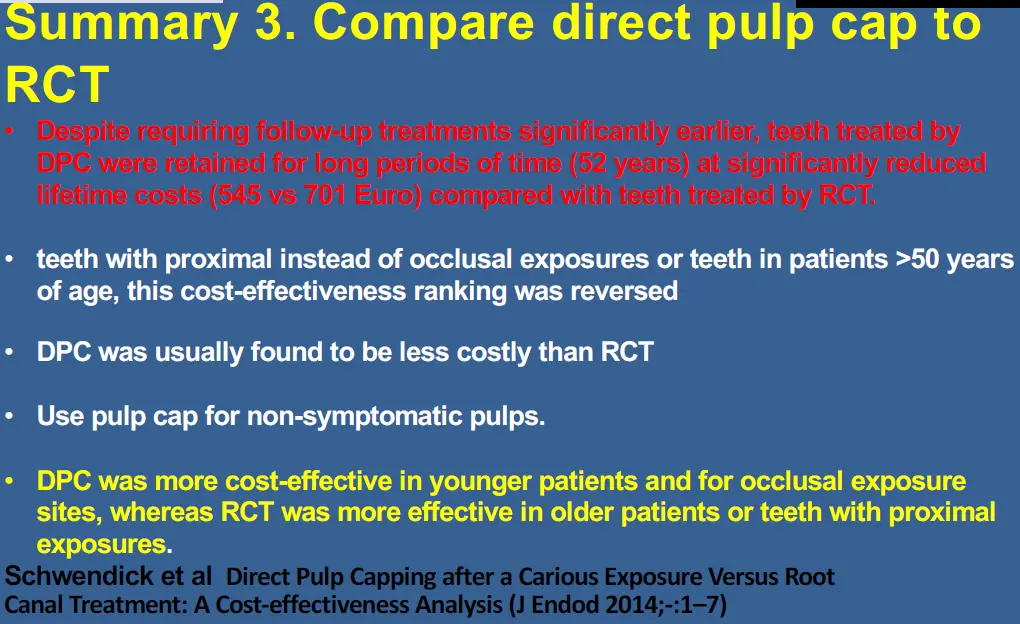
- DPC requires material placement directly over pulp to induce reparative dentine to protect pulp and reduce pulpal inflammation
- used for shallow carious lesions in outer 1/3 of dentine
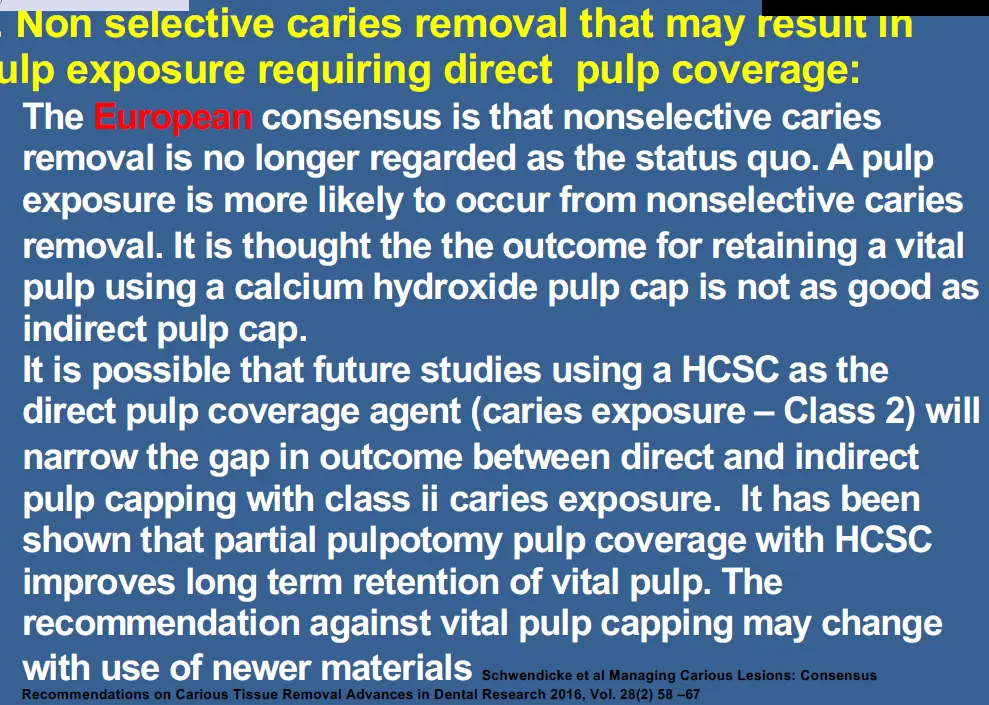
If pulp exposure arises
- all soft carious dentine must be removed before placing direct pulp coverage
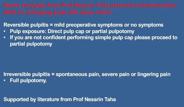
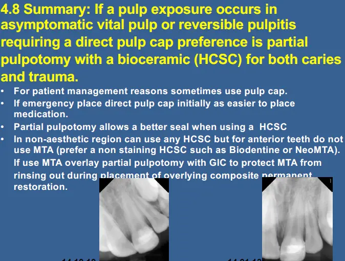
- partial (cvek) pulpotomy
- coronal/complete pulpotomy
- direct pulp cap
- miniature pulpotomy
Partial pulpotomy
- surgical removal of small portion of coronal pulp tissue 2mm into pulp to preserve pulp
- inflamed zone removed to level of healthy tissue
- may be better than direct pulp cap
- more conservative
- for trauma - better outcomes
- allows pulp sensibility testing
- not as predictable for irreversible pulpitis with mechanical allodynia
Materials used as direct pulp cappiing agents
- glucocorticoids combined with antibiotics
- ledermix
- not recommended as associated with high levels of pulp necrosis
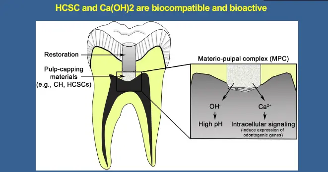
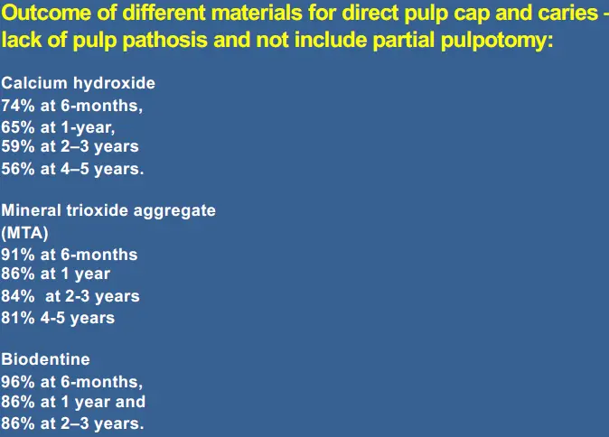
- calicum hydroxide
- creates some thin necrosis on layer but initiates dentine bridge formation, anti microbial
- lakcs inherent adhesion
- dentine bridges can form tunnel defects
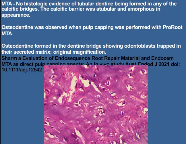
- MTA
- superior
- less alkaline than CaOH
- odontoconductive
How de we rationalise different treatment modalities to arrive at a treatment plan
Treatment decision - try to keep vital pulp is first consideration
- value of vital pulp is undeniable in permanent tooth no matter mature or immature
- keeping pulp will reduce loss of tooth structure and maintain defensive mechanisms
- pulpal infection and apical periodontitis is contraindication for vital pulp retention, must consider revitalisation process
Treatment decision - consider tooth restoraiton
- provide bacteria free environment using rubber dam
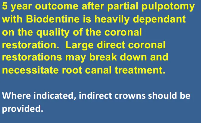
- ensure restoration over vital pulp therapy is approriate to minimise cusp fracture
WHat if a direct pulp cap due to pulp exposure is required
- varies depending on cause
- deep or very deep caries (class ii)
- if pulp exposure occurs remove all carious dentine
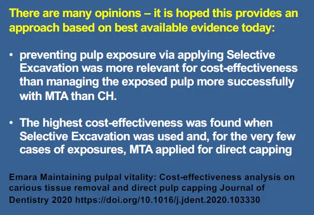
- partial pulpotomy best, about 2mm
- MTA best but long setting time, difficult to manipulate, some aesthetic concern
- trauma (class i)
- still partial pulpotomy
- more likely to retain vitality than carious
- discussion
- haemostasis after pulp exposure
- 5 min is cut off when bleeding should arrest
- when to use complete coronal pulpotomy?
- traditionally coronal pulpotomy used in cases of irreversible pulpitis as emergency procedure
How to place a partial pulpotomy direct pulp cap
- wash with sterile water or saline
- 2mm pulpotomy with diamond bur and water
- wait for bleeding to arrest
- wash with sterile water or saline
- place with biocompatible pulp capping agent
- cover with GIC or RMGIC
- restore with well sealing coronal restoraiton