Restoration of endodontically treated teeth
Tooth is never compromised
- options are
- restorable
- excellent
- good
- fair
- borderline
- end stage (unrestorable)
Permanent Coronal Restorative Step
Myth vs Reality
- RCT teeth are not brittle
- survival of root canal treated teeth is dependant on remaining amount of tooth
Aim
- manage coronal aspect of pulp space to maintain tooth as integral member of dental arch and oral environment
Objective of permanent coronal restorative step
- preserve RC filling to prevent coronal leakage - heal AP
- replace lost tooth substance - function
- protect and reinforce remaining tooth - tooth survival
- provide appropriate aesthetics - appearance
Risk management
- overall survival at 4 years following root canal filling is 96%
- depends on
- endo prognosis
- marginal perio prognosis
- structural prognosis
- strategic prognosis
Principles of Root Canal Material
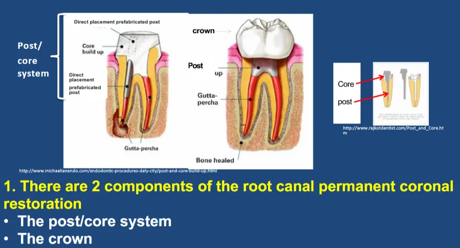
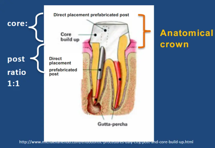
Post/core system
- core
- aids in replacing lost dentine inside an anatomical or prosthetic crown
- not supported by dentine
- post
- aids in replacing pulp space and is supported by dentine
- connects unsupported core to surrounding dentine
- may be direct or indirect
- direct post may include direct placement prefabricated post DPPP
- provides added stability to post core
- aids in stress distribution to tooth
- together post and core work synergistically and interdependently with the remaining tooth to support both the remaining tooth substance and the prosthetic restoration
- Prosthetic crown
- overlays anatomical crown
- essentially replaces enamel with cusp coverage
- may be direct or indirect
Survival of root canal treated tooth
- complex geometry of tooth and periodontium
- remaining anisotropic tooth structure
- post/core system
- crown, occlusal forces
- periodontal structures (marginal and apical)
Coronal and apical seal continuum - heal AP
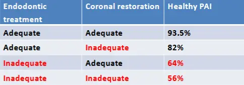
- quality of restoration and root canal filling is more important than the type of restoration (including post) for healing apical periodontitis
Replace lost tooth substance - function
- occlusal management
- cusp management
- interproximal contact area
Protect and reinforce remaining tooth - tooth survival
- ensure anatomical crown protected by changing the stressed placed on anatomical crown
- protect cusps with appropriate cusp overlay to minimise cusp flexure
- the more coronal tooth substance the less flexure
- cusp flexure results in caries, cracked cusp, crack or split
- ensure stress distribution through crown of tooth
- coronal stressed accumulate and are distributed through CEJ, known as
- peripheral rim
- peri cervical dentine
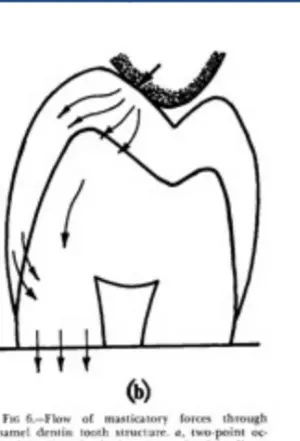
- compression dome effect
- distribution of compressive forced through enamel and or cornw
- absorbed by cervical margin (pericervical dentine/ peripheral rim theory)
- coronal stressed accumulate and are distributed through CEJ, known as
- importance of peri-cervical dentine
- area roughly 4mm above alveolar bone crest and 6mm below
- supports coronal compression of bio dome
- acts as medium to aid in transmission of forces between occlusal aspect and roots
- critical factor in regards to long term survival of tooth and optimum tooth function
- area roughly 4mm above alveolar bone crest and 6mm below
- partial cover restoration relies mainly on compression dome complex
- tangential forces are created into axial forces through the dentine into cervical rim
- may be direct or indirect cusp coverage restoration
- full coverage indirect restoration
- more pericervical dentine the better the tooth can absorb and redirect occlusal forces creating occlusal compressive axial not oblique forces
- if an indirect full crown is present, the supragingival dentine is the ferrule
- Crown to root ratio
- anatomic ratio - clinical average crown:root 1:2
- clinical ratio - amount of alveolar support clinical crown to clinical root intra-alveolar 1:1.4
- Restorative ratio - amount of unsupported core to supported post ratio, prefer about 1:1 + apical GP
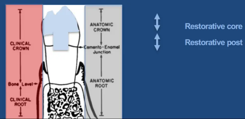
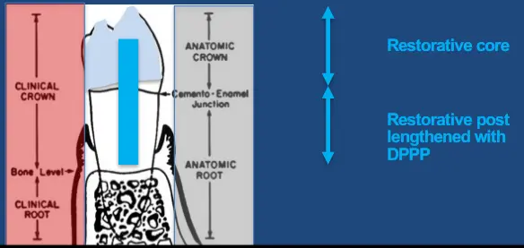
- DPPP or indirect post
- width
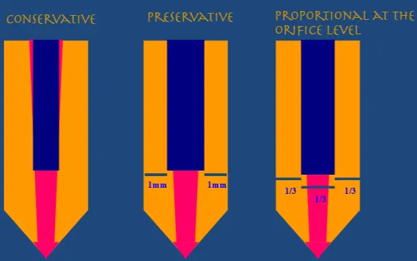
- no correlation between crowned root canal treated teeth and vertical root fracture
Provide appropriate aesthetics - appearance
- composite very aesthetic
Implementation of Restorative principles during RCT
Differential diagnosis
- informs risk assessment prognosis in 4 dimensions
- structural - amount of tooth
- endodontic - pulpal/periapical
- strategic - stability/deterioration
- Marginal periodontal - supporting
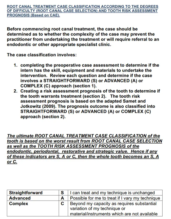
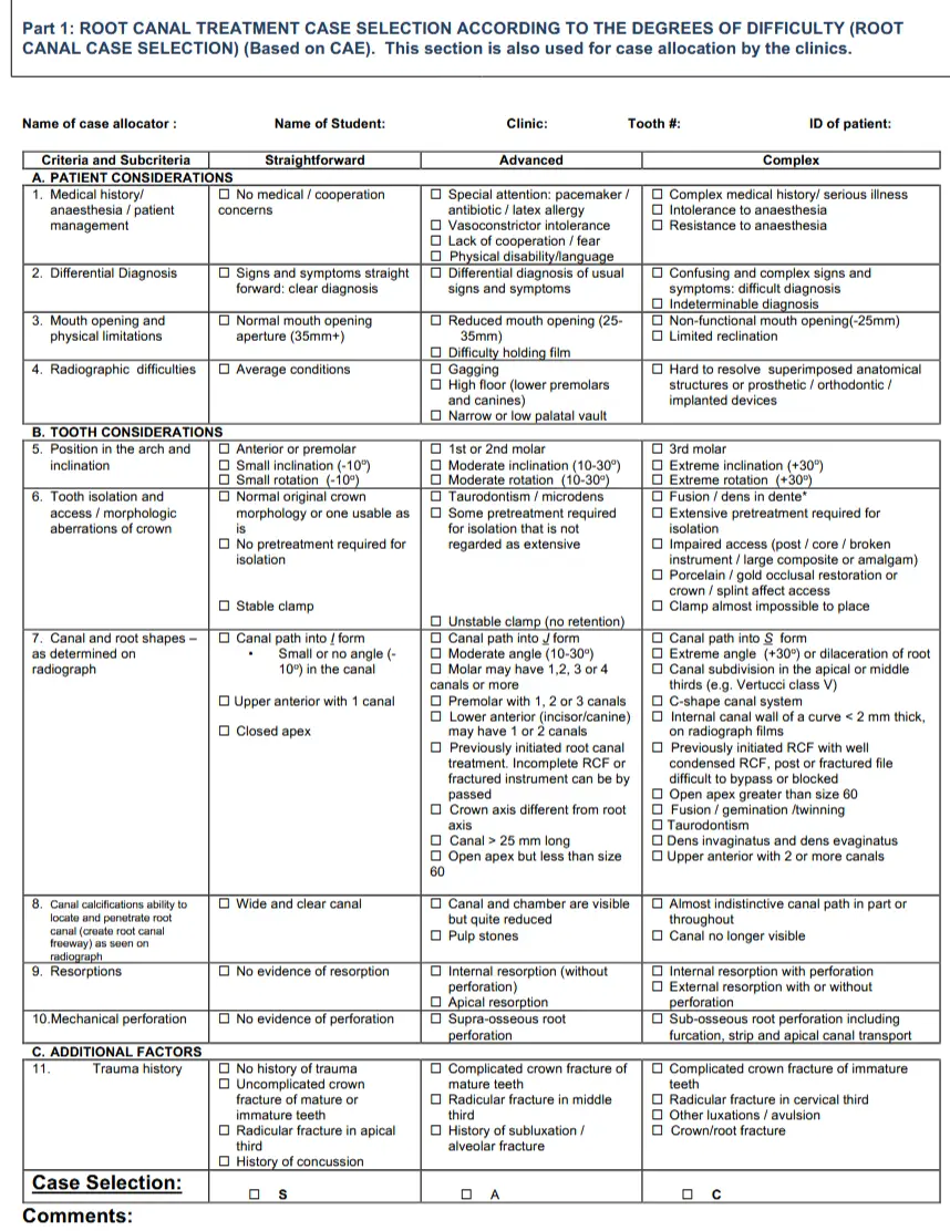
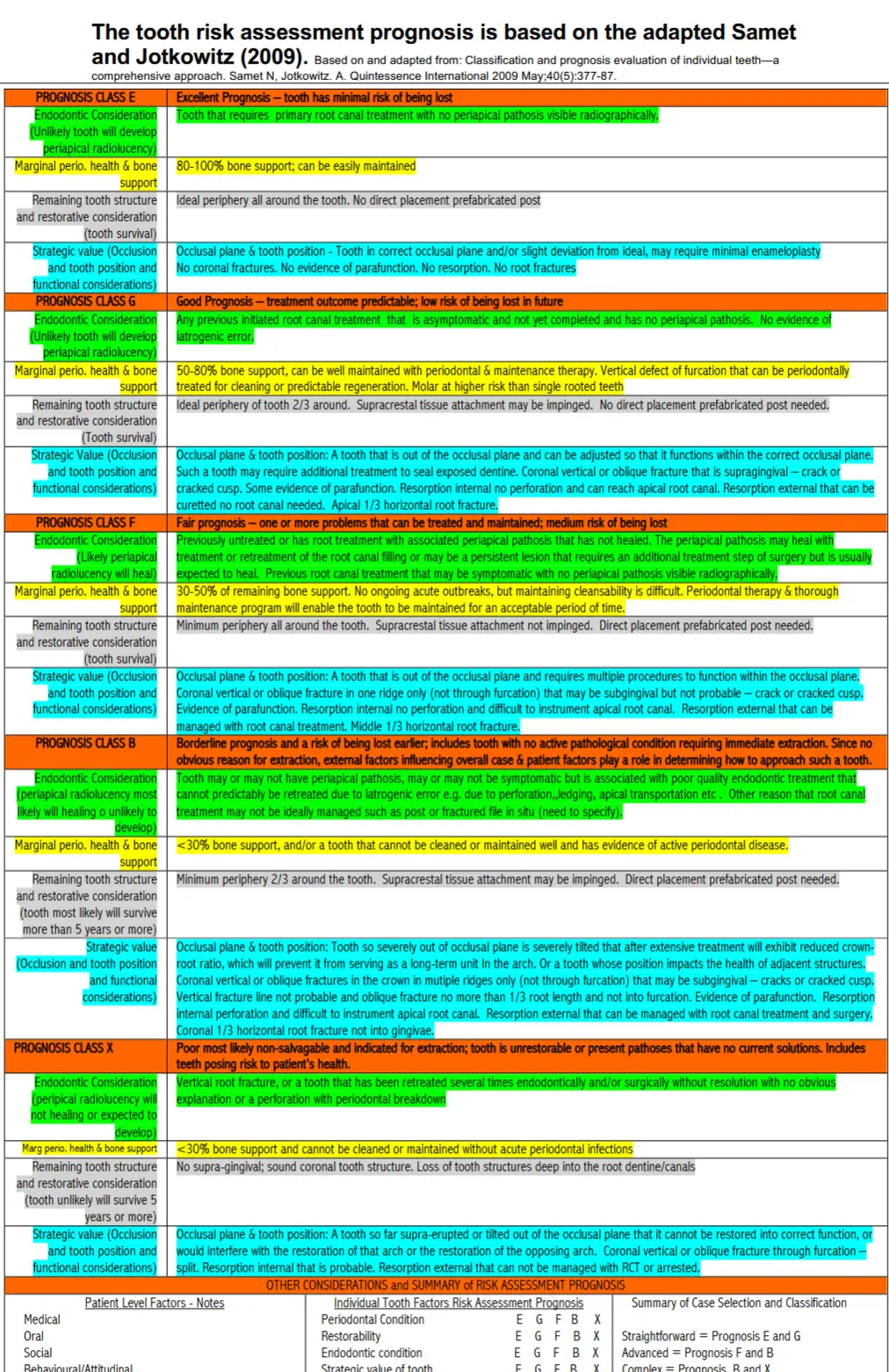
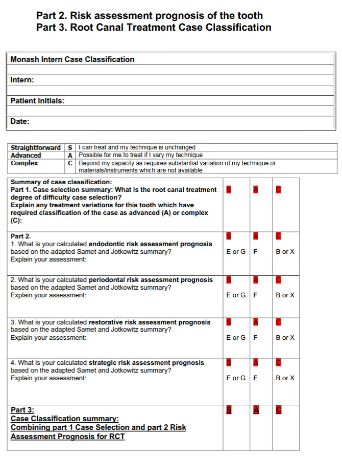
- Case classification part 1
- skill experience
- equipment available
- instruments available
- materials available
- case classification part 2
- tooth risk based prognosis for each tooth
- part 3
- summary
- straightforward
- advanced
- complex
- summary
- prognosis guides treatment plan - defined by risk
- excellent
- good
- fair
- borderline
- extraction
Risk assessment prognosis
- guides treatment plan
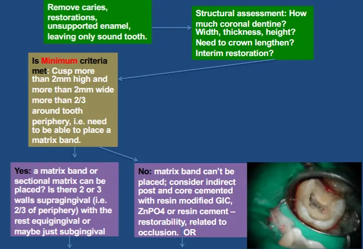
- **Structural assessment
- preferred minimum
- tooth remaining supragingival is 30% tooth substance
- cervical rim - 2mm wide and 2mm high 2/3 around tooth
- ideal amount
- supragingival dentine wall more than 1/2 occlusal height, 2mm wide, 2/3 around the tooth
- what material?
- conserve as much cervical tooth as possible
- partial coverage direct/indirect
- inlay - no cusp
- onlay - 1 or more cusp
- overlay - all cusp
- full coverage - indirect
- therefore permanent coronal restoration related to
- amount of supragingival tooth
- need for cusp coverage
- patient risk factors
- terminal/catastrophic failure of tooth not more prevalent in any cusp coverage group
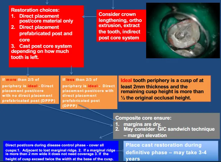
- When to place DPPP
- when 2mm thick cusp wall is less than half the height of the anatomical crown and at least 2/3 around the tooth
- restorative core:post ratio minimum 1:1 and need apical gp
- Choosing DPPP
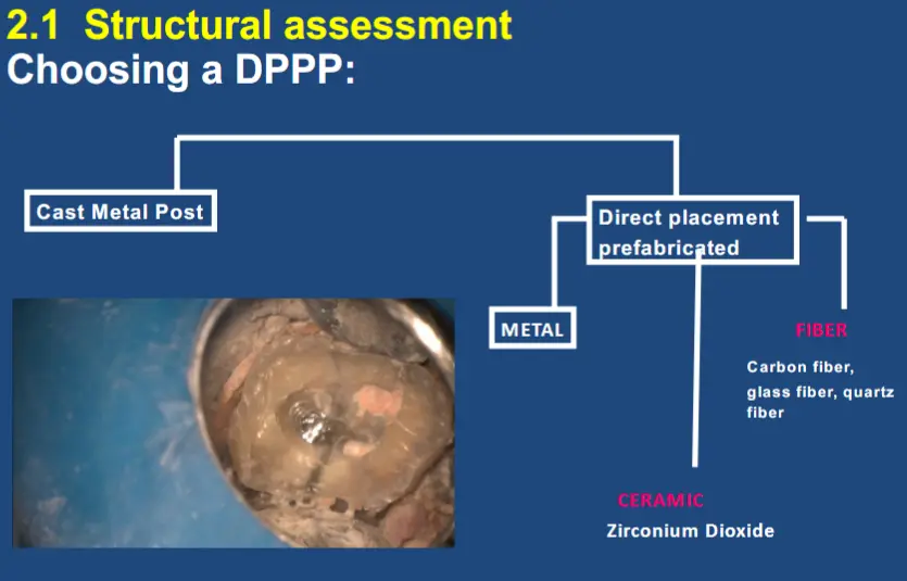
- outcome for diverse choice of DPPP is minimised when suitable supragingival coronal dentine is present
- bonded direct placement prefabricated fibre posts do not reinforce tooth restoration complex
- bonding of fibre DPPPP still remains a challenge
- DPPP fibre posts may degrade in contact with fluid
- cusps should be covered - direct or indirect
- adjacent to lost marginal ridge
- if a marginal ridge is less than 2mm wide
- if height of cusp excess twice the width at the base of the cusp
- direct cusp coverage should be at least 2mm thick
- preferred minimum
- Endodontic assessment
- use direct resto during disease control phase while reviewing PA healing
- Strategic assessment
- note any pre-existing fractures - coronal, radicular
- avoid tooth fracture or restoration fracture - material synergy - direct post core restoration function initially independently of a cusp coverage indirect restoration
- post core system may be required to support an indirect restoration in the future
- place indirect restoration as the crown
- may be deterioration of the direct coronal restoration or colour changes after 2 to 3 years
- indirect for functional or aesthetic reasons
- may need to improve interproximal contact or embrasure with indirect restoration
- Marginal Periodontal Assessment
- after RCT ideally want 2/3 of root embedded in bone, may be an issue with crown lengthening
- what about DME? does supracrustal tissue attachment matter?
- after 12 months with resin and GIC subgingival restoration, no further periodontal bone loss with no progressive clinical attachment loss and negligible inflammation
Treatment plan
- details anticipated outcome
- patient centred