Endodontic Radiography and Working length determination
#D3/S2/W2
#Endo
Importance of diagnosis
- data gathering
- clinical
- history taking (general and focused)
- examination (objective and subjective)
- radiographic
- definitive diagnosis
- formulation of appropriate treatment plan
- clinical
Bare minimum radiographic sequence
- preop radiography
- working length radiograph
- mastercone radiograph
- mid-obturation radiograph
- post-operative radiograph
- recall
Endodontic radiology
- use largest size film possible
- use film holder for reproducibility
- centre film on tooth of interest
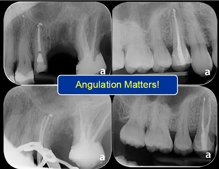
- paralleling xray beam
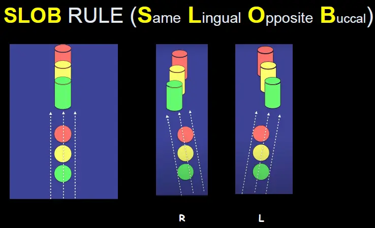
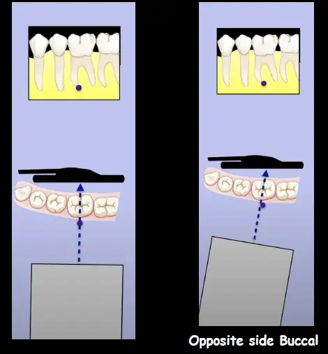
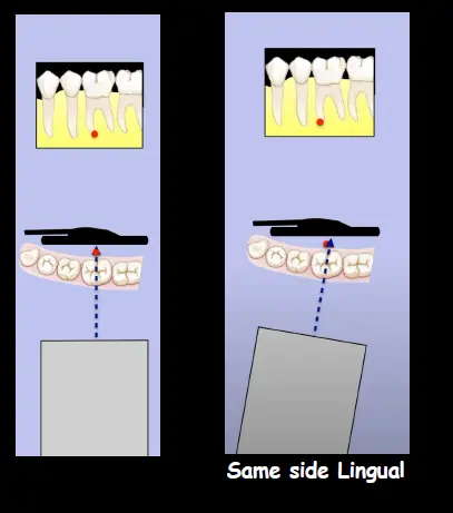
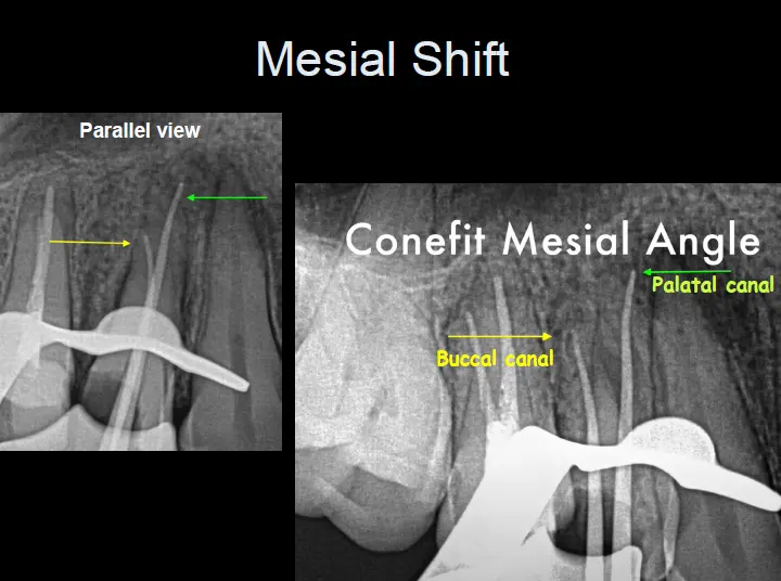
- shift radiographs
Endo ray
- helpful to keep out of way while clamp and files are there
Other products - specialty holders
- truview PSP
- XCP-DS Fit
Paralleling technique
- more accurate length determination
- less superimposition
- reproducibility
Radiographs are not optional
- delphin v martin 2012
- failure to take a preoperative radiograph is negligent
Multiple films
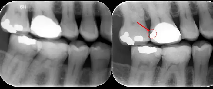
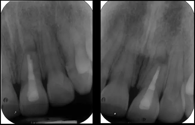
- bare minimum = 1PA + 1 shift PA (10-15 degrees) and BW
Example cases
-
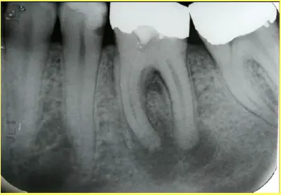
- no tender to percussion
- pain disturbing sleep
- normal response to ice, pulp response normal
- chronic infection
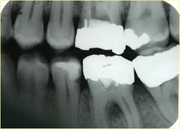
- chief response was not even tooth in question, was maxillary tooth, hard to localise
-
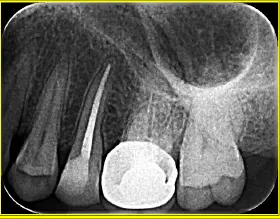
-
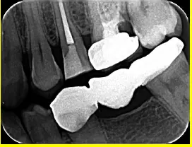
-
shift views
Trauma
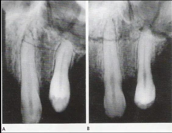
- shift in beam shows the previously thought horizontal root fracture has magically moved, therefore it is not a horizontal root fracture but rather an alveolar fracture
Diagnosis
- tracing GP using sinus tract
Pre-op assessment
- take your own bitewing and PA to assess
**Cracks **
- reactions to cracks (J shaped radiolucency)
- large cracks
Mid treatment radiograph
- essential to
- confirm depth of cavity
- confirm working length
- confirm anatomy
- confirm disinfection
- confirm obturation length and density
Working length film
- minimum size 15K
- if longer or short by 2mm retake radiograph
Mastercone radiograph
- may require more than one depending on number of roots and access
- confirms instrumentation level, obturation length and size
- can identify problems before its too late to pull out
MId obturation
- check length prior to melting
- check density prior to melting
- remedy defects early
post obturation
- identify extra roots etc.
- asymmetric radiograph
- off-centre ~85% have 2nd canal - Hoen et al 2002
Periapical index
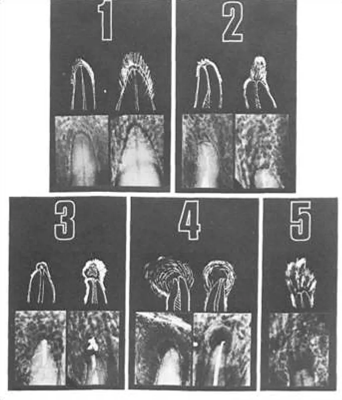
Tracking disease progression
- histological changes taken 2-4 months in infected cases
- radiographic change in relation to pulpal infection can take up to 6 months
Importance of recall radiographs
- ensure there is a change in radiographic healing (or absence of)
- reversal of healing
Digital radiography
- introduced in 1987
- good and bad
Limitations
- 2d film, cant see 3d
- double exposure
- moisture on fingers
- superimposition of structures
- supernumerary
- plates
- other teeth
- zygomatic buttress
- need a lot of bone mineral loss to see it on the radiograph (cortical plate involvement)
- anatomical stuctures
- mental foramen
- incisive foramen
gag reflex
- salt
- LA
- discussion
- referral
- cbct
- behavioural Mx
- sedation
Cone beam CT 3d
- endo indications
- morphological assessment
- difficult diagnoses
- pre-surgical assessment
- resorption
- cracks
- misadventures
- non odontogenic anomalies
- trauma
- unusual anatomy
- dens invaginatus
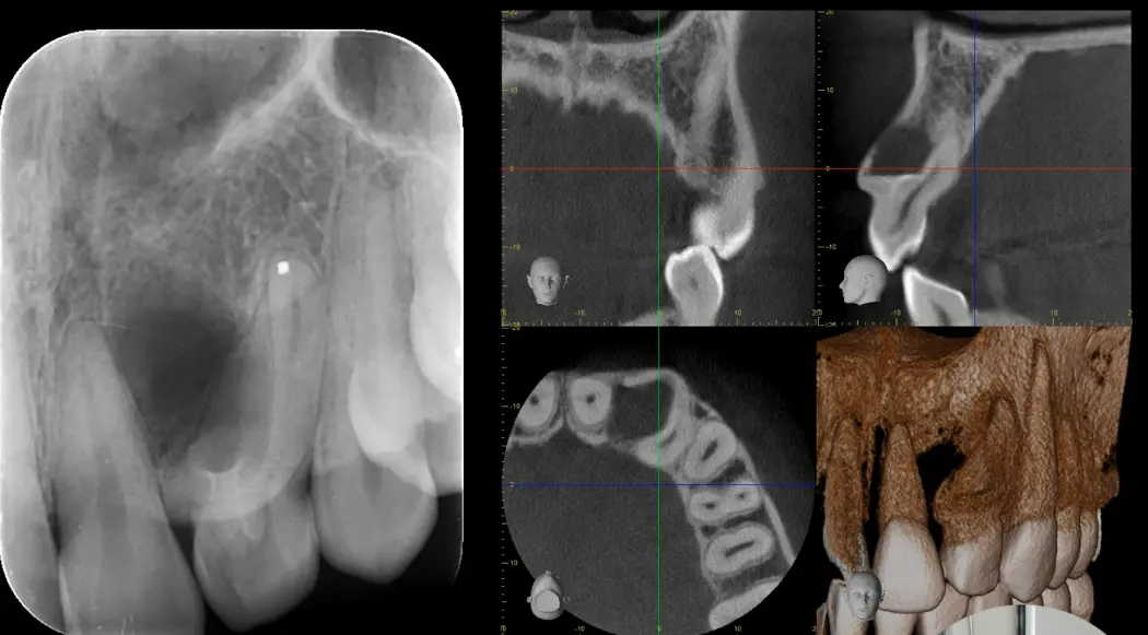
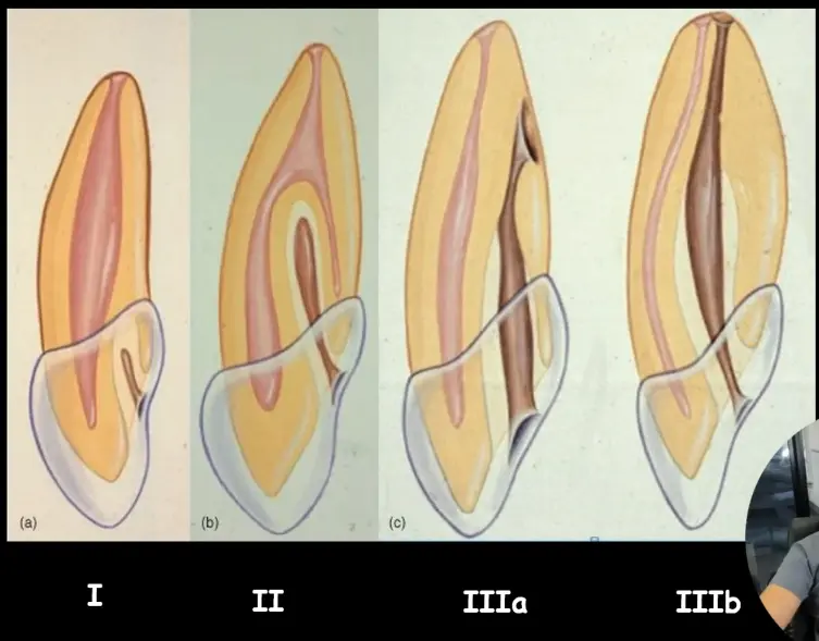
- Anatomy
- close proximity to inferior alveolar, mental nerve
- resorptive defect
- perforation
Dosages
- ALARA
- as low as reasonably achievable
- young patient
- thyroid cancer
- pregnant patient (especially 1st trimester)
Working length
working length
- distance from a coronal reference point to point which canal prepartion and obturation should end
rational for length determination
- optimise treatment effectiveness
- avoid problems of underextension
- avoid problems of overextension
Does it matter?
- critically important for infected cases, undisturbed biofilm in untreated RC system
- 69-75% healing if underinstrumentation
- 90-95% healing if instrumented to optimal length
- overinstrumentation related to lower success rates (not always true)
Pulpal vs periodontal
- tissue that includes and supports odontoblasts is pulpal
- tissue icluding and supporting cementoblasts is periodontal
radiographs arent entirely reliable
- magnification/distortion
- lack of third dimension
- inability to see soft tissue
- anatomic superimposition
- inter observer disagreement
- intra observer disagreement
- Benefits
- documentation
- relative length
- determines degree of curvature/canal anatomy
- 3d approx.
- cant reach constriction or PDL
other means
- paper point
- not accurate
- relies on blood etc.
- tactile sensation
- unreliable
- operator dependant
- visually
- not reliable
- patient sensation
- if no vital pulp
- if PDL at apical foramen
Electronic canal length measurement
- how do they work
- landmark: conductive tissue
- dentine: good insulator
- different generators
- file is a conductor with lip clip (mucous membrane), as dentine stops insulating then a circuit is formed
- electrical resistance between instrument and oral mucous membrane is a constant (6.5kV)
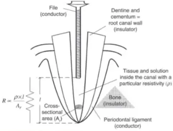
- difference in generations
- 1 resistance only
- 2 impedence only
- 3 two frequency, impedance ratio
- 4 two frequency, impedance difference
- Contraindication
- pacemakers or other electrical implanted devices (not really)
- allergic to metal
- in children
-
- Inappropriate signal paths to periodontium
- metal restoration
- caries
- gingival tissue
- perforations/fractures (horizontal but not vertical)
- large accessory canals
- conductive fluid i.e. NaOCl, blood, pus, saline, LA
- Inappropriate signal paths to periodontium
- Trouble shooting
- check connections
- check for haemostasis
- no fluid in pulp chamber
- water flush
- lubricants (RC prep)
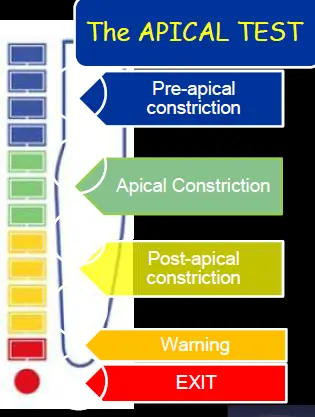
- apical test
- Method
- do a few passes back and forth during test to make sure you get the same reading at same point each time
- Safety nets
- anatomical statistics
- radiographic reasonableness
- paper point/bleeding
- apical stop/tug back
- tactile reporting
- other uses
- detection of perforations, root fractures
Apex
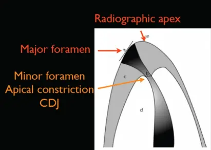
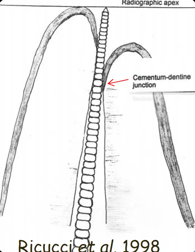
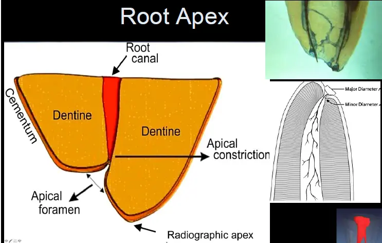
- not anatomic apex
- not radiographic apex
- physiologic apex or apical constriction
- constriction varies 0.2-3.8mm from radiographic anatomic apex
- can be different for same tooth different roots as well
Changes to apical anatomy
- deviation dependant on age and tooth
- non-uniform position of AC
- pathological changes (resorption)
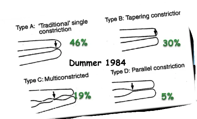
- not always a constriction