Module 3
#DEN4002
#D4/S2/W3
#Periodontology
Mucogingival Conditions
Normal mucogingival condition
- absence of pathosis
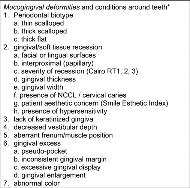
Periodontal biotype
- based on components of masticatory complex
- gingival biotype / thickness and keratinised tissue width
- bone morphotype
- tooth dimension
- three biotypes
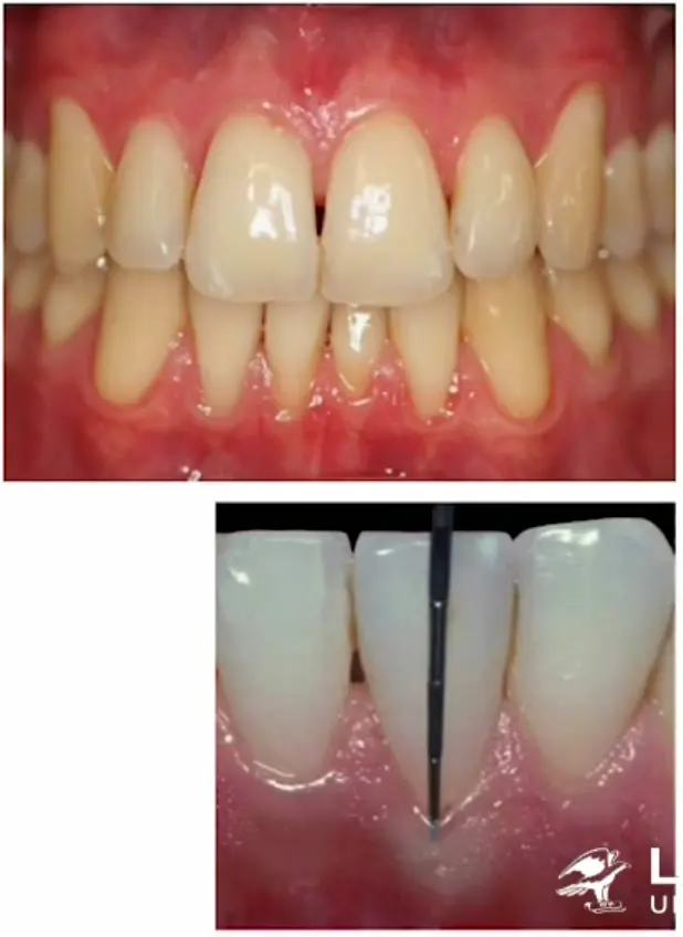
- thin scalloped , slender triangular crown, subtle cervical convexity, interproximal contacts close to incisal edge and narrow zone of keratinised tissue, translucent thin delicate gingiva, thin alveolar bone
- thick flat, square shaped tooth crowns, pronounced cervical convexity, large interproximal contact located more apically, broad zone of keratinised tissue, thick fibrotic gingiva and thick alveolar bone
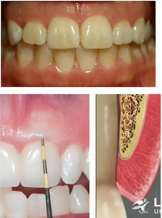
- thick scalloped, thick fibrotic gingiva, slender teeth, narrow zone of keratinised tissue, pronounced gingival scalloping
- THICK more common than THIN
- thin more gingival recession than thick
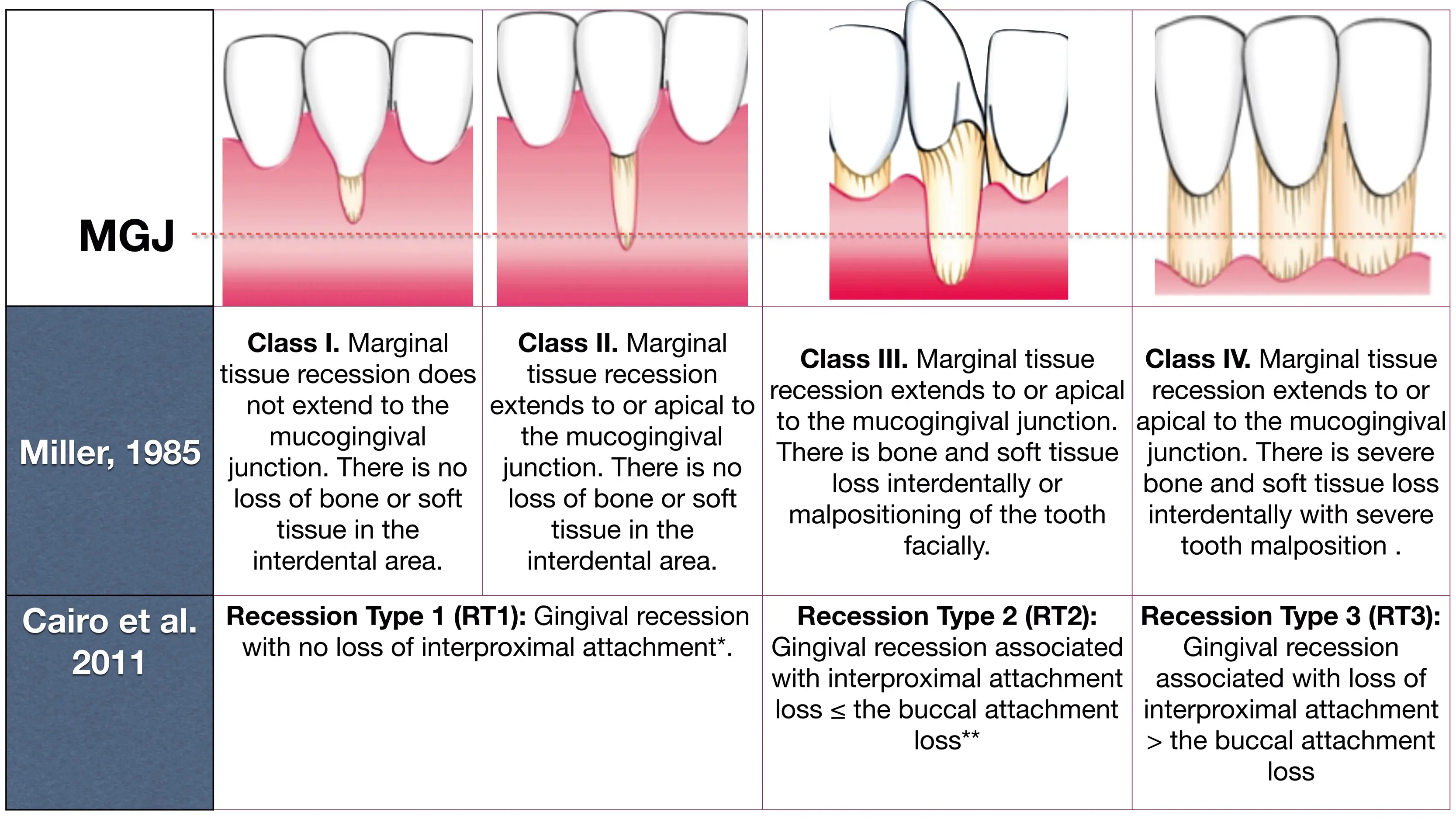
Cairo et al 2011 recession types RT1 2 and 3
- RT1
- gingival recession with no loss of interproximal attachment
- interproximal CEJ not clinically detectable at mesial and distal
- RT2
- gingival recession associated with loss of interproximal attachment
- interproximal attachment loss is less than or equal to buccal attachment loss
- RT3
- gingival recession associated with loss of interproximal attachment
- interproximal attachment loss greater than buccal attachment loss
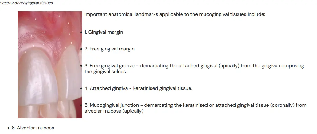
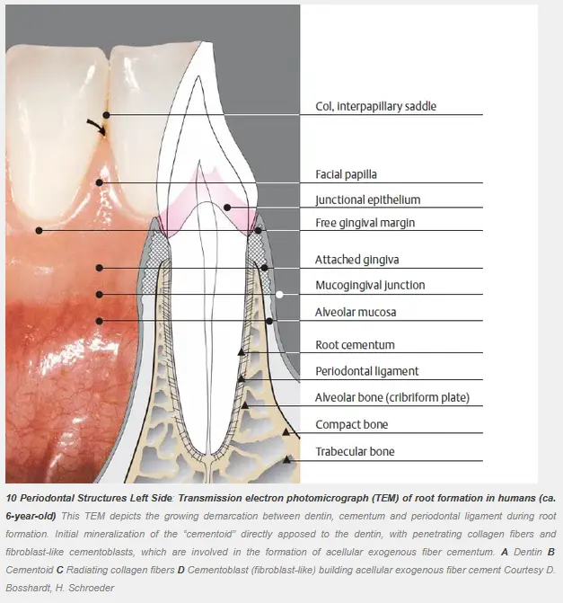
Col-interpapillary saddle
- apical to contact area, interdental gingiva assumes a concave form in labiolingual section view
- col will be of varying depth and breadth depending on expanse of contacting tooth surfaces
Attached gingiva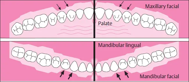
- facial: narrowest at canines and first premolars
- lingual: posteriors > incisors (mandibular)
- different between patients and phenotypes
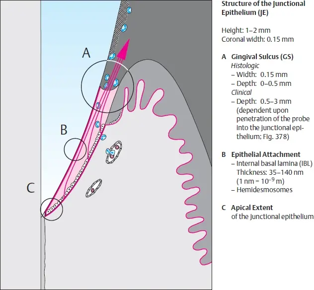
Objectives of periodontal plastic and aesthetic surgery
- maintain healthy and stable mucogingival complex
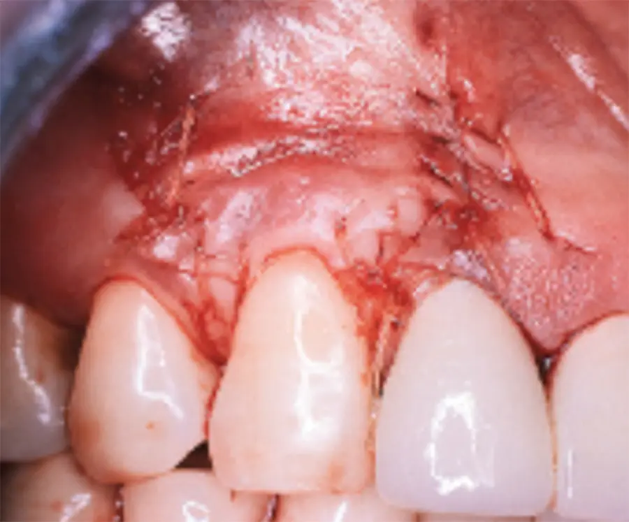
- cover exposed root surfaces to tackle aesthetic or hypersensitivity concerns
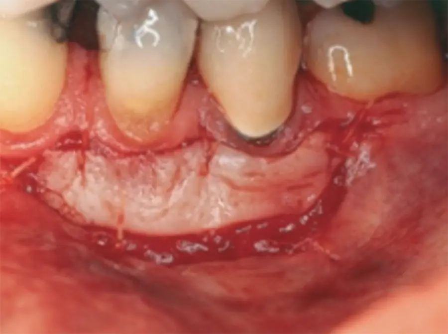
- establish optimal zone of attached keratinised gingiva
- provide adequate vestibular depth
- eliminate aberrant frenal pull
- overcome complications of subgingival margins in prosthetic dentistry
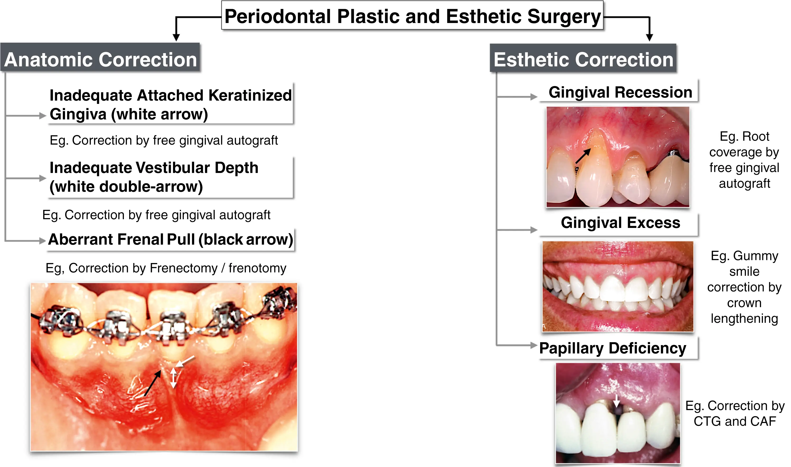
- CAF coronally advanced flap
- CTG connective tissue graft
Donor graft tissue
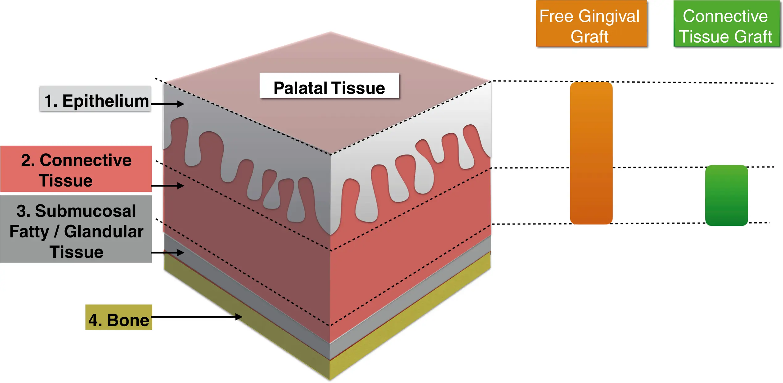
- various sites, edentulous ridge, tuberosity, gingivectomy, palatal tissue
- usualy area of choice is palatal tissue distal to anterior rugae in relation to premolar and first molar areas
- has widest gingival zone with least amount of submucosal tissue (fatty anteriorly and glandular posteriorly)
Gingival recession
recession
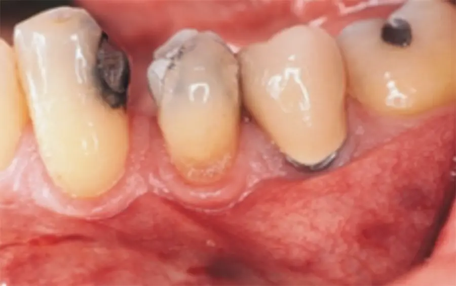
- apical migration of junctional epithelium with exposure of the root surface
- aetiology
- perio
- toothbrush abrasion
- buccally positioned teeth (especially ortho)
| Root Coverage Procedures | Procedures to Augment Keratinized Attached Gingiva | |
|---|---|---|
| Area of correction | Soft tissue augmentation above existing gingival margin | Soft tissue augmentation below existing gingival margin |
| Primary objectives | - Recession coverage for esthetics - ↓ dentin hypersensitivity in exposed root surfaces |
- ↑Tissue thickness - ↑ Keratinized gingiva width |
| Goals achieved | Aesthetics Hypersensitivity Treatment of NCTL improve thickness/biotype |
improve thickness/biotype |
| Procedures | Pedicled grafts - Laterally and coronally displaced flaps Free grafts - FGG - Free CTG Combinations - CAF + SCTG - CAF + GTR - CAF + biological mediators |
Pedicled grafts - apically displaced flaps Free grafts - FGG - Free CTG Combinations - Vestibuloplasty |
- FGG free gingival graft
- SCTG subepithelial connective tissue graft
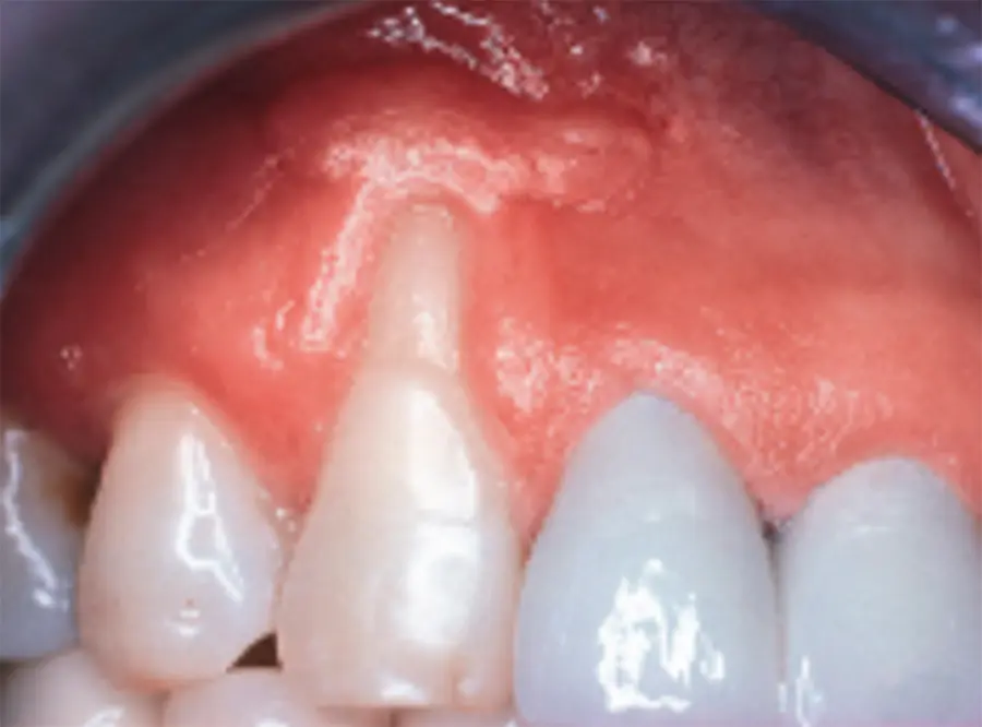
Dehiscence and fenestration
- both can cause recession, typically post ortho and dependant on phenotype
- dehiscene, v shaped linear defect continuous from coronal to apical portion, causing exposed root
- fenestration, window defect of exposed root in apical region
Lack of keratinised tissue and reduced vestibular depth
Lack of keratinised tissue
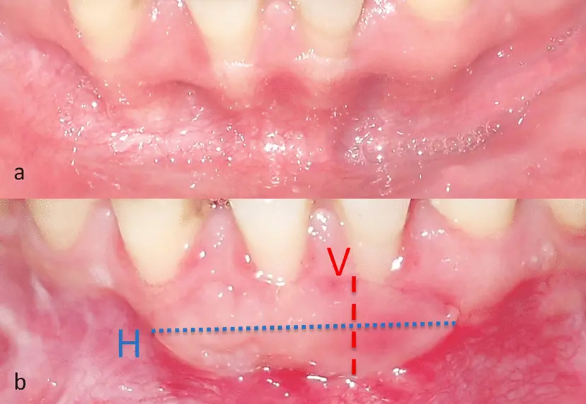
Reduced vestibular depth
- short vestibule frequently associated with lack of keratinised tissue and is difficult for patient to comfortably clean the site
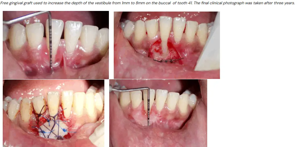
Aberrant Frenae
Frenectomy vs Frenotomy
- relocation of frenum - frenomoty
- removal of frenum - frenectomy
Frenum - thin fold of mucosa enclosing muscle fibres which attaches to mucosa, underlying periosteum and bone
Typical location - maxillary buccal midline and premolars
- mandibular buccal midline and premolars
- mandibular lingual midline
Aberrant attachments
- cause tension/pull on gingival margin causing recession or pull on gingival margin of pocket causing ingress of plaque and progression of perio
- aberrant lingual frenum causes ankyloglossia
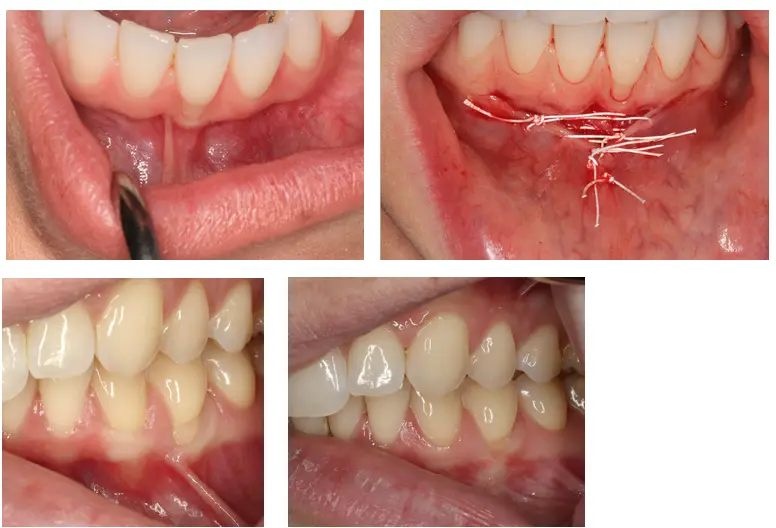
Gingival excess and abnormal colour
Gingival excess
- reduction of pseudo pockets and gingival overgrowth
- uneven gingival margins
- gummy smiles
- all will require surgical crown lengthening [[]]
Abnormal colour
- pigmentation can range from solitary lesion (macula, amalgam tattoo, melanoacanthoma, naevus, vascular lesion, melanoma) to racial pigmentation involving most buccal tissue
- can be addressed using variety of modalities
- excision (scalpel, periodontal knife, bur)
- abrasion
- electrosurgery, cryosurgery, laser
- excision and grafting using a free gingival graft or a matrix
Furcation Lesions and Root anomalies
Definitions
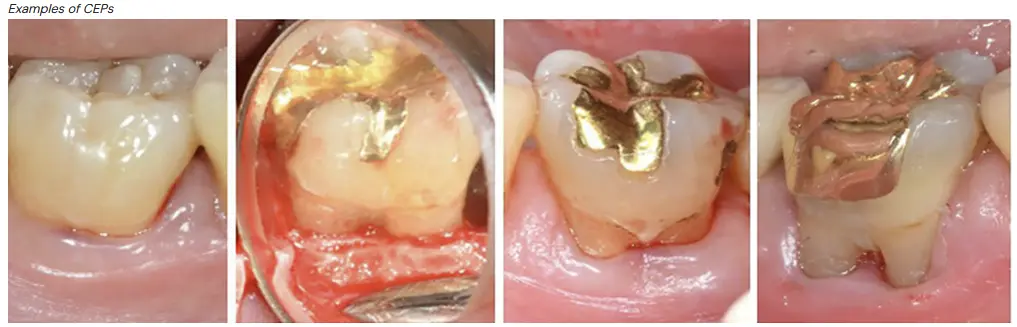
- cervical enamel projections
- developmental anomaly characterised by extension of enamel toward furcation entrance, hinders plaque removal, eventually leading to furcation involvement, more common in 2nd molars
- CEP classificaiton Masters and Hoskins 1964
- grade I - cep from CEJ to furcation
- grade II - cep approaching entrance to furcation
- grade III - cep extending horizontally and entering into furcation
- furcation arrow
- radiolucent triangle showing m or d furcation involvement in maxillary molar
- negative predictive value 85%, 42% of degree II and III has furcation arrow present Hardekopf et al. 1987
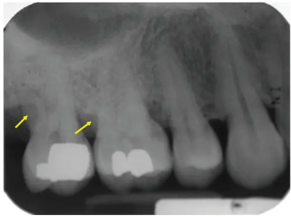
- fornix
- roof of furcation
- intermediate bifurcation ridges
- ridge of primarily cementum, common in mandibular molar hinder plaque removal
- runs from mesial surface of distal root via bifurcation to high on the mesial root
Furcation
- diagnostic challenges
- initial furcation involvement can go undetected in radiographs (superimposition of structures)
- initial furcation involvement usually depends on probing for detection, but entry of probe depends on absence of obstructing soft tissue, width of osseous defects, root divergence, furcation ridges, furcation may also not be accessible to probing if too narrow/tortuous
- therapeutic challenges
- majority of walls in a furcation defect are nonosseous in nature (root surfaces and furcation dome), usualyl covered by cementum dentine or enamel defects, reduced area of vascularity with limited source of bone precursor cells is a challenge for periodontal regenerative therapy
- cervical enamel projections have shown great complexity in recent electron microscopic evaluations, can be pouch like openings which can resist stringent oral hygiene measures
- sometimes furcation fornix is so narrow its virtually impossible to keep oral hygiene once exposed to oral environment
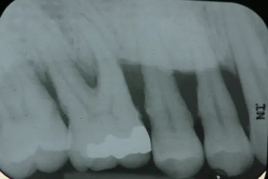
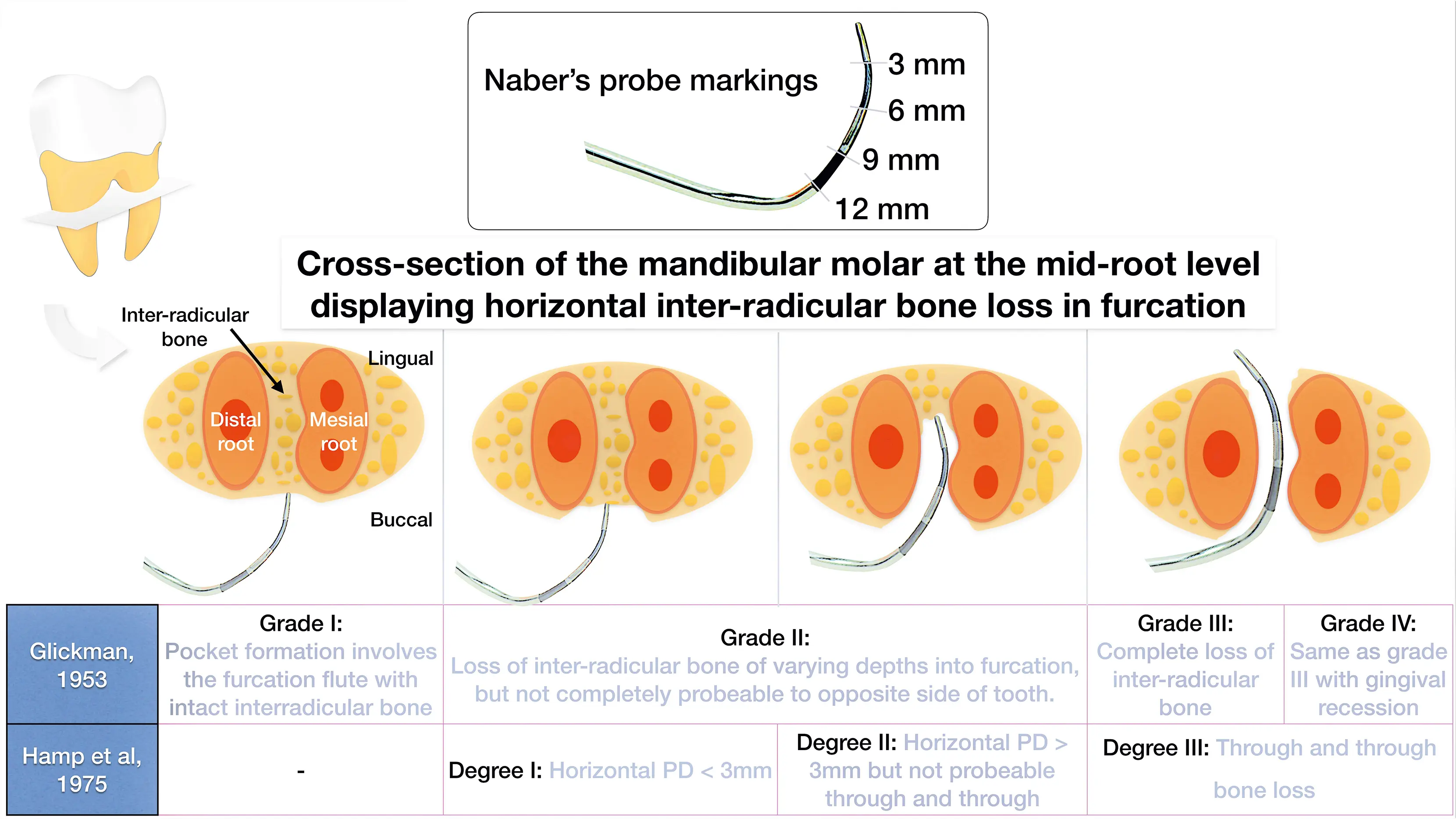
- Hamp et al is horitzontal only, each degree is 3mm increments essentially
- tarnow and fletcher is vertical only, A B C
approximate probing depth of furcations
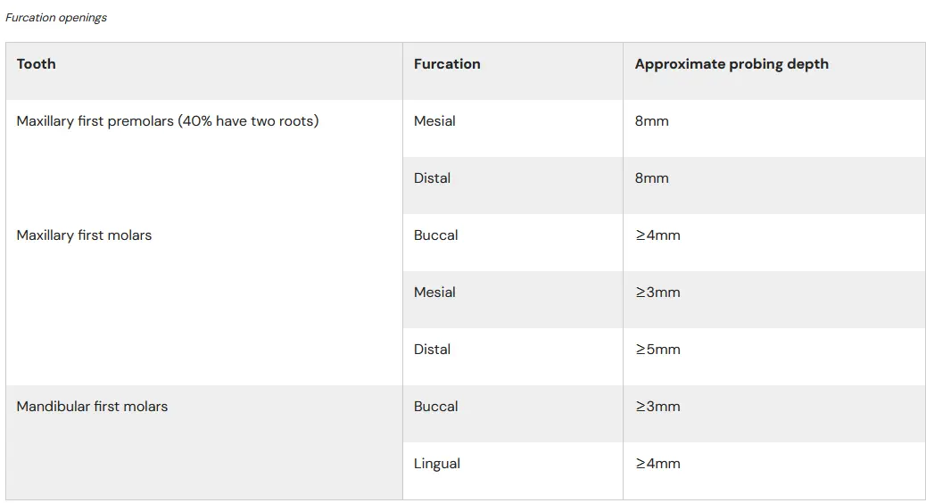
Etiology
- local anatomic factors
- enamel pearls
- cervical enamel projections
- root concavities in furcal areas
- trauma from occlusion
- endo perio
- root fractures extending into furcation
- iatrogenic cofactors
- endo perf
- overhanging resto
- biologic width violation
Treatment considerations in furcation management
- root divergence
- closely approximated or fused roots can prevent adequate instrumentation, widely separated roots are readily treated
- Bower 1979, 58% of maxillary first molar furcation entrances can not fit a standard curette
- root trunk length
- easier access for maintenance
- root length and remaining bone
- is boneloss progressing to furcation the start or end of the issue, e.g. long root trunk short roots
- root proximity to adjacent teeth
- similar issue to root divergence
- pattern of bone loss
- deep multiwalled bony defects may be candidates for regenerative therapies
Treatment
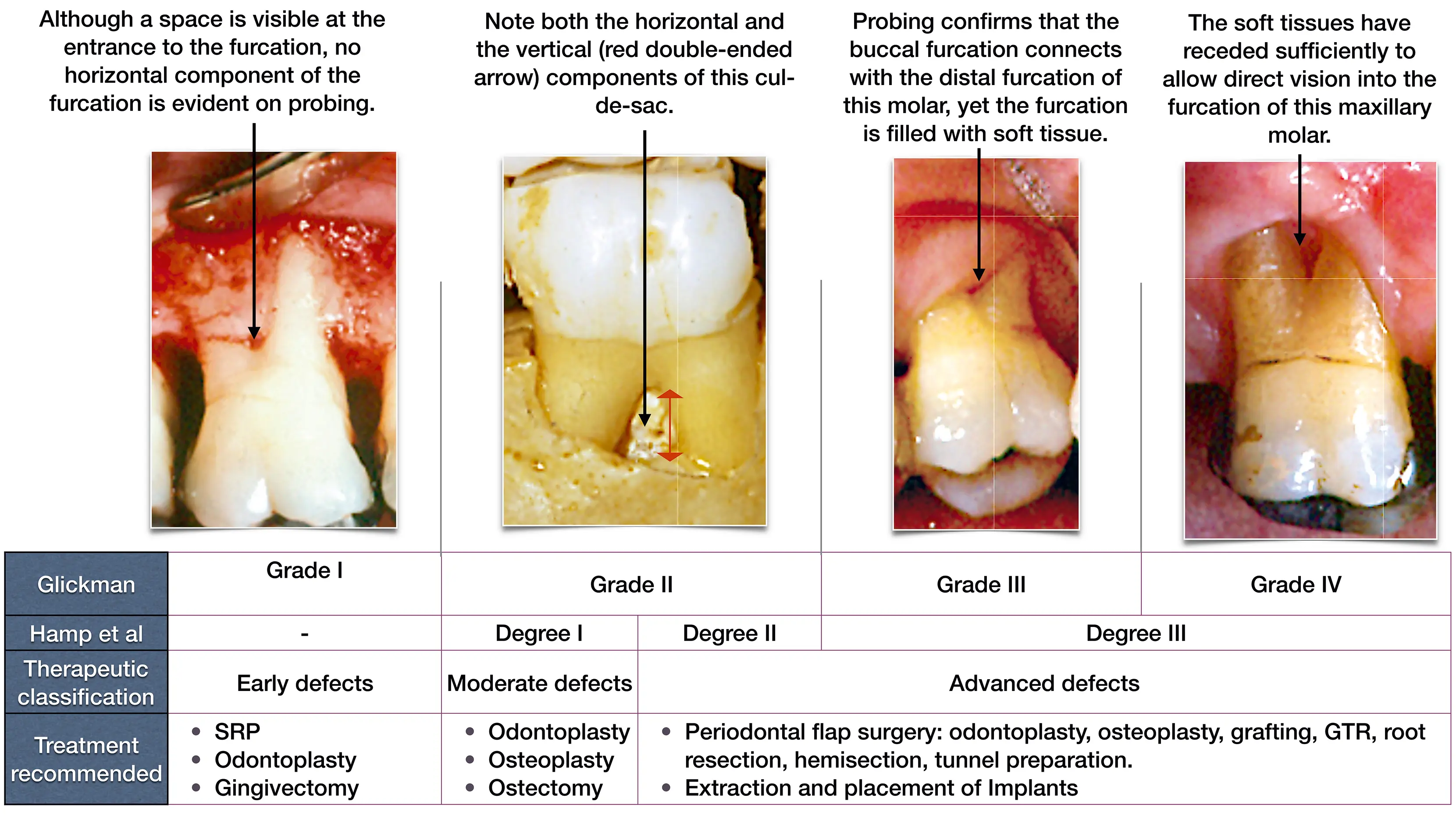
- glickman, hamp
Therapeutic classes of furcation defects
- early defects
- amenable to conservative periodontal therapy
- resolve overhanging margin, facial grooves or CEP by odontoplasty/recontouring
- resolution of inflammation and repair of PDL and bone should be sufficient to restore periodontal health
- moderate (early grade II)
- shallow horizontal involvement without significant vertical bone loss usually responds favourably to localised flap procedures with odontoplasty, osteoplasty, ostectomy
- advanced (late grade II, III and IV)
- significant horizontal component or deep vertical component added more problems
- nonsurgical treatment is usually ineffective because the ability to instrument the toot hsurfaces adequately is compromised
- perio regen or resective surgery, endo therapy, hemi section, root resection or tunnel preparation may be required
- GTR regen in grade II mandibular molar is well indicated
- sometimes treatment is more predictable with exo and dental implant
Prognosis of furcaiton involved teeth
- Hirschfield and wasserman 1978
- 7% overall tooth mortality in 600 patients over average 22 years
- 31% tooth loss in multirooted teeth with furcation involvement
- Mcfall 1982
- 10% overall tooth morality in 100 treated patients over 19 years
- 57% tooth loss in multirooted teeth with furcation involvement
- i.e grade 2 furcation is poor prognosis and furcation involved teeth are hard to treat
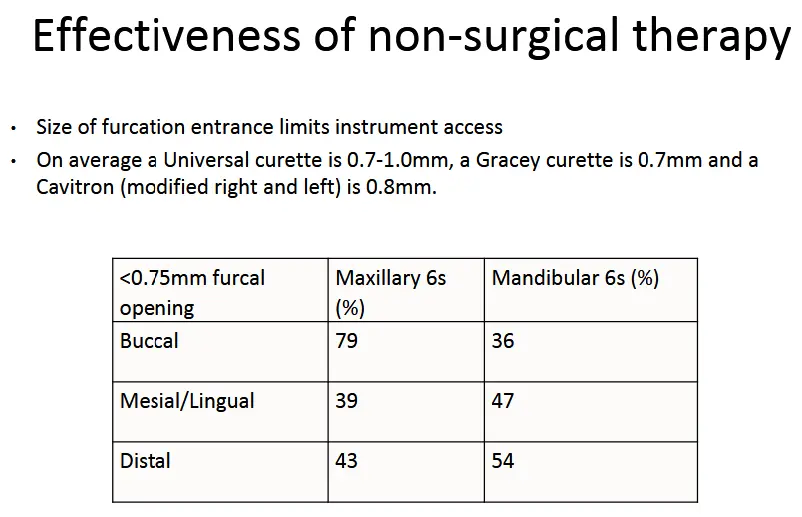
Loos et al 1989
- 24mo observational study of molar furcation, molar flat and nonmolar sites
- 1682 sites, 139 moalr furcation sites
- sites <3.5mm no change
- sites 4-6.5mm,
- non molar 1.5mm reduction after 3months
- molar furcation sites 24 months similar to baseline (therefore relapse is common)
-
7mm sites
- moalr furcation less reduction than others, less than 2mm with tendency to rebound
- conclusion
- furcation sites are true compromised healing response due to anatomy of furcation
- complicated by poor accessibility for instruments to furcation roof which have concavities and other surface irregularities
Odontoplasty/Furcationplasty
- resective technique to eliminate interradicular defect
- mainly buccal furcations class I, early II
- involves odontoplasty + osteoplasty
- technique
- full thickness flap
- granulation tissue removal + SRD furcation area
- odontoplasty -> remove crown and root tissue from furcation area, decrease horizontal component of defect and widen furcation entrance
- osteoplasty -> recontour alveolar bone crest to lessen buccolingual dimension of bone defect
- reposition and suturing of flaps at level of alveolar crest to cover furcation entrance
- should result in papilla like tissue close to entrance of furcation
- risk of sensitivity caries with extent of tooth structure removal
Tunnelling procedure
- coverts grade III and deep grade II furcations into grade IV furcations to improve access for oral hygiene
- indication
- deep horizontal defect
- short root trunk
- wide separation angle
- long divergence between roots
- appropriate teeth
- majority of mandibular first molar
- some mandible 2nd and maxillary 1st molar
- technique
- B and L flap retraction
- granulation tissue removal + SRD
- furcation area widened by removal of some interradicular bone
- tooth recontouring to establish enough space
- apical positioning of flaps
Root resection
- root amputation
- removal of root from multirooted tooth
- hemisection/bicuspidisation
- surgical separation of a multirooted tooth with removal of a root and associated portion of the crown
- preferred roots to amputate
- DB root maxillary first molar
- d root mandibular first molar
- indication/contraindications
- root trunk length, shorter root trunk more favourable
- distance between roots/divergence makes separation easier
- length and shape
- fused roots
- access

- technique
- endo
- provisional
- root separation and resection
- definitive restoration
Root anomalies
- CEP
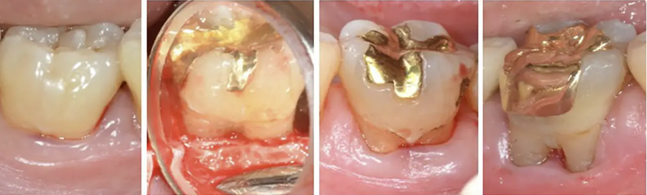
- lack of connective tissue attachment to enamel surface
- mandibular moalrs 25-35.5%
- maxillary molars 8-17%
- Md 2 > Mx 2 > Md 1 Mx 1
- enamel pearl
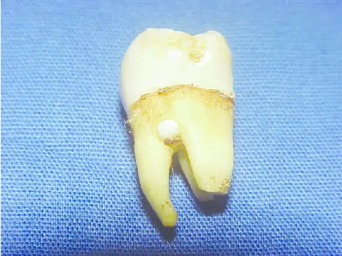
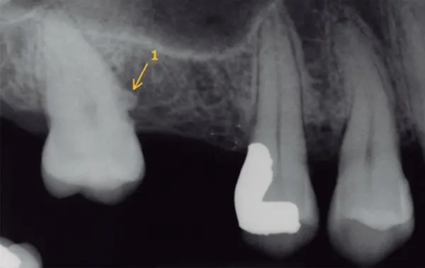
- .3-2mm dimension, plaque retentive factor
- 1-5.7% of all molars
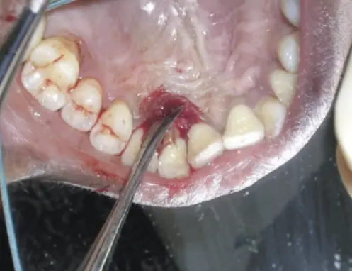
- developmental grooves (usually maxillary lateral incisors)
- 2.2% prevalence
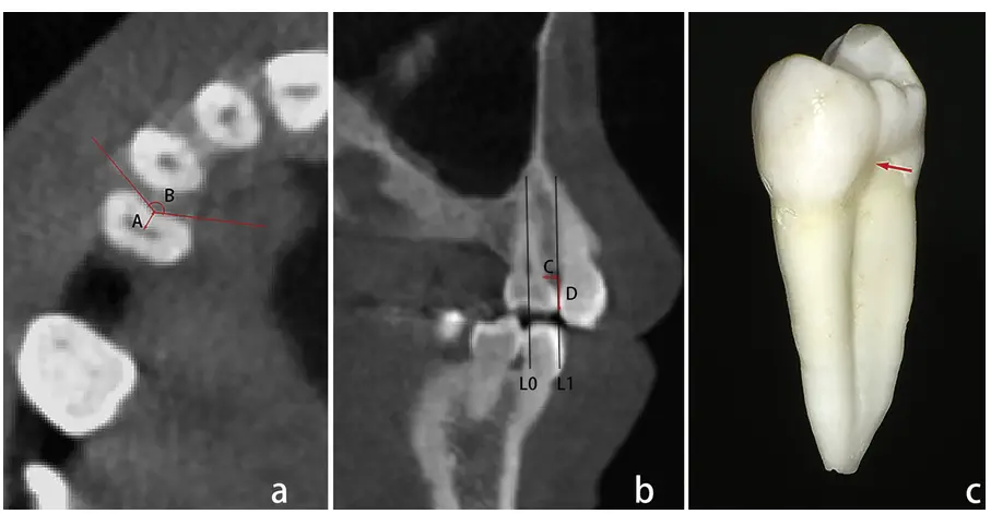
- root concavities
Traumatic Occlusal forces
Definitions
- traumatic occlusal force (TOF)
- any occlusal force resulting in injury of the teeth and or periodontal attachment apparatus (exceeding reparative capacity of PDL)
- occlusal trauma OT
- histologic term
- term used to describe the injury to the periodontal attachment apparatus including PDL, cementum, adjacent bone
- fremitus
- palpable or visible movement of a tooth when subjected to occlusal forces
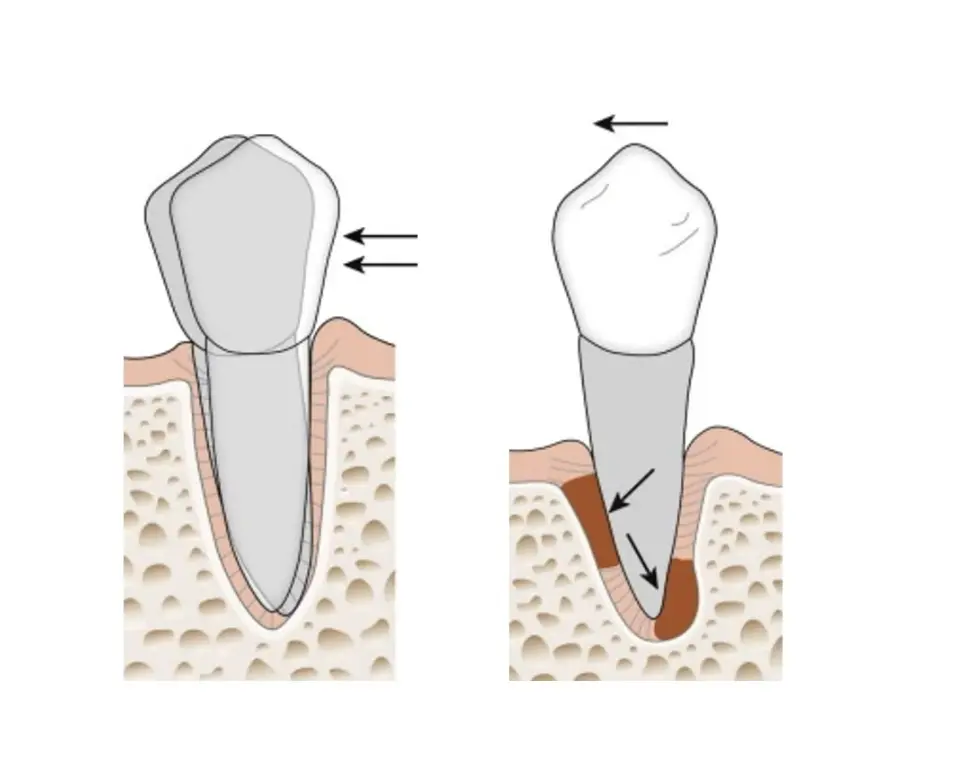
Non axial forces
- same as ortho, bone under compression resorb and bone under tension deposit
Mobility
- adaptive (physiological)
- destructive (pathological)
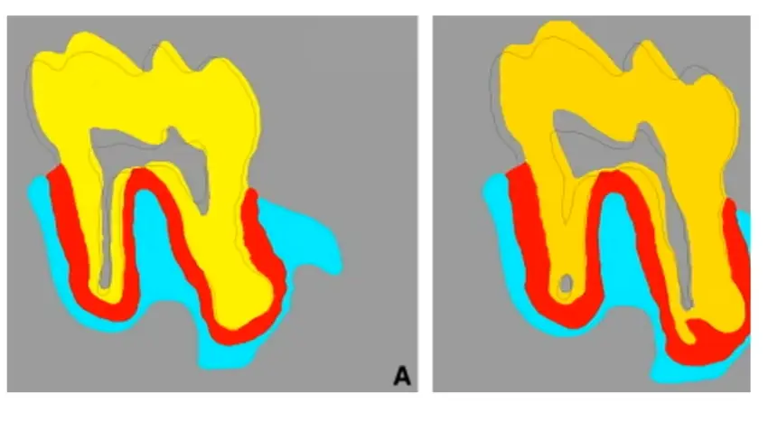
- depends on
- how much force
- and how much tissue is there to support
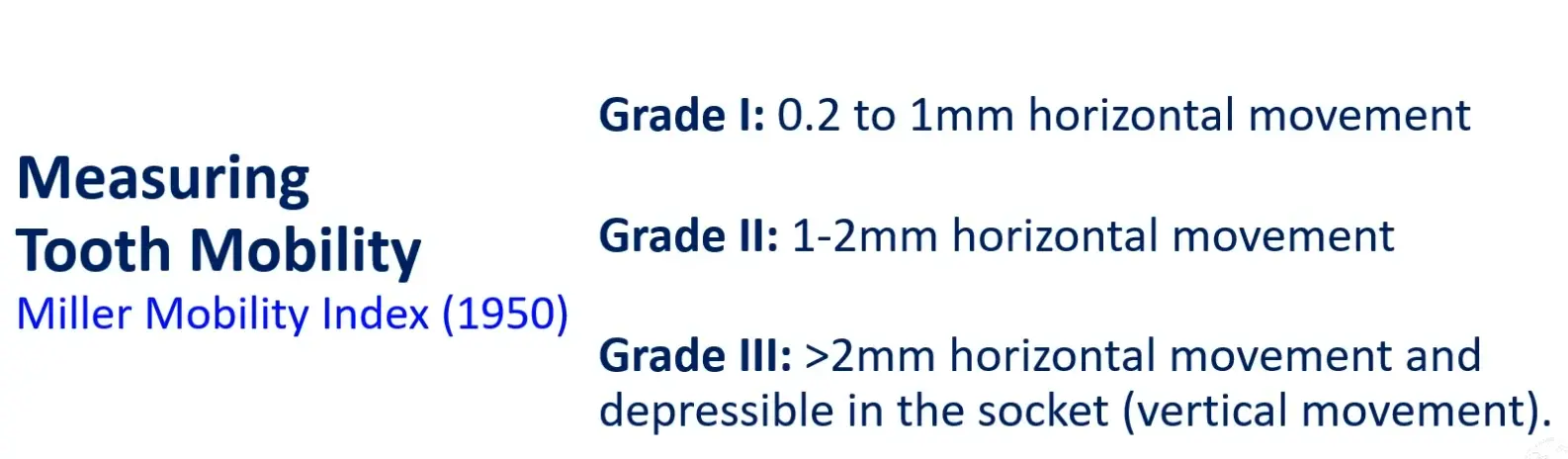
DDx mobility
- perio
- occlusal trauma
- trauma
- crown root fracture
- infection
- root resorption
- neoplasia
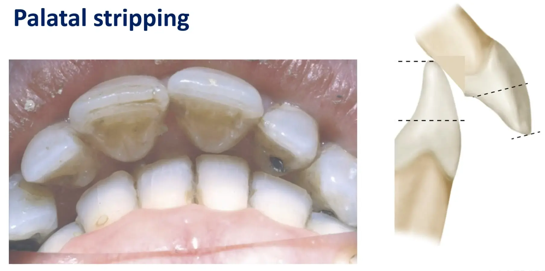
Jespen et al 2018 Classificaiotn of traumatic occlusal forces
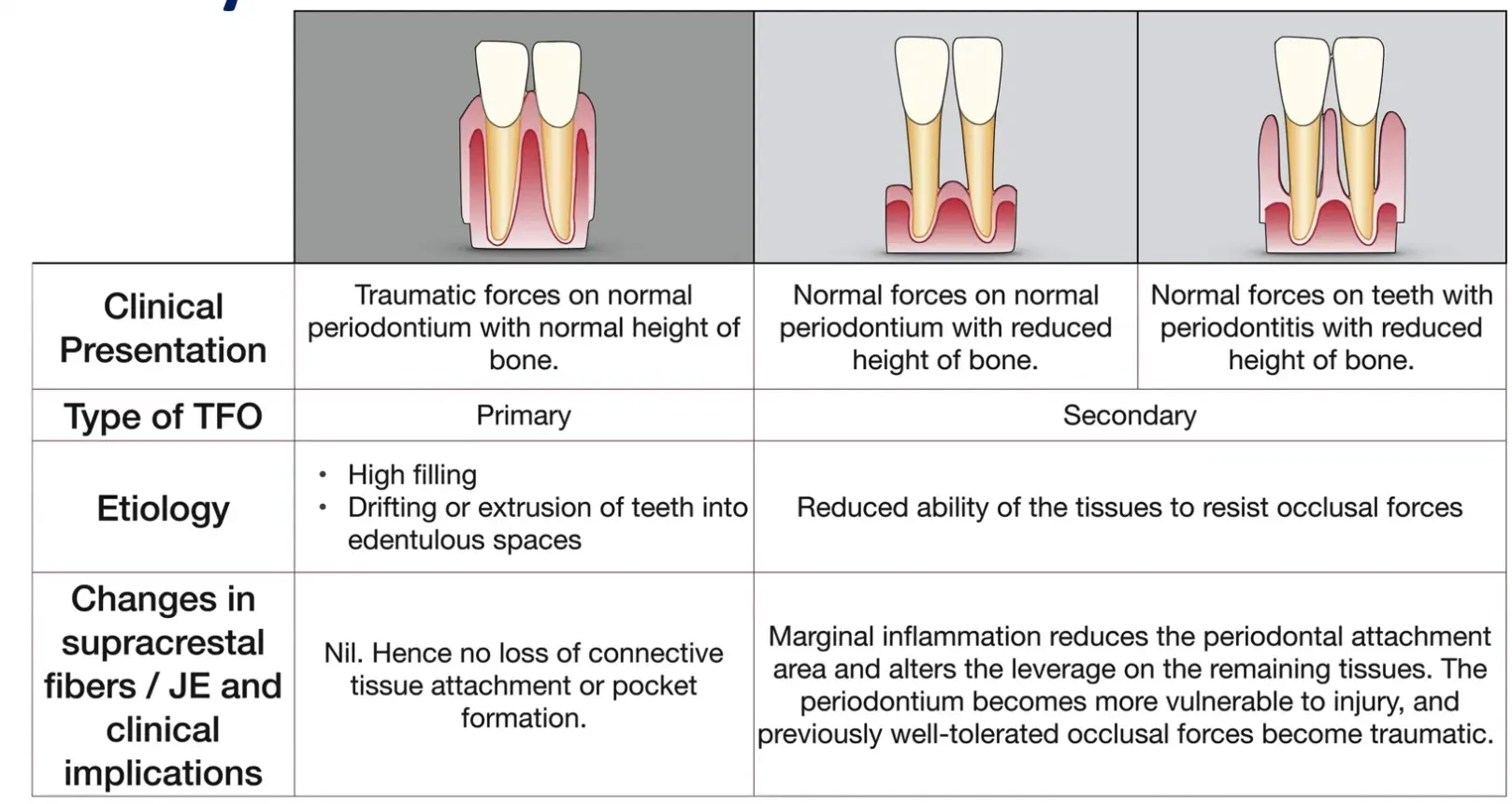
- primary occlusal trauma
- injury to periodontal tissue from traumatic occlusal forces applied to tooth with normal periodontal support
- manifests as adaptive, non progressive mobility
- secondary occlusal trauma
- injury resulting in tissue changes from normal or truamatic occlusal forces applied to a tooth or teeth with reduced support
- teeth with progressive mobility may also exhibit migration and pain on function
- orthodontic forces
Signs and symptoms
- fremitus
- tooth mobility
- thermal sensitivity
- excessive occlusal wear
- tooth migration
- discomfort/pain on chewing
- fractured teeth
- radiographically widened PDL
- root resorption and or hypercementosis
- cemental tear
Tenuous evidence
- no evidence that traumatic occlusal force or occlusal trauma causes periodontal attachment loss or an increase in inflammation in humans
- however evidence exists of traumatic occlusal forces associated with severity of periodontitis
- reducing tooth mobility through occlusal adjustment/splinting may improve treatment outcomes
Occlusal interferences as a risk factor
- despite conflicting evidffdence, occlusal interference have a negative effect on periodontium and tend to cause more rapid pocket formation and poorer prognosis when compared to teeth that do not have occlusal intereference
- confusion stems from past history of research attempting to define occlusal forces as having causative role rather than risk factor for perio destruction
Assessment
- ask if any loose teeth or difficulty during chewing/function
- check for fremitus during centric occlusion and protrusive/excursive movmenet
- signs of pathologic tooth migration
Management
- periodontal therapy - spontaneous correction of tooth migration
- occlusal adjustment
- reshaping surfaces to make harmonious contact
- splint mobile teeth - for patient comfort and or aesthetics
- occlusal splint therapy for parafunctional habits
- orthodontic correction if periodontal health is stable
Supracrestal tissue attachment
- also known previously as biologic width
- histologically composed of junctional epithelium and supracrestal connective tissue attachment
- infringement within the supracrestal connective tissue attachment of restorative margins is associated with inflammation and loss of periodontal supporting tissue