Module 6
#DEN4002
#D4/S2/W6
#OralSurgery
Third Molar Surgery
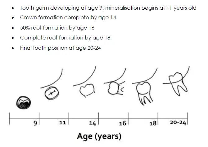
Wisdom teeth Epidemiology
- 96% of people develop at least one wisdom tooth
- 72% develop all 4
- 10% develop only 3
- 9% develop 2
- 5% develop only 1
- 4% develop none
- variably present depending on populations studied and are the most common congenitally absent tooth
Theories of impaction
- predominantly based on third molars being last to erupt and subsequent lack of space for eruption
- differential growth of mesial/distal roots
- inadequate arch length/space by the time third molars are erupting
- third molar development lagging behind jaw growth
- modern diet consists of highly processed food therefore teeth are less likely to wear
- 46% impaction rate for upper wisdom
- 72% have at least one lower wisdom impacted
- if left alone between 18 and 26, 55% of non impacted teeth will erupt and 33% of impacted teeth will erupt showing significant shift through the bone (some studies show movement still occurring between 20 and 32)
Indications
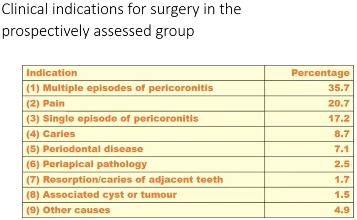
indications
- typically some type of progressive and non irreversible pathology
- caries
- periodontal disease
- damage to adjacent tooth (root resorption, caries, perio)
- pericoronitis
- odontogenic cyst or tumour
- less common
- impaction under dental prosthesis
- reduce mandibular fracture
- facilitate orthodontic healing
- optimal periodontal healing
Pratt et al 1998
Pericoronitis
- acute localised soft tissue infection associated with impacted tooth
- pain, oedema, erythema, purulence, halitosis, trismus, dysphagia
- infection may spread to head and neck, posing risk to airway
- causative organisms
- obligate and facultative anaerobes
- mostly red and orange complexes
- commonly, alpha-haemolytic streptococci, genera prevotella, veillonella, bacteroides, caphnophaga
- Nonsurgical treatment options
- LA / debride / Oral hygiene
- pain management
- systemic antibiotics if at risk
- surgical treatment options
- operculectomy
- pericoronal ostectomy (removing bone to aid eruption)
- extraction
- opposing tooth, maybe in advanced case with long wait time for surgeon
- involved tooth, definitive solution
Other reasons for removal
- other indications
- PPD >5mm on adjacent 2nd molar
- orthodontic/orthognathic surgery
- interference under denture
- food impaction discomfort
- medical reason, organ transplant, radio, chemo, joint prosthesis, preventing potential pathology which may be more risky in certain groups
- prevention of mandible fractures, can reduce 2.8X for high impact contact sports angle fracture
- occupational indications
- tooth which lines in line of an existing mandibualr fracture
AAOMS 2007 white paper on third molar data (Pogrel)
is it possible to predict the course of third molars?
- not possible in all cases, but adequate space between anterior border of ramus and 2nd molar distal appears necessary for successful eruption
- eruption to occlusal plane does not imply good state of health, as adequate osseous space is not the same as physiologic space
- third molars may still change in position after age of 25
periodontal considerations
- presence of impacted wisdom
- adversely affects perio of adjacent sound 2nd molars as reflected in disruption of PDL, root resorption, PPD/CAL
- removal of impacted wisdom
- can negatively impact periodontium of 2nd molars, preop assessment of intrabony defect, age level of plaque control can help to predict adverse outcomes
- reduction in postop CAL
- no surgical approach can minimise loss more than others
- GTR and/or DBP may be beneficial where there is already pre-existing CAL
- NSPT and good OHI have potential to reduce postop CAL
- perio disease severity
- patients with visible third molars have greater overall levels of perio severity
- perio disease progression
- presence of wisdom associated with 5mm or greater PPD on 2nd molar distal, including progressive PPD even with asymptomatic wisdoms
- bacteria and mediators of inflammation
- visible wisdoms are associated with bacteria that are responsible for severe and refractory periodontitis, and biochemical mediators of inflammation
- treatment
- poorer treatment response when wisdoms are associated with perio, symptoms may reduce but microbial counts will not reduce to level of control
- NSPT less effective when third molars present
- overall perio and thirs molar
- wisdom associated with overall elevated perio in immediately adjacent teeth
- adjacent teeth perio and wisdoms is progressive and only partially responsive to therapy
Microflora
- absence of symptoms does not indicate absence of disease or pathology
- red and orange complexes in clinically significant numbers can exist in and around asymptomatic wisdoms
- perio disease progresses in absence of symptoms
Age and wisdoms
- perio defects deteriorate with increasing age in presence of retained third molars
- caries in erupted third molars increases in prevalence with increasing age
- incidence of postop morbidity following wisdom removal is higher after 25 years
- germectomy (removal of wisdom <1/3 root formation) associated with lesser post morbidity
Is wisdom associated with crowding
- Likely that wisdom play role in etiology of crowding, but only one factor
- crowding is multifactorial and cannot easily predict with regards to wisdoms
- state of knowledge does not allow us to identify with accuracy who is at risk
To remove asymptomatic impacted third molars under rempros?
- position and disposition of unerupted teeth is dynamic and unpredictable
- ultimate decision must be under clinical exam and review of age, position of tooth, anticipated difficulty of removal, type of prosthesis and risk associated with removal
CT and wisdoms
- in 2007, unable to tell
Coronectomy
- should be considered in cases where tooth must be removed and IAN close association
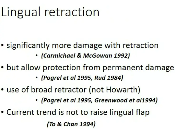
Lingual flap elevation and retraction
- raising and retraction lingual flap is acceptable where clinicians see fit
- periosteal elevator must remain subperiosteal at all times, and no sharp edges on lingual retractor
Should anything be placed in the socket following third molar removal
- GTR, DBP, PRP work in high risk or near high risk third molars
Nerve damage
- occasional damage to IAN and lingual nerve occurs following lower wisdom removal
- at least 50% of cases recover spontaneously
- nerve surgery results are variable, 50% improvement when performed between 4.5-7months
- later repairs can still show some recovery
Contemporary Management of Third Molars ADA Hyam 2018
Classification
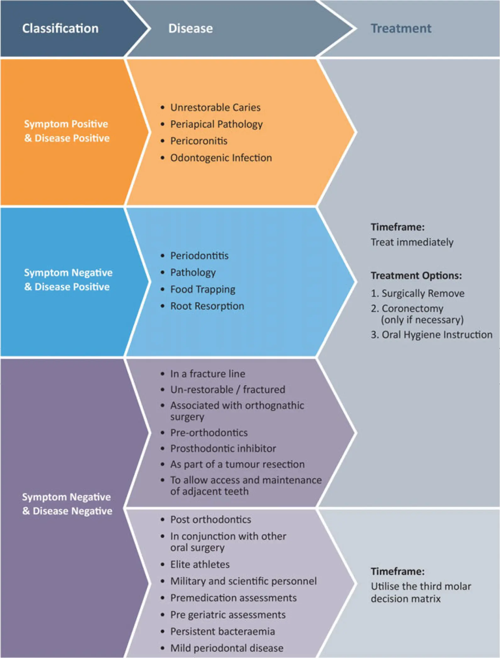
Decision matrix
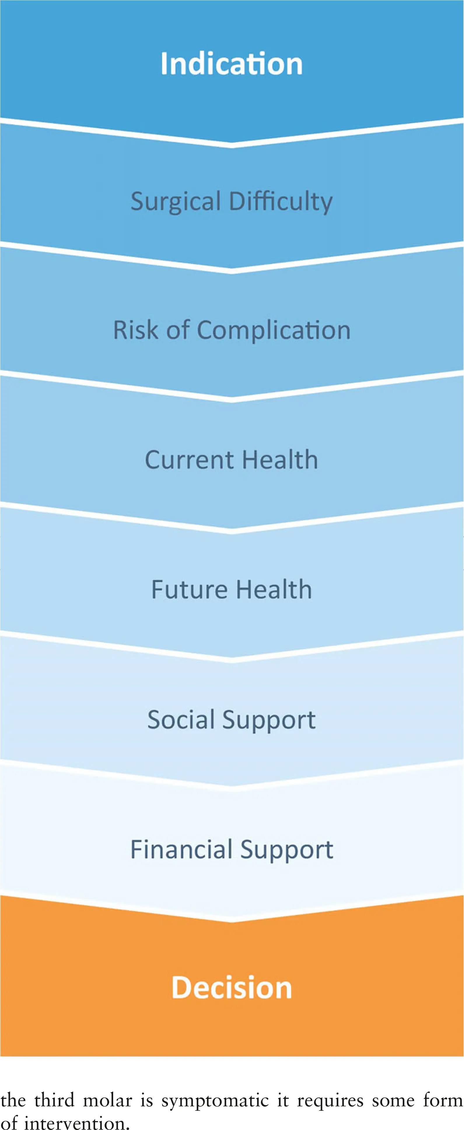
Prophylactic removal (six main groups)
- sport or military
- contact sport
- peak performance
- reduce physical dexterity
- tremors
- neuromuscular disease
- altered mental capacity
- dementia
- cognitive development
- pre medication
- bisphosphonates
- anticoagulants
- pre treatment
- radiotherapy
- chemotherapy
- immunomodifier therapy
- bacteraemia
- tissue transfer patients
Considerations of prophylactic removal
- judge likelihood of tooth needing removal will acquire disease/symptoms in particular future period, balance this risk against surgical removal
- must feel that the patient will be better for having had surgery at this time versus surgery at a later time where procedure may be more complex, recovery more tenuous and complication rate higher
Surgical assessment
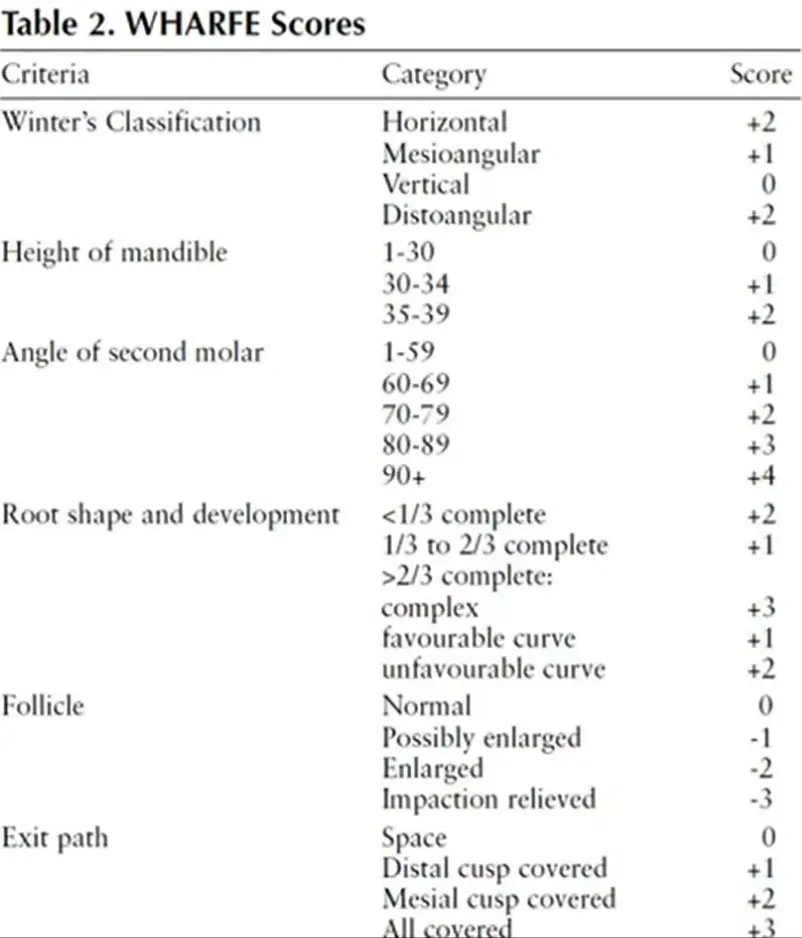
- WHARFE
- winters lines
- height of mandible
- angulation
- root form
- follicular sac size
- exit pathway of tooth
risk of complication
- experience, IAN proximity, lingual nerve, age, recovery
Current health modifiers
- cardiac state, immunocompromise, coagulation function,
- bisphosphonates and denosumab
- respiratory function
- fitness for type of anaesthesia and pregnancy status
Future health modifiers
- commence medication, tissue transfer procedure, become pregnant
Social modifiers
- access to care and support post surgery
- transport/home help
Financial Modifiers
- insurance plans
- loss on income due to recovery period
Complications
Contraindications
- patient too young <12yo, difficult to predict future eruption potential
- older patient group, >40y, risk potentially outweighs benefits
- medically compromised patients who are unfit for surfery
- extreme limitation in mouth opening
- unacceptable risk of IAN injury
- lack of surgery experience
Types of complications
- inadequate assessment
- inadequate premedication/preparation
- inadequate anaesthesia
- incorrect surgical techniques
- immediate postop complications
- delayed postop complications
Minimising complications
- proper patient preparation
- asepsis
- meticulous management of hard and soft tissue
- controlled force when applying instruments
- haemostasis
- adequate postoperative instruction
Patient Assessment
Patient level factors
- age, sex, race
- radiograph, CT scan
- presence of infection, swelling, lymphadenopathy, trismus, purulent discharge
- pt anxiety
- access, degree of mouth opening, size of tongue, oral cavity, crowdinging
- pericoronitis and soft tissue
- anatomy
Radiographic Assessment/ Classification
Elements of difficult surgery
- distoangular
- class 3 ramus (tooth completely in ramus)
- class C depth
- long, thin roots
- divergent curved roots
- narrow PDL
- thin follicle
- dense, inelastic bone
- contact with second molar
- close to IAN
- complete bone impaction
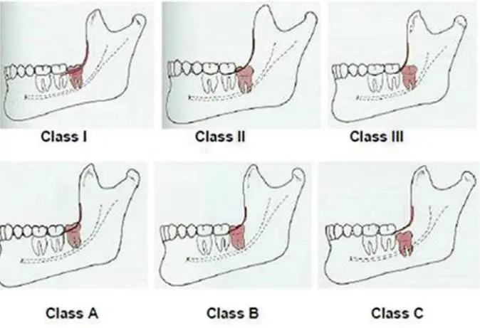
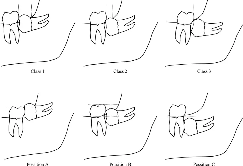
Gregory and pell
- degree of impaction of third molar in vertical (ABC) and horizontal dimensions (123)
- vertical/ABC, comparing occlusal surface to occlusal plane and neck of 2nd molar
- A: at or above occlusal plane
- B: between occlusal plane and neck of 2nd molar
- C: below neck of 2nd molar
- horizontal/123, comparing distance from distal2nd molar to anterior ramus, to AP size of wisdom
- 1: ramus to 2nd molar is larger than wisdom
- 2: ramus to 2nd molar is smaller than wisdom
- 3: no space between ramus and 2nd molar (wisdom completely in ramus)
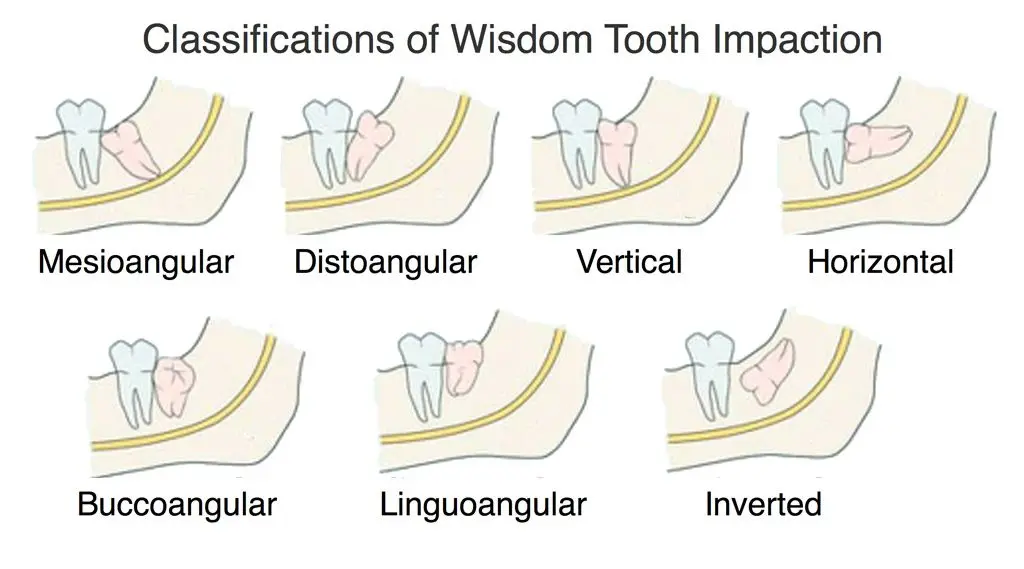
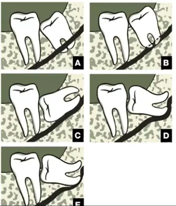
Winters classification of angulation impaction
Relationship to the inferior alveolar nerve
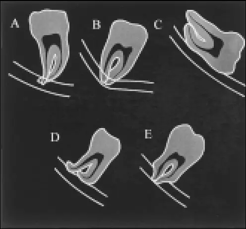
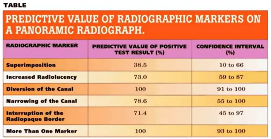
- 5 main radiographic markerrs on OPG
- radiolucency across roots
- deviation of mandibular canal
- interruption of canal cortication continuity
- deflection of roots by canal
- narrow of roots
Factors associated with less complicated surgeries
- mesioangular
- class 1 ramus
- class A depth
- roots 1/3-2/3 formed
- fused conical roots
- wide PDL
- large follicle
- elastic bone
- separated from 2nd molar
- separated from IAN
- soft tissue impaction
FOR MAXILLARY WISDOMS
- 1st level: fully or partially erupted
- 2nd level: crown margin below apices of adjacent 2nd molar
- 3rd level: crown above apices of adjacent second molar (high risk of displacement into infratemporal fossa or antrum)
Surgical Process
recipe
- adequate prep
- patient prep
- anaesthesia
- positioning
- prepare instruments
- raise flap
- bone removal
- tooth sectioning, delivery
- wound toilet
- wound closure
Raising a flap
- 4 types of incision
- distal reliving incision
- envelope incision
- limited access for deep impaction
- buccal extension flap
- increased access
- potential perio issues
- triangular flap
Bony removal
- high power airless handpiece, protect flap with Minnesota
- round or flat fissure surgical bur
- gutter around tooth
- expose entire crown/furcation point/ height of contour
- create purchase point
- removal of distal bone (if ramus is capturing)
- make sure lingual flap retracted
Sectioning tooth
- depends on angulation of impaction and depth of impaction
- section of crown in horizontal impaction
- division along longitudinal axis in mesioangular impaction
- sectioning distally impacted tooth with caution
Wound toilet
- may be filled with bony debris, cystic lining, dental follicle lining, granulation tissue
- irrigation with normal isotonic saline
- special attention to space under flap
- removal of all surgical debris and loose bony dust
- adequate haemostasis so no postop haematoma
Wound closure
- key suture directly behind second molar
- accurate apposition of wound edge
- second suture across distal relieving incision
- avoid water tight closure
- resorbable sutures
Minimising complications
- antibiotics placed in alveolar socket to decrease bacterial plaque (Olech 1953, Verbic 1953)
- use of preop antibiotics
- little gain with postop ab alone martin et al 2005
- use of chlorhexidine mouth rinse prior to surgery (Rango and Szkutnik 1991)
- Improving oral hygiene preoperatively (Penarrocha et al 2001)
Maxillary molars
- flap design
- distal incision sagittally along maxillary tuberosity and diagonally across tuberosity
- mesial incision across buccal attached gingival extension according to heigh of impaction
- removal of buccal bone
- should be quite membranous and easy to remove
- elevation
- fine curved elevator
- elevate buccodistally
- protect with retractor to prevent displacement
- wound toilet
Prophylactic corticosteroids
- to be effective at minimising oedema steroids must be given in doses exceeding physiological levels
- decrease levels of lymphokines, prostaglandins, bradykinin, serotonin, cortisol and perhaps histamine
- using a steroid with maximum antiinflammatory and minimum gluco and mineralocorticoid properties seems best
- fluorinated steroids such as dexamethason are well fit, with dexamethasone having only minimal immunosuppressive functions
- contraindications
- active or treated TB, fungal or viral infection (especially ocular herpes), glaucoma, psychosis
- peptic ulcer disease, cushings, diabetes, hypertension, pregnancy, myasthenia gravis
- Pederson et al, Bierne et al support significantly decreased swelling without increased infection or orthoer complication, dexamethasone has a long half life and is useful as prolonged courses up to 1day postop are recommended to reduce rebound swelling
Dry socket
- chlorhex rinse for streptokinases
Nerve Injuries
Causes
- inadequate assessment
- incorrect surgical techniques
Seddon 1943 classification
- neuropraxia
- axonotmesis
- Neurotmesis
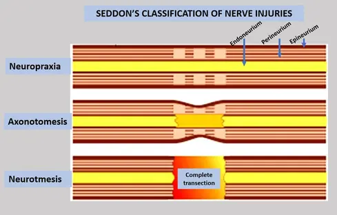
Pattern of sensory loss Merrill 1979
- hypoaesthesia - decrease sensitivity
- hyperaesthesia - increase sensitivity
- paraesthsia - abnormal sensation (pins and needles/tingling)
- dysaesthesia - unpleasant abnormal sensation (burning, sharp, aching pain)
- anaesthesia - total loss

Treatment options
- in case of witness transection
- immediate referral for surgical repair is necessary
- if anaesthesia post wisdom teeth surgery
- referral for assessment and possible nerve repair
- altered sensation in lip and chin
- just monitor and map for 6months
-
- if anaesthesia persists >6months
- further XR to assess continuity of mandibular canal
- if anaesthesia persists >6months
- consider surgical exploration and decompression or repair of nerve
Nerve repair options
- 3 types
- perineurial repair
- group funicular repair
- epineural repair -> most logical choice
- trigeminal branches are polyfasicular without grouping
- using 8-0 to 11-0 monofilament nylon, with minimal number (3-6 optimal) and close approximation
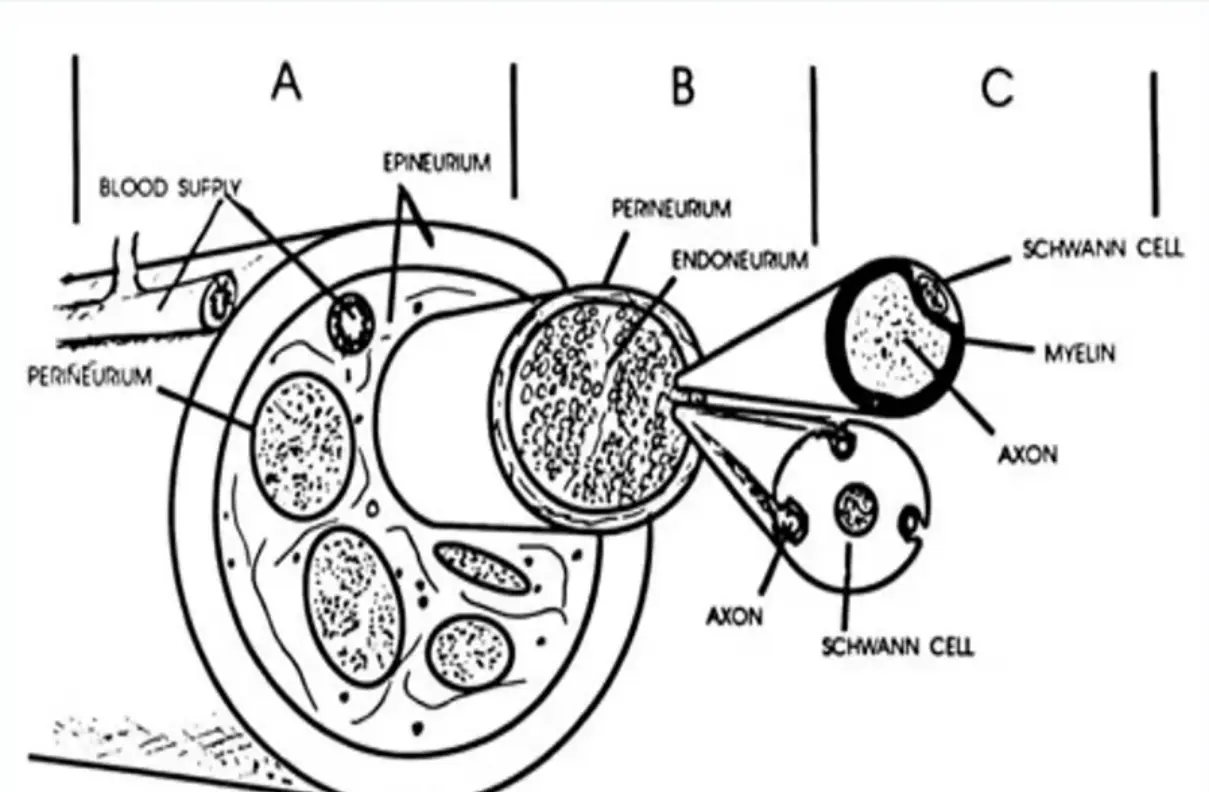
Lingual nerve paraesthesia
- ranges from 0.278%-13%
- clinically there is sensory disturbance
- hypoesthesia, hyperesthesia, anaesthesia, dysesthesia
- mechanisms of injury controversial