Diagnosis and Management of Cracked & Fractured Teeth
Biology of the dental hard tissues and fractures
Myth vs fact
- fractures are not actually a point of weakness, rather a strategy of damage containment for tooth survival
How do teeth protect themselves
- resilent, can absorb energy before suffering complete failure
- individual toot hstructure are brittle, but together can absorb and distribute energy before fracture
- have inbuilt mechanism to distribute and disperse forces and limit fracture propagation
- enamel tufts and lamellae provide compliance allowing distortion of enamel, tufts provide stress shielding
- strctural integrity
- biocomposite material - variable distribution of force
- force distribution is not even - ansiotropic
- fractures in teeth occur when biologic tolerance is exceeded
- stress can concentrate in different part of tooth
- depending on where stress accumulate will determine outcome
- can accumulate at
- cervica lrim (NCTL, abfraction)
- cusp tip (shovel shaped depression)
- marginal ridge (fracture)
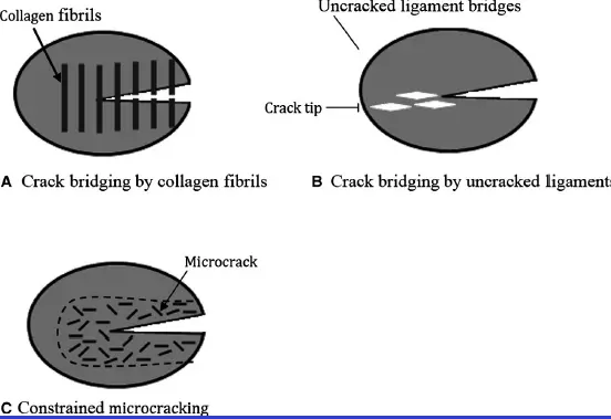
- fracture toughening in dentine to blunt fracture progression by deflection of fracture and distribution of stress concentration
- biomineralisation of dentine, preferable to retain pulp
- teeth with RCT lower survival than teeth with vital puolp
- teeth with vital pulp undergo continuing changes in mineralisation of dentine - biomineralisation
- dentine can adapt to forces and age changes that are placed on tooth
Epidemiology of fracture
FOrces may
- be absorbed and distributed within tooth hard tissue
- result in wear of the tooth
- result in fatigue deformation and fracture
- affect pulp with traumatic hyperocclusion
Classification of fatigue fractures in teeth
Diagnostic nomenclature
- abfraction noncarious cervical lesion
- infraction
- cracked tooth
- fracture
- split tooth
- root fracture
- cracked tooth syndrome
Classify fatigue fractures based on origin
- fracture
- broad term which means to break
- used to define separation of hard tissue
Fatigue fractures due to stress accumulation
- enamel origin
- enamel lamellae
- craze line
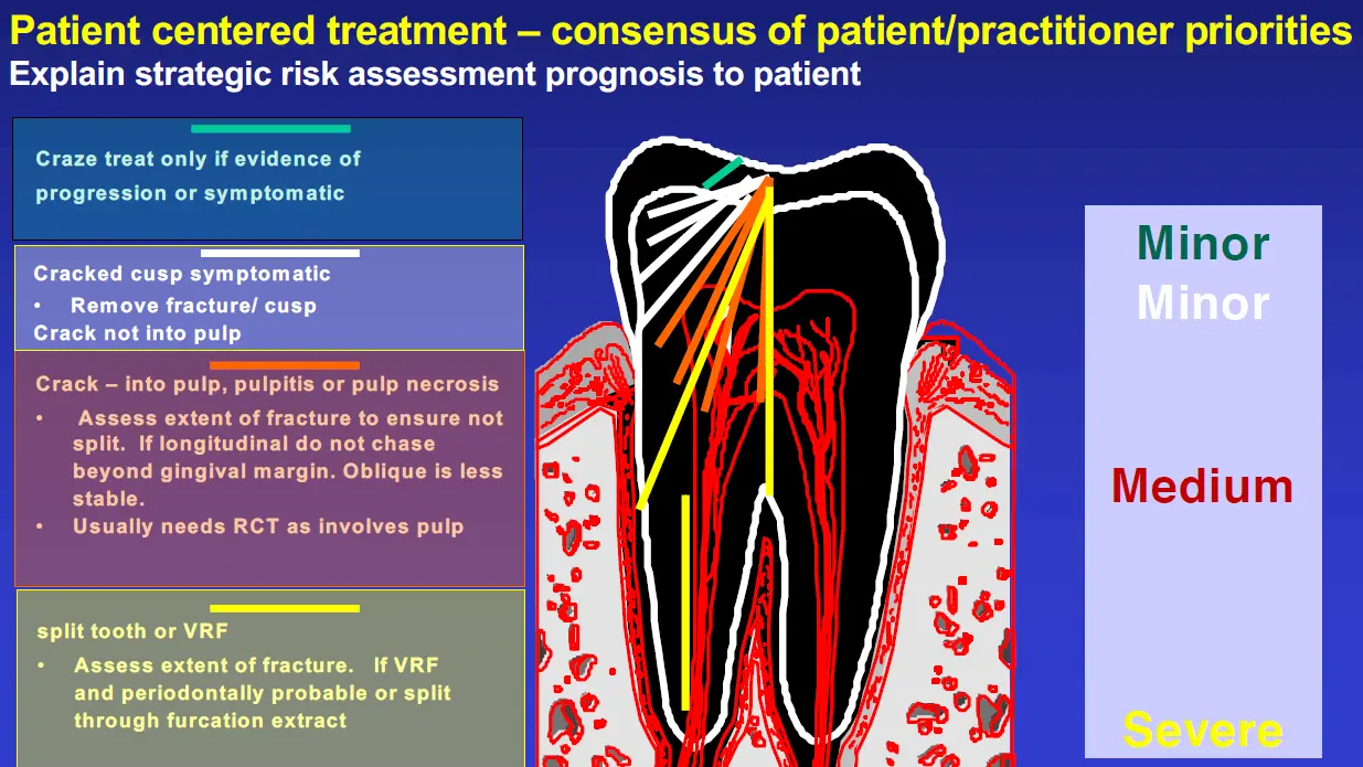
- crack (cracked tooth)
- split
- dentine origin
- cracked cusp
- cementum origin
- vertical root fracture (root origin fracture)
- cementodentinal tear
Acute Traumatic
- acute trauma induced crown fracture
- acute trauma induced crown - root fracture
- acute truama induced horizontal root fracture
Enamel Lamellae origin
- developmental defect
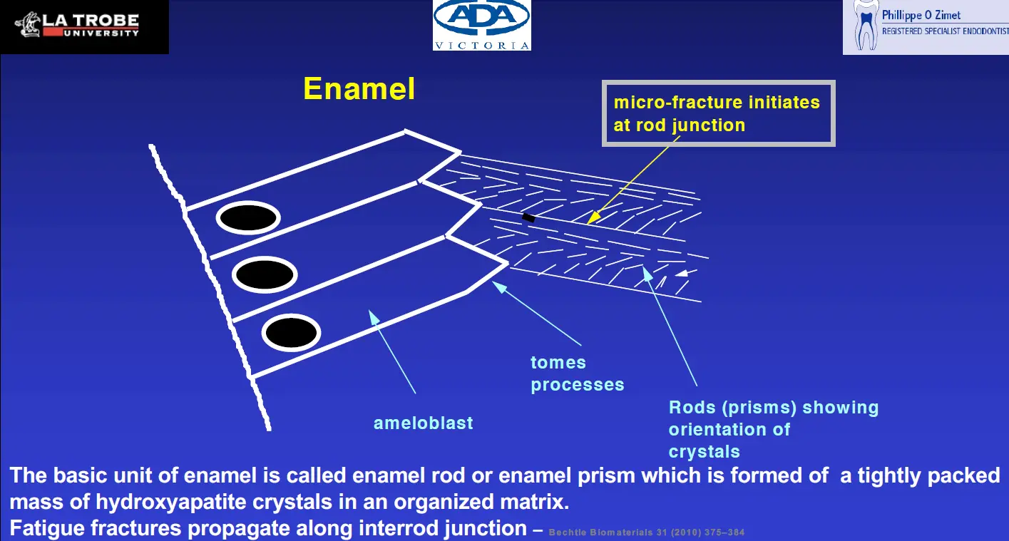
- developmental defects in surface of enamel that are visible to the naked eye
- enamel tufts and lamellae act to provide enamel compliance
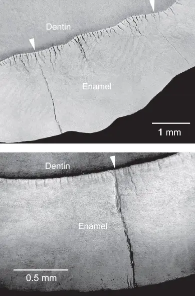
Craze or enamel infraction
- confined to enamel due to parafunciton or acute trauma
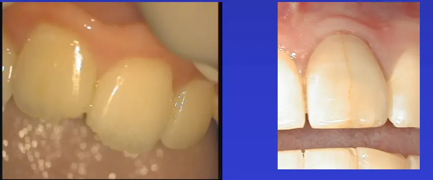
Crack
- incomplete fracture beyond enamel
- extends usually longitudinally from enamel wall
- may or may not be symptomatic
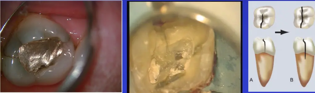
- may have associated probing defect
Split
- through and through fracture
- complete separation of fragments
- begins in crown
- may be associated with periodontal probing defects
- may be longitudinal or oblique
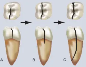
Cracked cusp
- begins in dentine under cusp
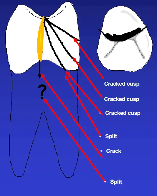
- extends laterally under one cusp extending no more than coronal 1/3 of root
- initially may be incomplete
- crack becomes a split when the fracture extends through the furcation
- cracked cusp becomes a split when cracekd cusp extends beyond coronal 1/3
Usually only detect and manage coronal fractures when it becomes symptomatic
- cracked tooth syndrome
- perio defect
- tooth chipping evidence
- fracture progression
- evidence of pulpal necrosis and infection
Cracked tooth syndrome
- not a fracture type
- defined set of symptoms from vital pulp inside a tooth with a fracture in hard tissue
- may be due to cracked cusp, crack, split tooth
- how is pain felt
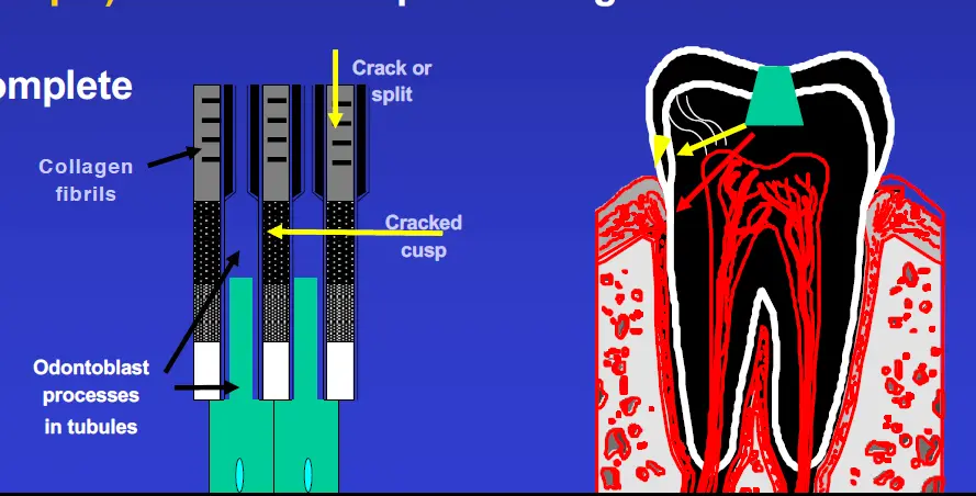
- pressure change in fracture - bidirectional fluid movement in pulpodentine complex - hydrodynamic or transduction theory where odontoblastic processes have a mechanosensory role
- direct stimulation of pulp by fracture
- inflammation of pulp from microorganisms in fracture line
Vertical root fracture
- root from RCT treated tooth is fractured
- begins in root
- rare without RCT
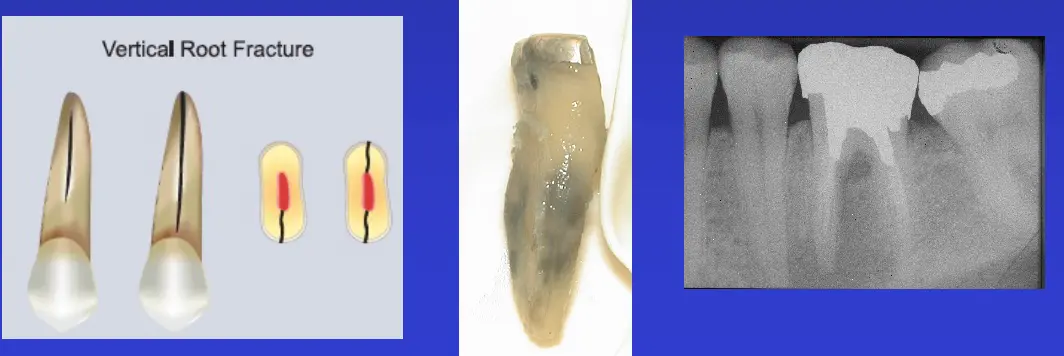
- may have narrow isolated probing
- can confuse with endo probing, perforation, split, palatal groove
- J shaped radiolucency
- most occur 2-5 years after RCT
- many explanation
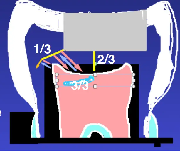
- parafunction - the occlusion and force accumulation in the root. Inability of root filled tooth root to adapt to stress concentration with calcific change.
- lack of coronal and cervical dentine allowing absorption of forces from direct crown limiting cusp flexure (crown helps absorb and distribute forces).
- lack of binding effect (ferrule) limiting root flexure as well as stress distribution through the cervical of the tooth
- method of obturation – use of SS spreader and not NiTi.
- method of instrumentation: apical preparation size, flare, type of instrumentation
- Width of direct post
- type of post system
- Over instrumentation of apical foramen causing apical crazing
- Use of expanding amalgam in canals – Nayyar technique
- Expanding root canal filling materials
- Lack of proprioception of teeth with RCF
- Loss of protective effect of pulp
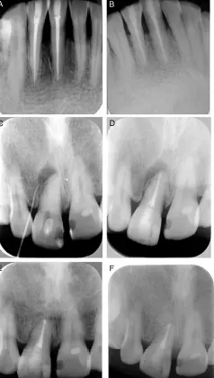
Cementodentinal tear
- tear of surface cementum with resorption of overlying bone
Clinical Management
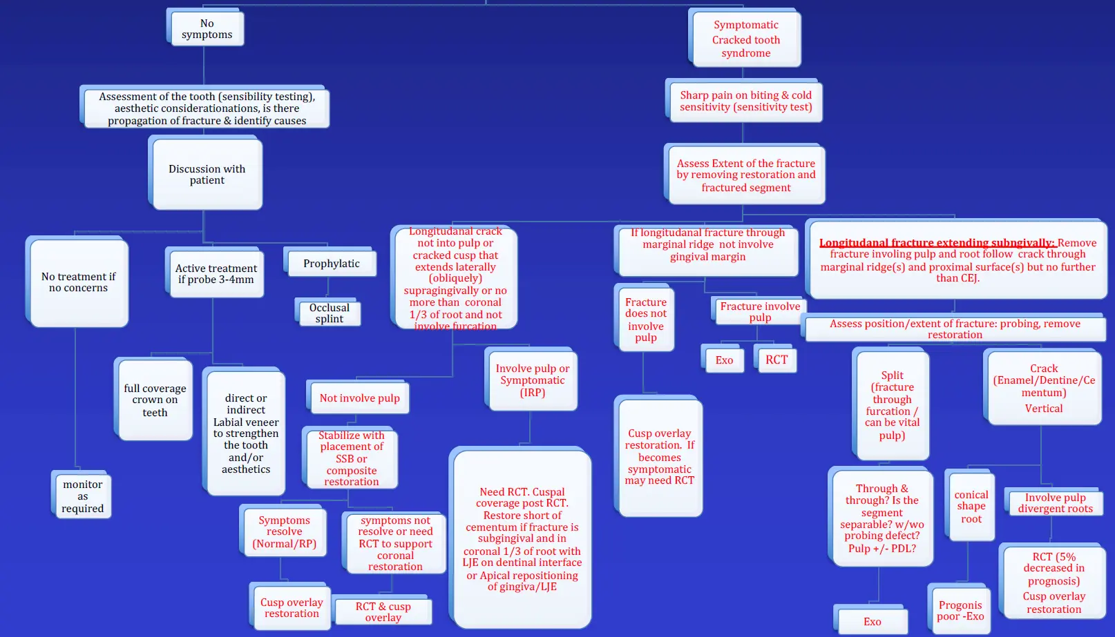
treatment plan based on signs and symptoms
- differential diagnosis
- informs risk assessment in 4 dimensions
- risk assessment prognosis
- guide treatment plan
- treatment plan
- details anticipated outcome
Differential diagnosis
- state of health of each tooth in 4 dimension
- endodontic - pulpal periapical
- strategic - stability/deterioration
- marginal periodontal - supporting bone
- structural - amount of tooth
Endo assessment and differential
- symptoms of CTS?
- pain on off biting at a particualr angle
- may respond to thermal change with allodynia and thermal hypersensitivity
- pathognomic for coronal fatigue fracture with vital pulp normal or pulpitis
- is there pulpitis as well as classic CTS
- thermal hypersensitivity and allodynia use pulp sensitivity test
- is there pulpal infection
- all tooth is TTP vs CTS only one cusp responsive usually
- no response to pulp sensibility
Strategic assessment
- bite sticks to locate fracture if CTS and vital pulp
- visual assessment (direct and indirect)
- PA radiograph may note shallow restoration in teeth with unusual pulp/periapical symptoms/signs
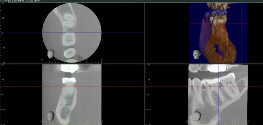
- fractures poorly detect in tooth on CBVT
- fracture needs to clear voxel size in width
- Voxel size = .08mm
- fracture must be atleast .15mm
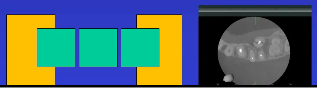
- CBVT may display pattern of bone loss to indicated crack of split or VRF in tooth
- not all marginal bone loss is due to root fracture
Perio assessment
- bone loss extent
Structural assessmentt
- how much tooth remains
- Risk assessment prognosis guides treatment plan
- excellent
- good
- fair
- borderline
- extraction
Endodontic management
- if tooth has symptomatic CTS don't just place band or crown
- 29% of symptomatic CTS required RCT after band placement (some had crowns placed)
- as part of endo management need strategic assessment of extent of symptomatic coronal fracture before stabilise the cusps as fracture may be into pulp
- any fracture that extends into pulp as longitudinal crack = RCT
- if not longitudinal coronal crack do not chase beyond gingival margin
- pulp infection or irreversible pulpitis = RCT
- survival outcome does not change for pulp necrosis and infection
- even 7mm probing results in 2-4 year survival
- remove resto and visual fracture
- remove fracture, is pulp exposed?
- ss if pulp not exposed and no irreversible pulpitis and restorable
- bands used for temporary restoration of teeth, not used for diagnosis
Strategic Management
- survival of RCT with longitudinal cracks
- coronal cracks predictable treatmetn - 99% survival at 5 years
- extension of fracture onto pulpal floor 88% survival at 5 years
- extract
- if longitudinal fracture through furcation - split
- if oblique fracture laterally beyond apical 1/3 - split
- if any fracture involves a conical shaped root
Perio Management
- maintain when lateral coronal fragment of cracked cusp extends less than 1/3 into root and not involve furcation = split
- prognosis reduced when fracture associated with more than 5mm pocket
- asymptomatic coronal fracture - crack + vital pulp
- if crack with 4-5mm probe and responsive to pulp sensitibility but not pulpitis treat with cusp coverage
- asymptomatic coronal fracture - crack + vital pulp
- if crack and probing 4-5mm and responsive to pulp sensibility but not pulpitis treat with cusp coverage
- no cusp coverage may result in fracture propagation
Structural management
- assess amount of remaining tooth after management of fracture especially if cusp lost
- usually place cusp overlay direct during disease control
Treatment Plan