Module 2
#DEN4002
#D4/S2/W2
#Periodontology
Acute Periodontal Conditions
American academy of periodontology classification
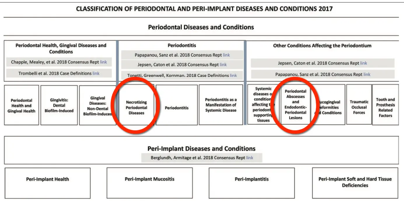
3 common features
- rapid onset
- rapid tissue destruction
- acute pain or discomfort
Necrotising Ging, Perio and Stomatitis
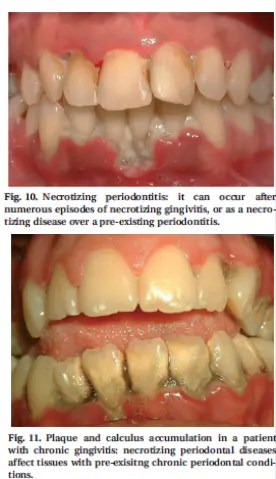
Necrotising periodontal disease
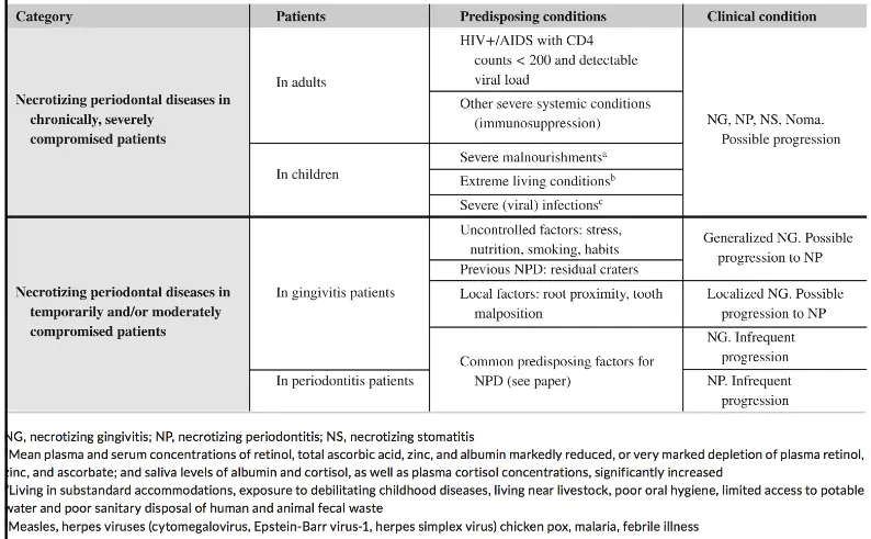
- diagnosed under many names for over 100 years
- vincents disease, trench mouth, ANUG etc.
- nectrotising periodontal diseases (AAP 1999)
- latest classification (Herrera et al 2017)
- necrotising gingivitis -> marginal gingiva
- necrotising periodontitis -> loss of attachment
- necrotising stomatitis -> beyond mucogingival junction
- cancrum oris / noma -> facial tissues
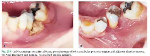
- 3 common typical features
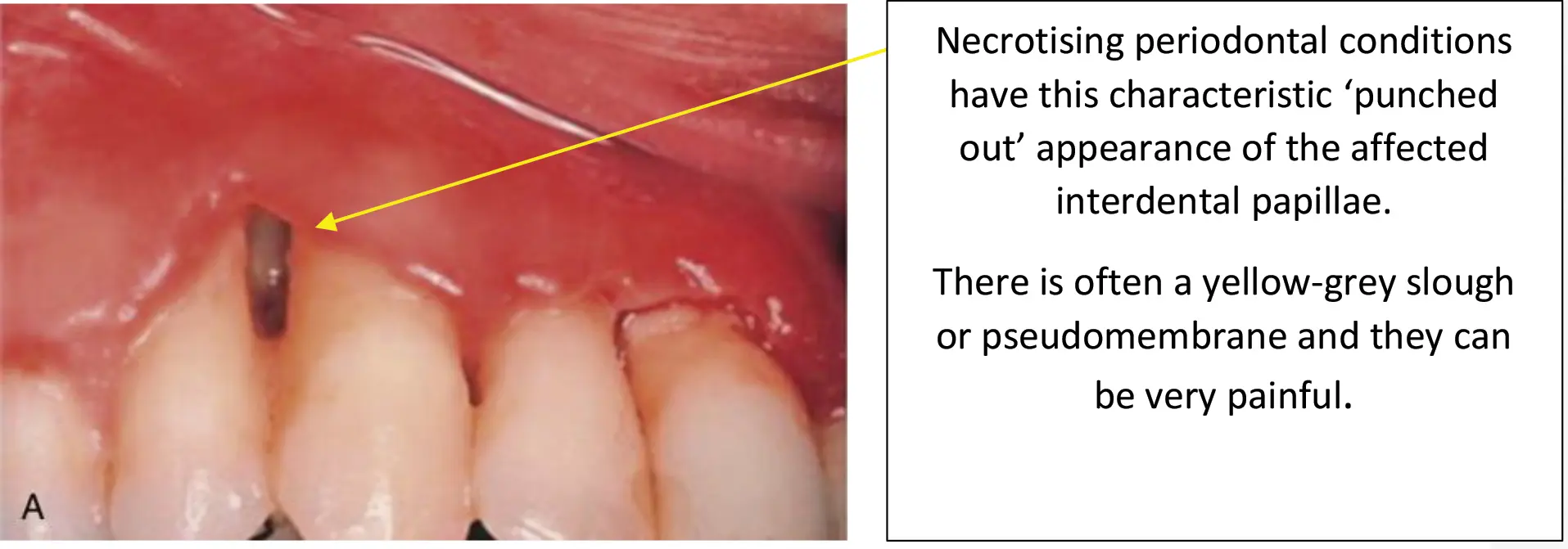
- necrosis of papilla
- bleeding
- sudden onset of pain
- pathogenesis
- opportunistic infection of commensal bacteria within plaque coinciding with a temporary suppression in host immunity
Necrotising gingivitis
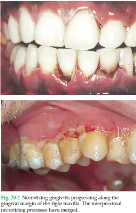
- clinical features
- pain, especially on probing
- gingival ulcers
- punched out craters of papillae
- may involve marginal gingiva
- anterior mandible most common
- spontaneous or readily bleed
- secondary features
- halitosis
- whitish/yellow pseudomembrane
- lymphadenopathy
- fever, malaise
- sialorrhea (excessive salivation) in children
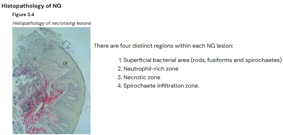
- typically due to specific bacteria
- spirochaetes e.g. treponema spp, fusobacterium spp, prevotella intermedia (orange complex)
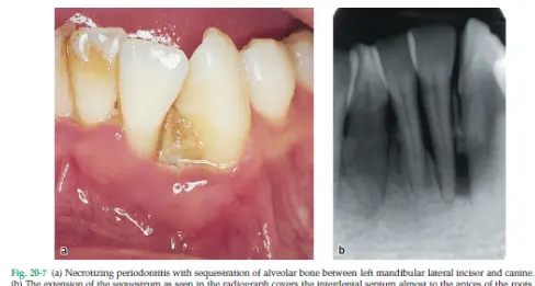
Necrotising periodontitis clinical features
- loss of attachment
- rarely deep pocketing as gingival necrosis accompanies bone loss
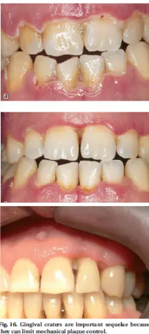
- deep interproximal craters divide papilla into facial and lingual
- denudation of interdental alveolar bone
- sequestration of necrotic alveolar bone
Necrotising Periodontal Disease
- local factors
- poor OH
- plaque retentive factors
- pre existing gingivitis
- history of NPD
- systemic factors
- immunocompromised host (e.g. HIV)
- stress
- poor nutrition
- smoking
- alcohol
- young age
- acute phase treatment
- pain relief and prevent disease progression
- gentle debridement under LA
- 3% H2O2 recommended by Robinson 2002
- CHX rinse, care with mechanical OH, remove plaque but no further trauma to soft tissues
- metronidazole 400mg bid 5 days + analgesia
- prevent dehydration
- subsequent maintenance phase
- treat pre existing periodontal disease
- corrective therapy of gingival craters
- HIV status if undiagnosed
- address underlying cause of immunosuppression: i.e. stress, sleep nutrition, smoking, systemic health. for Necrotising perio: HIV, organ transplant, chemo etc.
- early review
Necrotising stomatitis
- necrosis extending past perio into alveolar mucosa
- can present without prior NG/NP
- severely immunocompromised
- bone sequestra, large areas of osteitis (inflammation of the bone)
- becomes a noma if perforates cheek
differential diagnoses
- vesicullobullous disease (lichen planus, pemphigus, pemphigoid)
- herpetic gingivostomatitis
- toothbrush abrasion
Herpetic Gingivostomatitis
Disease
- etiology
- epidemiology
- clinical signs
- pathophysiology
- management
Herpetic gingivostomatitis (HGS)
- aetiology
- initial exposure and systemic infection with herpes simplex virus (HSV)
- epidemiology
- most frequent in infants, febrile illness with intraoral sequelae
- most primary HSV are asymptomatic, HGS is most common manifestation and seen in 15-30% of cases
- clinical signs
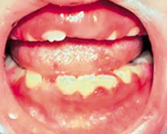
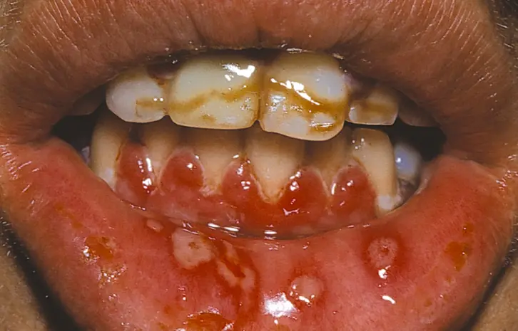
- one or two ulcerations +- widespread intraoral vesicles and ulceration
- sudden onset
- high fever, cervical lymphadenopathy
- sore throat and mouth, fiery red gingiva
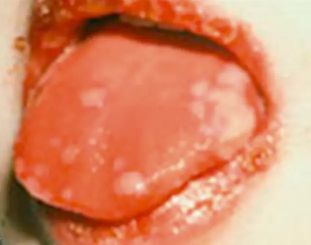
- intra-epithelial vesicles can form within 24h of infection, typically tongue buccal mucosa palate and gingiva
- vesicles rupture early to give numerous small round/irregular superficial greyish yellow ulcers with red halo
- lesions take 10-14 days to heal (sometimes up to 21)
- highly contagious (occurs 1 week after contact)
- pathophysiology
- management
- supportive/ palliative care (soft diet, fluids, rest, analgesia, topical anaesthesia)
- chlorhexidine rinse
- antiviral agent
- if immunocompromised child, IV acyclovir 10mg/kg 8 hourly until there are no new lesions
- if immunocompetent but severe pain/dehydration within 72h onset of disease, acyclovir 10mg/kg (max 400mg) oral 5 per day until no new lesions
- children who do not have control of oral secretions, should be excluded from childcare/school until sores have dried
- differential
- Hand food mouth
- herpangina
- RAS
- SJS
- behcet
Reactivation of HSV
- primary illness leadss to infection of trigeminal ganglion
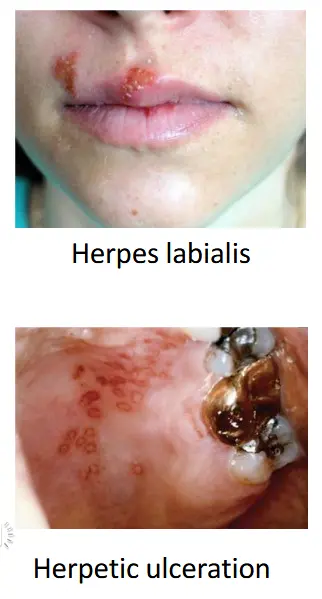
- can reactive as herpes labials (cold sore)
- herpetic ulceration
- intraoral reactivation may occur following trauma e.g. surgery or local anaesthesia (palatal infil)
- reactivated intraoral lesions, multiple clusters of small punctate ulcers
- reportedly associated with COVID19 infection causing immunosuppression and respiratory distress
Periodontal Abscesses
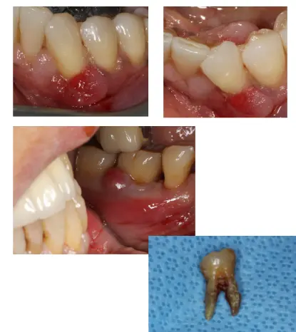
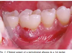
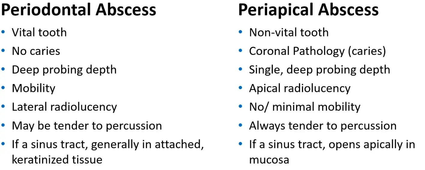
- acute lesions characterised by localised accumulation of pus within the gingival wall of the periodontal pocket/sulcus, rapid tissue destruction and are associated with risk for systemic dissemination
- results in significant tissue destruction
- primary signs/symptoms
- ovoid elevation in gingiva along lateral root
- BOP, deep periodontal pocket, suppuration on probing (pocket or fistula)
- Pain, swelling, redness
- tooth feels higher than usual when biting (PDL is inflamed)
- increase mobility
- lymphadenopathy (10% of cases)
- usually vital tooth but can be nonvital
- radiology might not reveal anything
- can cause rapid severe bone loss
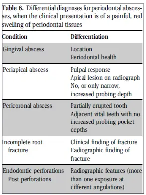
- previously classified by location
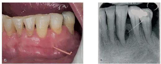
- gingival (foreign body in gingiva or sulcus)
- periodontal (infection adjacent to pocket)
- pericoronal (partially erupted tooth)
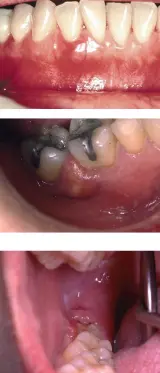
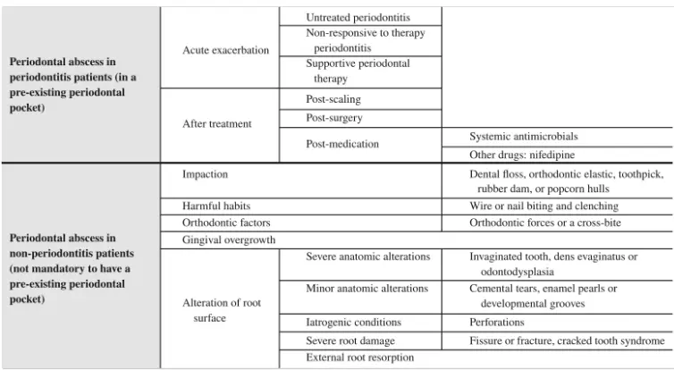
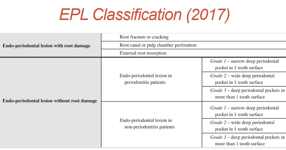
- 2017 classification (Herrera et al., 2017)
- **Etiology
- in perio patients
- represents period of disease exacerbation
- presence of torturous pockets, furcation involvement, vertical defect
- marginal closure of pocket - lack of coronal damage (leaving apical calculus but cleaning more coronally so gingiva heals and seals over the calculus)
- changes in subgingival microbial composition
- decrease in host defence
- non perio patients
- previously healthy site
- foreign body (floss, fingernail, food particle, popcorn kernel)
- harmful habit
- ortho factors
- alterations to root surface
- can occur directly after debridement, due to dislodgement of calculus fragment into gingival wall
- in perio patients
- epidemiology
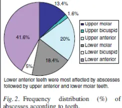
- 3rd most frequent reported dental emergency
- most occur in perio patients
- 60% untreated
- 14% active treatment
- 37% maintenance phase
Pathogenesis
- exact is unclear
- majority of abscesses occur in pre existing perio pockets
- inflamed pockets normally drain continuously
- occlusion of pocket prevents dranage
- supragingival S/C
- incomplete removal of calculus
- altered root morphology (invaginations, furcations, ERR, cemental tears, endo perforations)
- bacteria remain at base of pocket
- AB resistant strains (Topoll et al 1990)
- Inflammatory infiltrate destroys pocket wall + CT
Histopathology
- DeWitt et al 1985
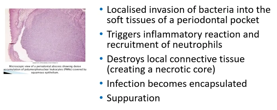
- bacterial invasion of soft tissues surrounding the periodontal pocket
- attraction of inflammatory cells and products eg cytokines
- leads to destruction of connective tissues, encapsulation of bacterial infection and production of pus
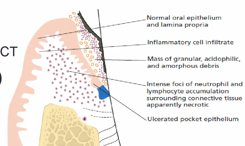
- outside -> inside
- normal oral epithelium and lamina propria
- acute inflammatory infiltrate
- intense focus of inflammation
- presence of neutrophils and lymphocytes in an area of destroyed and necrotic connective tissue
- destroyed and ulcerated pocket epithelium
Microbiology
- no different to untreated chronic periodontitis
- non motile, gram -ve, strict anaerobic rods
- Porphyromanas gingivalis 50-100%
- Prevotella intermedia,
- Prevotella melaninogenica
- Fusobacterium nucleatum
- Tannerella forsythia
- Treponema species
- Campylobacter species
- Capnocytophaga species
- Aggregatibacter actinomycetemcomitans
- or gram‐negative enteric rods

Acute Management
- drain abscess
- SRD
- +- occlusal adjustment
- Systemic antibiotics if signs of fever malaise
- can be controversial
- Antibiotics on their own to resolve acute symptoms first (Herrera et al 2000)
- poorly localised infection or cant establish drainage
- Antibiotics + supragingival scaling to avoid soft tissue trauma (Hafstrom et al 1994)
- metronidazole best profile (400mg BID 3-5 days, 6-10 tabs) NEVER MIX WITH ALCOHOL OR YOUR STOMACH WILL BE VERY SORE
- Azithromycin/amoxicillin work aswell
- Amoxicillin 500mg orally, tid for 3-5 days
- only for duration of acute phase
- still need mechanical debridement
- Extraction if hopeless
- mouthwash to reduce plaque formation short term
- hydrogen peroxide 3% 5ml mixed with 5ml water or 1.5% 10ml, rinse in mouth for 1 minute, then spit out, twice a day
- chlorhexidine 0.2% mouthwash 10ml or 0.12% mouthwash 15ml rinse in mouth for 1 minute then spit out, 2-3 times a day
- follow up 24-48 hours
Differential
- other acute periodontal lesions
- periapical abscess
- root fracture
- pericoronitis
- lateral periodontal cysts
- perio as a manifestation of systemic disease
- postop infection
- self inflicted gingival injuries
- neoplasms
Endo-periodontal lesions
EPL
- clinical conditions involving both pulp and periodontal tissues and may occur in acute and chronic forms (Herrera et al 2017)
Anatomical pathways
- apical foramen
- dentinal tubules - patent through no overlying cementum
- lateral canals, accessory canals
- developmental defects
- cracks or resorptive lesions
- DeDeus 1975
- 1140 adult human teeth studied, 313 had lateral, secondary and accessory canals
- molar and premolar mostly, incisor least ramifications
- majority apical
Signs and symptoms
- deep periodontal pocket reaching or close to tooth apex
- negative or altered response to pulp vitality tests
- bone resorption in apical or furcation region
- spontaneous pain or pain on palpation and percussion
- purulent exudate
- tooth mobility
- sinus tract
- tooth and/or gingival colour alterations
Aetiology
- endodontic and or periodontal infections
- carious lesions that affects the pulp and secondarily affects the periodontium
- 1st endo -> 2nd perio
- periodontal destruction that secondarily affects root canal
- 1st perio -> 2nd endo
- or both eveny concomitantly
- 1st endo and 1st perio (simultaneous but separate)
- "true combined" simultaneous and connected
- trauma and or inatrogenic factors
- perforations
- root fracture
- resorption
effect of endodontic pathology on the periodontium
- pathways of communication, apical foramen
- infected pulp leads to inflammatory response in PDL at apical foramen or small portals of exit
- inflammatory by-products permeate through apex or through smaller canals in apical third of the root canal system and exposed dentinal tubules
- can trigger an inflammatory vascular response in periodontium
- inflammatory by-products include living and non living pathogens
effect of periodontal disease on dental pulp
- conflicting evidence
- proposed that periodontal disease may have no effect on pulp until it involves apex
- other evidence suggest effect on pulp is degenerative including increase in calcification, fibrosis and collagen resorption, as well as direct inflammatory effect
- Czarneck and Schilder 1979
- perio vs healthy teeth
- histological study
- pulps were all histologically within normal limits
- Bergenholtz and Lindhe 1978
- 57% of teeth had mild pulp alterations
- secondary dentine formation +- inflammatory cell infiltrates
- SRD teeth, no increased incidence of pathologic pulp reaction
- Conclusion
- unless perio extends to root apex, pulp capable of surviving significant injury
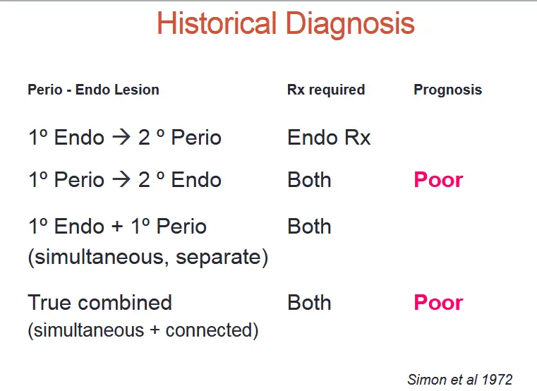
Historical classification
- simon 1972, Garguilo 1984
- primary endo
- chronic apical lesion with necrotic pulp
- primary endo secondary perio
- secondary involvement of periodontal breakdown due to plaque accumulation at gingival margin of sinus tract
- primary perio
- pulp test indicates clinically normal pulpal reaction
- primary perio secondary endo
- apical progression of periodontal pocket to invovle apical tissue
- infection enters pulp through lateral canals and apical foramen
- true combined
- endo lesion progression coronally joins with a periodontal pocket progressing apically
- least common, poor prognosis
- VRF may appear radio and clinically very similar, can be considered a true combined lesion
Diagnosis
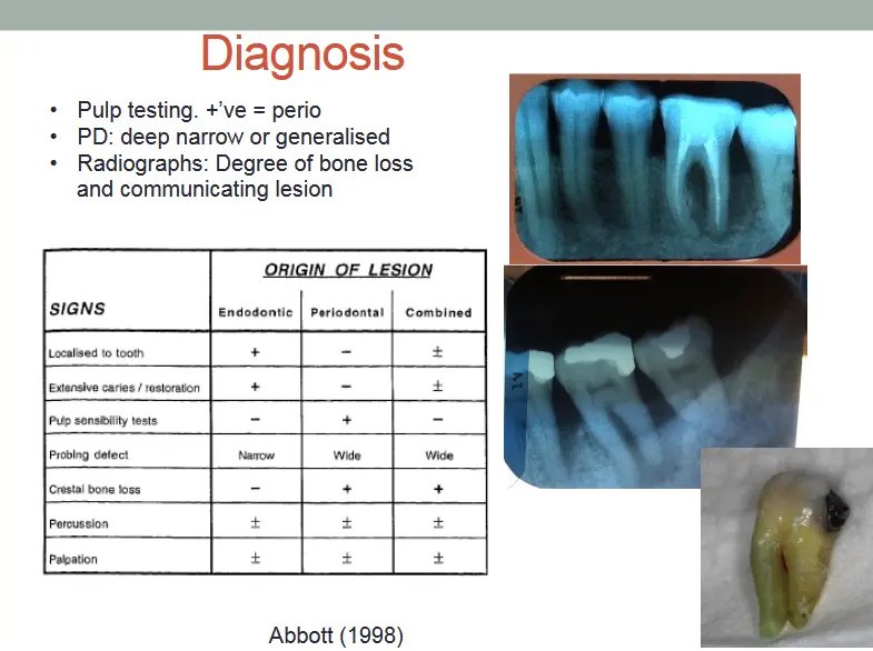
- pt histiroy
- truma/iatrogenic factors
- radio and clinical
- FM perio assess
- PPD
- CAL
- BOP/SOP
- mobility
- vitality/percussion
- endo perio lesions
- presence of perio pocket reaching close to apex combined with negative pulp vitality
Prognosis
- hopeless, poor, favourable
- prognosticating factors:
- extent of perio destruction
- presence/severity of periodontal disease affecting patients overall oral health
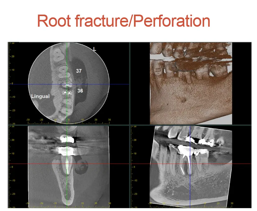
Root fractures
- cementum dentine pulp
- horizontal or transverse
- VRF
- longitudinal or transverse
- originate in crown or root
- common in RCT teeth
- clinically deep narrow periodontal pocket
- mild discomfort on function
- radiographic image varies, might be missed
- Siskin 1980 VRF
- N = 32
- 84% occurred during lateral condensation of gutta perfcha
- clinical signs
- 30/32 Deep narrow probing depths
- 9/32 recurrent periodontal abscesses
- 21/32 mild discomfort,
- 2/32 severe pain
- Radiographic signs 75% - diffuse widening of PDL
- treatment
- 21/32 required extraction
- 5/32 root amputation
- 4/32 ARF and osseous recontouring to expose fracture
Root perforations
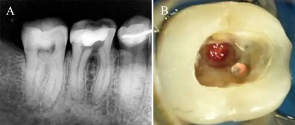
- may manifest as inflammatory lesions in marginal periodontium
- Alhadainy 1994, causes or perfs
- mainly iatrogenic
- straightening of endo fil in curved canal
- lessened with NiTi files
- strip perforation, removal of dentine from danger zone within furcal area of coronal third
- midroot
- transporation or zip perforation, excessive cutting of outer dentinal wal lcompared to inner dentinal wall
- clinical signs
- increased PPD
- suppuration
- increased tooth mobility
- periodontal abscesses
- symptoms
- sudden pain during endodontic treatment
- taste of irrigant during treatment
- diagnosis
- loos fit endo file
- bleeding into canal
- radiograph with endo file in situ
- prognosis
- coronal poorer, proximity to oral environment
- time lapsed before correction
- treatment
- nonsurgical
- MTA with CaOH2
- resto material to seal perforation
- surgical
- flap reflection to expose perforation
- sealing with appropriate restorative material
- nonsurgical
Root resorption
- physiologic or pathologic process resulting in loss of dentine cementum bone
- external root resorption (initiated in periodontium)
- progressive inflammatory (stimuli from pulpal infection and sulcular infection)
- Invase cervical resorption (non inflammatory)
- replacement resorption (non inflammatory)
- occurs following extensive necrosis of PDL with formation of bone onto denuded area of root surface
- complication of luxation injuries, especially in avulsed teeth
- internal root resorption
- chronic pulpal inflammation associated with an infected coronal pulp space
- multinucleated giant cell activity
- Heithersay 1999
- cervical root resorption
- cervical location, invasive nature
- pink discolouration of tooth crown
- surface irregularity might be probed, filled with vascular soft tissue
- pulp typically protected by a thin layer of predentine and dentine
- invaded at a late stage of process
- remains asymptomatic
- vital pulp
- fibrous tissue, numerous blood vessels and mononuclear clastic resorbing cells adjacent to the dentine surface
- Frank 1987
- case report seriers
- described treatment protocol
- confirm pulp vitality
- raise fla pto expose margins of defect
- spoon excavate fibrous tissue from lesion
- place hard setting CaOH
- restore with appropriate material
- suture flaps to desirable height to facilitate oral hygiene
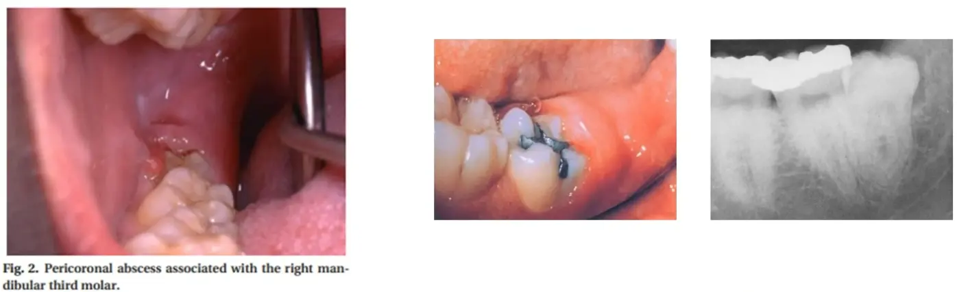
Pericoronitis
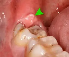
pericoronitis
- localised suppurative infection within the tissue surrounding the crown of a partially erupted tooth
- most commonly lower wisdom teeth retromolar pad angle of mandible
- associated with pain swelling trismus, occasional fever
- management
- exo
- operculectomy
- debridement and irrigation under la
- antibiotics if systemic signs of infection present
- review to ensure resolution of symptoms