Module 1
#DEN4002
#D4/S2/W1
#Periodontology
Periodontal Surgery
Objectives of non surgical therapy
- initial periodontal treatment (intial phase) is to restore biological compatibiltiy of periodontally diseased root surfaces to halt the process of the disease
- nonsurgical therpay aims to elimate both living bacteria in microbial biofilm and calcified biofilm microorganisms from the tooth surface and adjacent soft tissues
NSPT
- essential part of successful treatment
- gold standard to compare other treatment modalities to
- clinical considerations
- method of instrumentation
- removal of calculus
- root smoothness
- preventing bacterial repopulation
Tooth survival
- true endpoint
- long term tooth survival is most important end point for patinets
- surrogate endpoints
- PD, CAL, BOP and radiographic bone stability are important parameters, but not of direct interest to the patient
Planing vs debridement
- scaling
- removal of plaque and calculus from the surface
- planing
- softened cementum is remvoed to make root surface hard and smooth
- gingival curettage
- removal of inner surface of soft tissue wall of pocket with a curette
- root debridement
- removal of plaque or calculus from root surface without intentional removal of tooth structure
Instrument access
- very unlikely for any modality of non surgical therapy to reach apical extent of pocket
- Dragoo 1992, clifford et al 1999
- concept of critical mass
- reduce bacterial load to level resulting in equilibrium between residual microbes and host response, i.e. no clinical disease
Choice of debridement method
- hand, sonic, and ultrasonic scalers produce similar periodontal healing response with respect to probing pocket depths, bleeding on probing, and clinical attachment level
- Badersten et al. 1981, 1984; Lindhe & Nyman 1985; Kalkwarf et al. 1989; Loos et al. 1987
- debridement time spent per tooth may be reduce when using ultrasonic vs hand scaler
- Copulos et al. 1993; Boretti et al. 1995
- Sonic and ultrasonic scalers have been shown to produce less tooth surface loss compared to hand scalers
- Ritz et al. 1991; Schmidlin et al. 2001
Difficulties of thorough debridement
- increasing depth of periodontal pocket
- increasing width of tooth surface
- presence of root fissures, root concavities, furcations, defective subgingival restorative margins
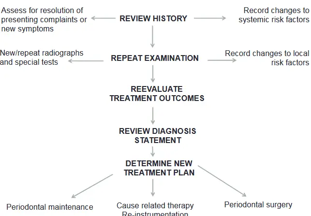
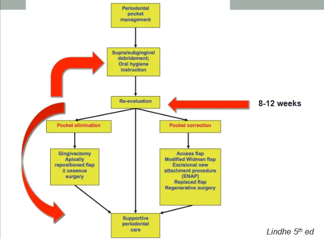
Treatment sequence
- systemic phase
- initial phase/cause realted therapy
- corrective phase
- maintenance phase (supportive periodontal therapy)
Clinical signs of periodontal healing
- visual signs
- resolution of redness erythema
- resolution of oedema - firm stippled tissue tone
- knife edge gingival margin
- possible recession of gingival margin due to resolution of swelling
- improvement in clinical parameters
- reduce PPD
- increase recession
- reduction in BOP scoring
When is best to re-evaluate?
- long junctional epithelium can be produced rapidly during wound healing, due to high proliferative activity of the epithelium
- underlying connective tissue continue to be remodelled for an extended period of time
- Badersten et al 1984
- even after 9months of periodontal therapy, there will still on going changes in periodontal probing depths
- Ciancio 1989
- american academy of periodontology world workshop agreed that a 4-6 week interval was usually adequate
- Waerhaug 1978
- reestablishment of attachment (reepithelialisaiton of junctional epithelium) occurred in 2 weeks, but granulation tissue was still immature and not yet remodelled
- Segelnick and weinberg 2006
- periods greater than 2 months may be too long due to the repopulation of pathogenic bacteria in periodontal pockets
- cobb 2002
- measurements taken prematurely may not be representative of completed healing and could therefore be misinterpreted as a poor clinical response
- CONSENSUS
- differing opinions in literature but generally 8-12 weeks re-evaluation following SRD
- consider 8 weeks for high risk patients
Factors affecting healing post SRD
- site level - initial PPD
- no magnitude of initial probing depth where non surgical periodontal therapy is no longer effective (Badersten et al 1984)
- tooth level
- tooth type, presence of furcations, grooves
- patient level
- systemic health - uncontrolled diabetes, smoking, HIV
- poor plaque control/ability to comply with oral hygiene instruction
What is periodontal surgery
- surgical manipulation of periodontal soft tissues and bone
- procedures to
- access
- resect
- regenerate
- access
- provide vision
- improve access for instrumentation of residual deep sites
- access to strucutres overlaid by mucosa, ie implant or orthodontic exposure
- resective
- hard
- osteoplasty, ostectomy, furcation plasty, root resection, hemisection and trisection
- soft
- pocket elimination, curettage, distal wedge, biopsy, frenectomy
- hard
- regeneration
- repair involves formation of LJE
- restoration of original structure of periodontium
- new cementum, inserting PDL fibres, alveolar bone
- GTR uses barrier to maintain space, which allows preferential accumulation of cells from PDL and bone, and excludes epithelial cells from lamina propria/epithelialisation
- true regeneration can only be confirmed histologically
**When is it best utilised
- only consider surgery as adjunct to cause related therapy
- must have good Oral hygiene
- inflamed tissue is more friable, bleeds more and harder to manage
- decision on type and number of sites after effect of cause related therapy (initial phase) evaluated
- may take up to 6-9 months for periodontal parameters to level out in deep sites (badersten et al 1984)
Surgical treatment objectives
- create accessibility for debridement (grooves, deep pockets, furcations, infrabony pockets)
- establish a gingival morphology which facilitates the patients self performed plaque control
- regenerate periodontal attachment loss due to destructive disease
- reduction or elimination of plaque retentive areas
- pockets (resective or regen techniques)
- furcae
- root irregularities, fusions, grooves (odontoplasty)
- elimiate inflammation
- enhance regeneration of periodontal tissues
- create physiologic architecture
- correct mucogingival defects
Indications
- impaired access for SRP
- PPD, anatomic, root fissure, concavities, furcation, defective margins
- help establish morphology which is conducive to good plaque control
- PPD reduction
When to decide
- after non surgical anti infective therapy has been accomplished
- if effective individual hygiene is provided
- site specific persisting periodontal pockets
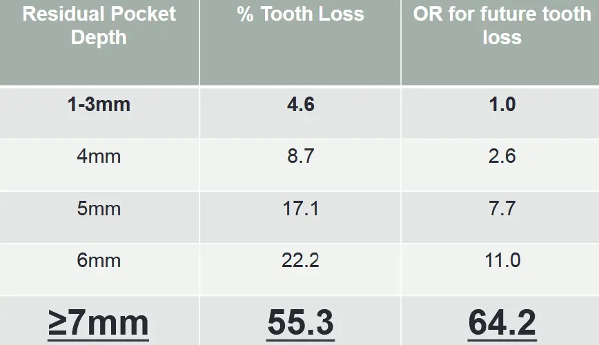
- threshold pocket depth??
- PPD >5mm
- Matuliene 2008 J Clin Periodontol
- influence of residual pockets on progression of periodontitis and tooth loss: results after 11 years of maintenance
- OR is odds ratio
Surgery aims to preserve the periodontium
- enhance access and vision for SRP
- remove or modify plaque retentive factors
- correct aberrant gingival morphology
- facilitate proper restorative therapy
- allow for periodontal regeneration to occur
- contraindications to surgery
- poor OH
- medical contraindications
- smoking - less improvement in PPD and CAL (Preber and bergstrom 1990)
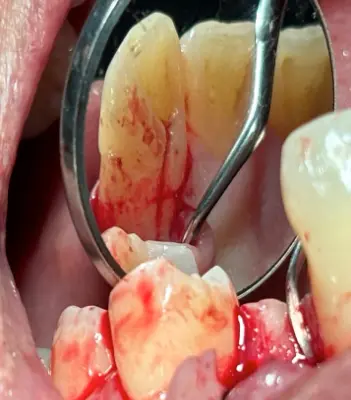
Open flap debridement case
- 2-3 rounds of NSPT and local maintainence
- upper lateral incisor palatal root groove
- residual deep pocket of 7mm with bleeding on probing
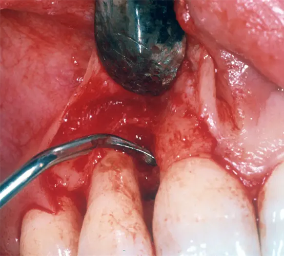
- mucoperiosteal flap raised to visualise and access depth of root groove
- extended 10mm from CEJ
- calculus detected at base of defect
- debrided using cavitron and hand instruments
- groove smoothened
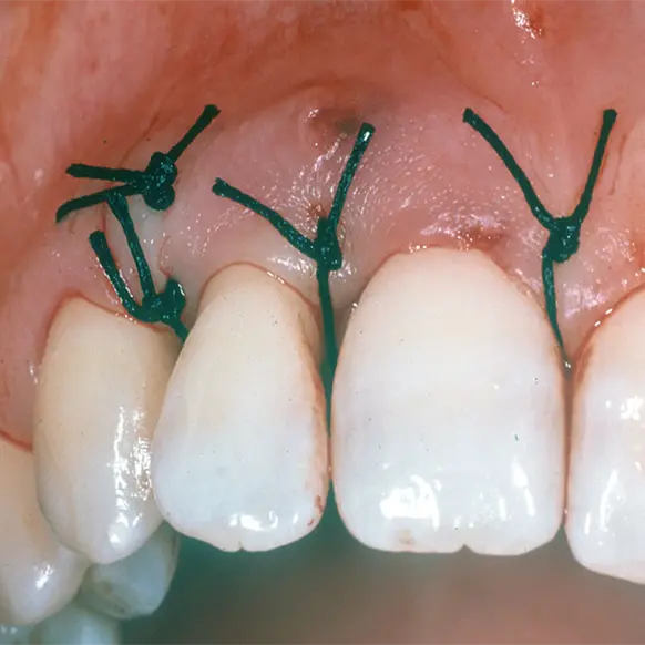
- flap replaced
EFP CLINICAL GUIDELINES 2020
European federation of periodontology
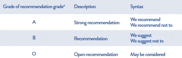
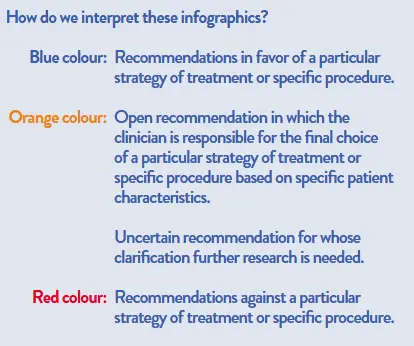
4 steps
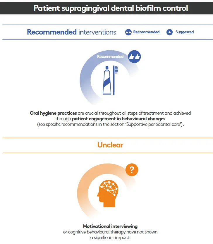
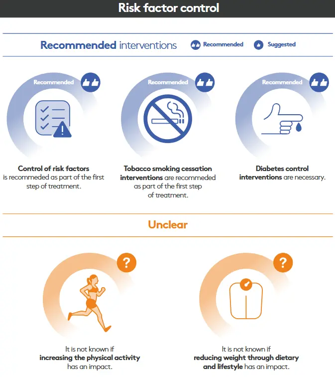
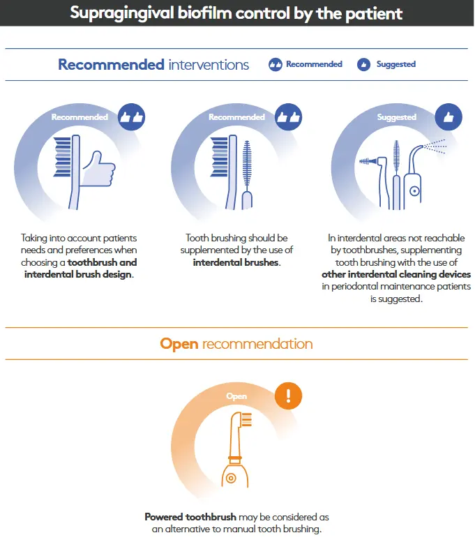
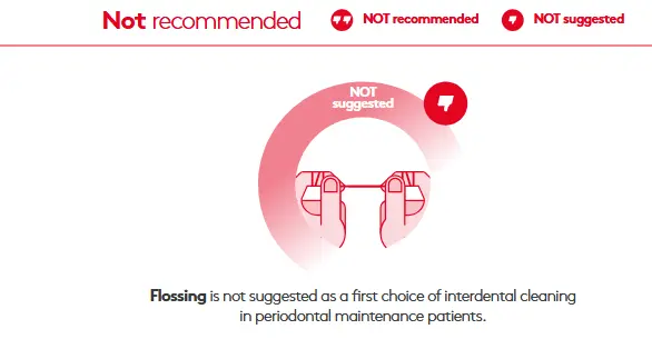
- step 1 patient OH and risk factor
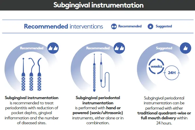
- step 2 NSPT
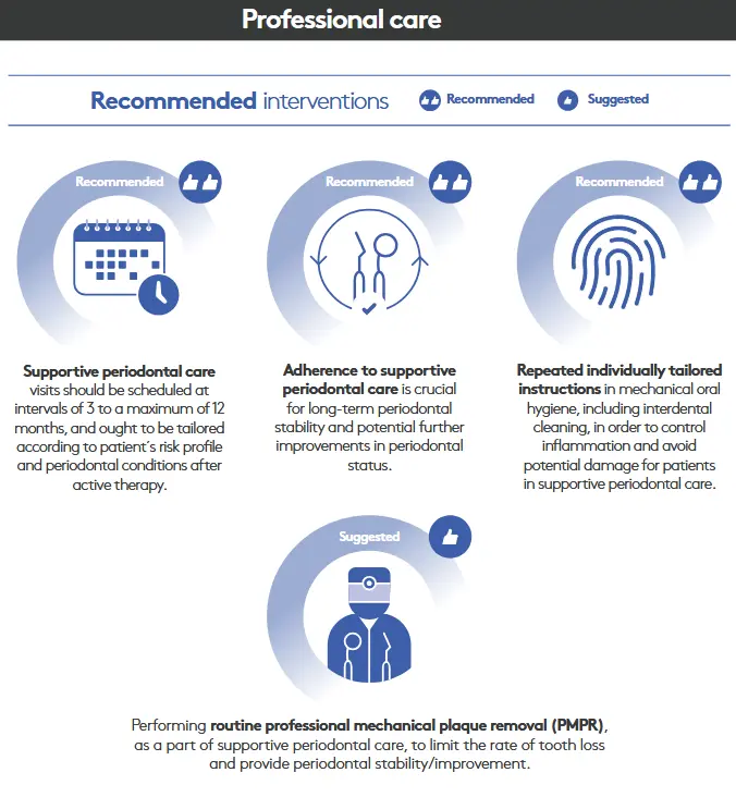
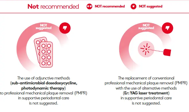
- step 3 SPT
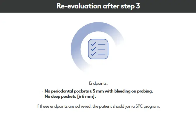
- step 4 supportive periodontal care (SPC)
Step 1
- guide behaviour change via motivation for patient successful removal of supragingival dental biofilm and risk factor control
- implemented in all perio patients
- frequently reevaluate
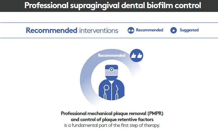
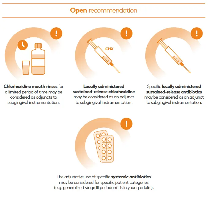
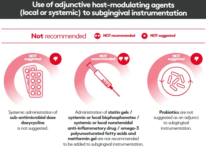
Step 2
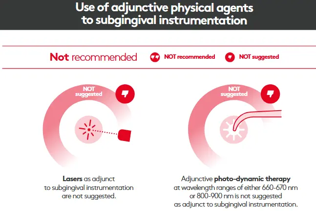
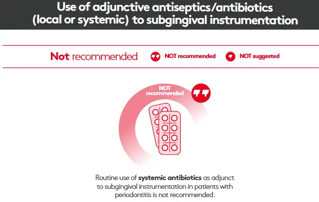
- control subging biofilm and calculus
- implemented in all perio patients
Step 3
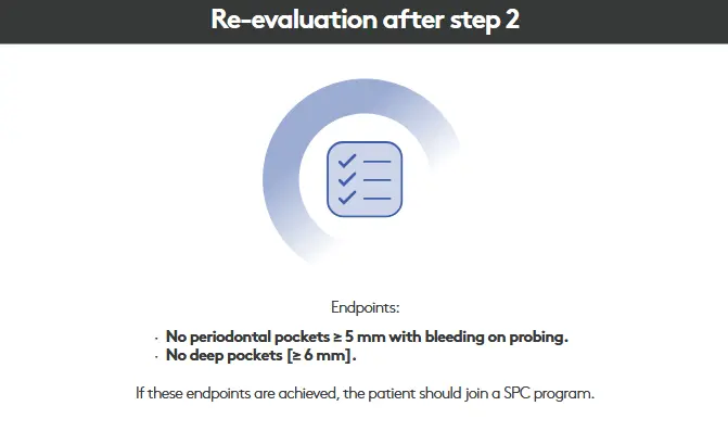
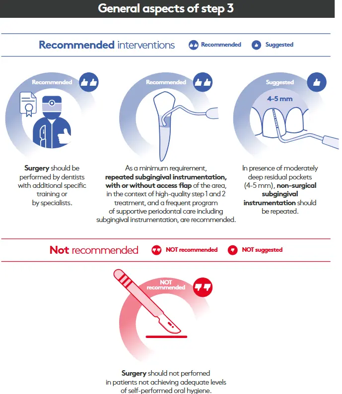
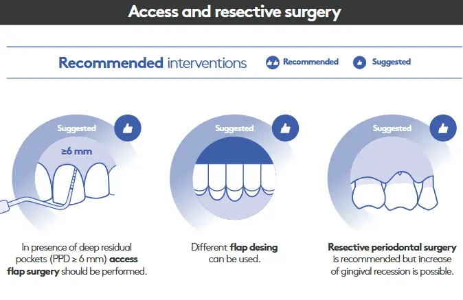
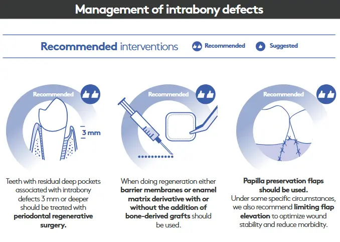
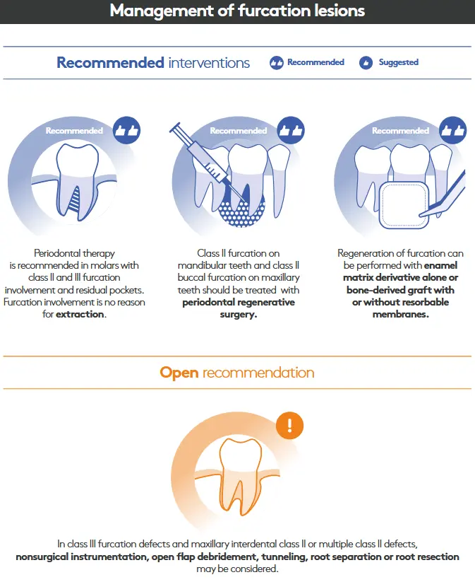
- treating sites which are not responding adequately to 2nd step, getting access to deep pocket sites, regen/resect lesions that add complexity to management of perio
- PPD >4mm with BOP or pockets 6mm or over after reevaluation
**Step 4 **
- no deep pockets 6mm or over and no pockets over 4mm which are BOP
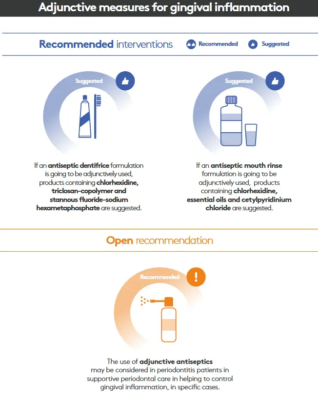
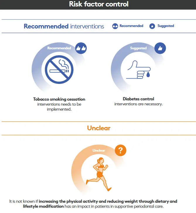
Plaque control
Nyman, Lindhe and Rosling 1977
- 25 patients, periodontal surgery in plaque infected dentitions
- 5 different surgical techniques used to reduce pockets
- no maintenance (patient returned to general dentist)
- results
- pocket depths rebounded to baseline values in the presence of poor oral hygiene
- regardless of the type of surgery performed
- maintenance and excellent plaque control is critical to prevent disease recurrence
- conclusion
- plaque is always going to cause a disproportionate host response, periodontal tissues healing is overwhelmed by inflammation
- maintenance and OH mandatory for any type or surg or nonsurg
Rosling 1976
- periodontal surgery in maintained (clean) dentitions
- patients after perio surgery split into two groups
- 2 weekly maintenance after surgery
- no maintenance - back to dentist
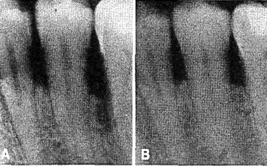
- maintain results
- 80-100% bone infill in all vertical defects
- mobile teeth firmed up
- excellent healing outcomes
- no maintenance results
- lesions deteriorated in presence of plaque
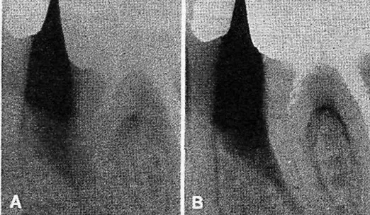
Principles of Periodontal Surgery
summary
- NSPT is effective for addressing perio
- NSPT completed before surgical to address bulk of periodontal disease and to optimise patient oral hygiene
- NSPT vs surgery
- similar attahcment gains in long term
- surgery quicker pocket reduciton
- surgery damaged shallow sites
- surgery generally reduces risk of breakdown over time (reduces need for retreatment)
- maintenance and plaque control is essential for maintaining attachment gains for surg and non surg sites
Access Periodontal Surgery
Access
- raising and reflecting back soft tissue overlying a periodontal defect to allow direct visual assessment of the root surface and within the periodontal defect
- known as open flap debridement / OFD
- provides direct access for removal or retained local factors (plaque/subging calculus)
- indicated for sites that have not responded favourably to SRD
Refractory sites
- unresponsive/failing to heal after perio treatment
- fail to heal due to retained local factors such as subging plaque and calc which are difficult to fully access during NSPT
- examples
- proving depths exceeding 5mm
- vertical bone loss
- furcation lesions
- root surface anomalies (cervical enamel projections, enamel pearls, root grooves, concavities)
- defective subgingival restorative margins
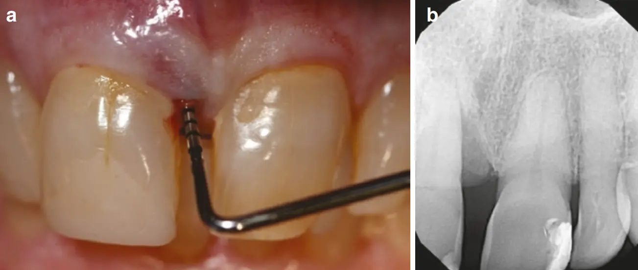
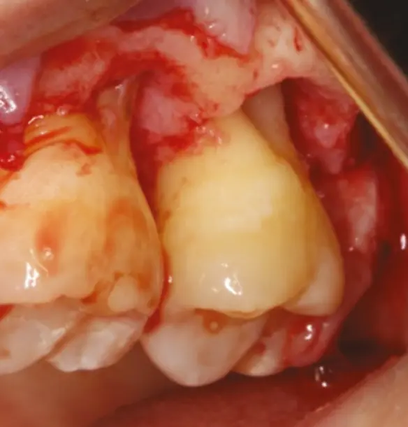
vertical dfefect buccal furcation lesion 27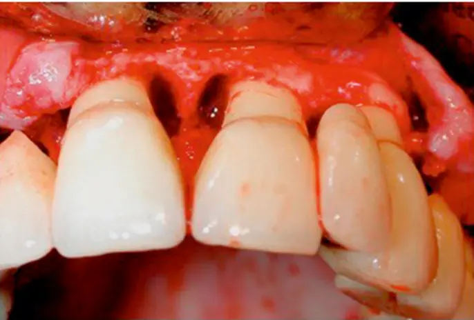
deep vertical defects
OFD
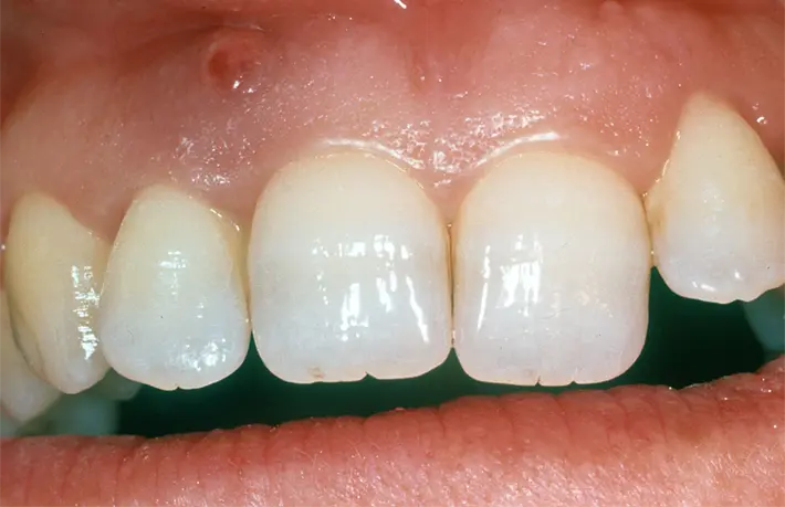
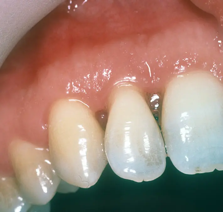
- open flap for perio abscess caused by retained subgingival calculus at mesial 12, note recession common
Resective Periodontal Surgery
resective
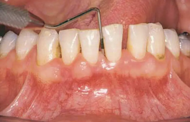
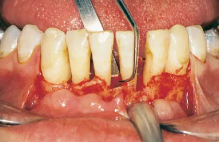
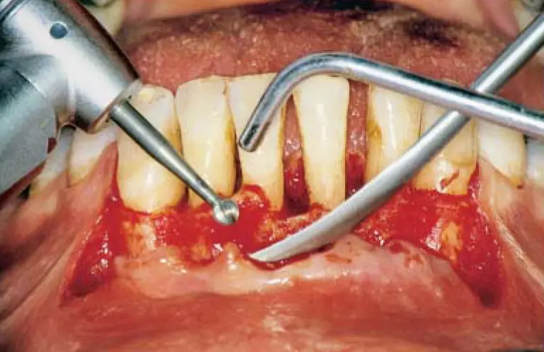
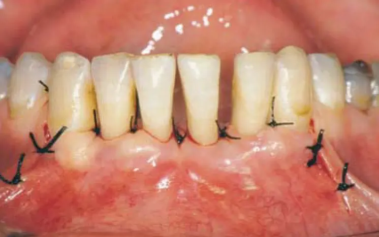
- removing bone and soft tissue to improve access for cleaning or to retain peridontally compromised teeth
- example
- pocket elimination, ostectomy, tunnelling, root resection
- osseous resection and recontouring during open flap debridement
- **Indications
- multiple deep probing depths 5mm+ in quadrant after phase I therapy with horizontal loss
- furcation involved molars with defects not amenable to regeneration
- gingival pseuopockets
- contraindications
- multiwalled intrabony defects or furcation defects amenable to regeneration
- severe bone loss / tooth mobility
- if resection may compromise perio support of adjacent teeth
- expected adverse postop outcomes following resective approach
- attachment loss
- dentinal hypersensitivity
- increase in tooth mobility
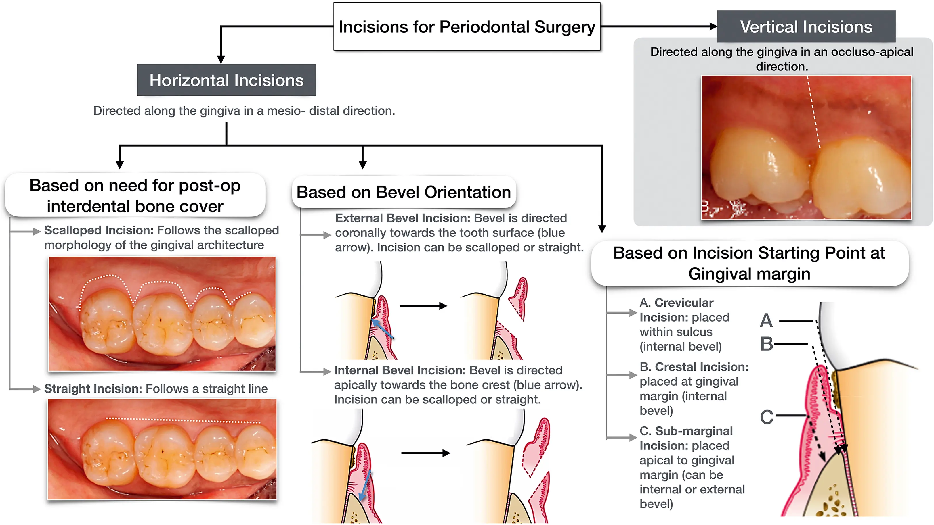
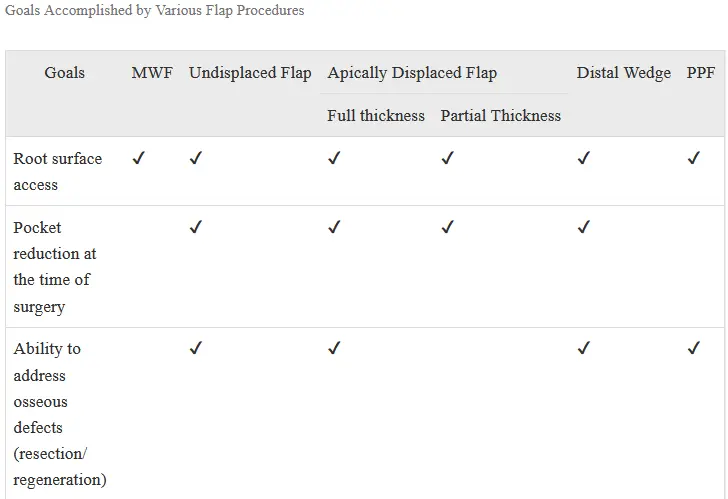
Flap procedure classifications
- bone exposure afte flap reflection
- full thickness/mucoperiosteal flap, cut directly to bone with entire thickness reflected using blunt dissection using periosteal elevator
- partial thickness flap/mucosal/split thickness flap, incision stops before periosteum, therefore only mucosa is elevated off underlying connective tissue and periosteum with no bone exposed, indicated for apically positioned flap, thin crestal bone margin, dehiscence/fenestration present
- placement of flap after surgery
- undisplaced, placed and sutured original position
- displaced flap, apically, coronally, laterally to original position
- management of papilla
- conventional flap, papilla thinned or split beneath contact point while placing incision
- papilla preservation flap, in regenerative therapy and aesthetic cases, retains entire papilla, requires adequate width of interdental space to allow intact papilla to be reflected with one side
Definitions
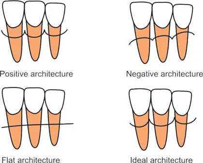
- flat architecture
- interdental bone remains at same level as the buccal or lingual/palatal bone
- ideal bone architecture (positive/scalloped)
- interdental bone is coronal to buccal or lingual palatal bone
- negative/reversed architecture
- interdental bone is apical to the buccal or lingual/palatal bone
- widows peaks
- peaks of bones that remain on the facial and lingual palatal aspectof teeth and interproximal line angles during osseous surgery procedure
- peaks of bones that remain on the facial and lingual palatal aspectof teeth and interproximal line angles during osseous surgery procedure
Regeneration Periodontal Surgery
regeneration
- to restore lost periodontal tissues to improve prognosis and aesthetics of a tooth
- using barrier membrane, bone graft, biologic matrix derivative
- combinations of materials and techniques are common
Definitions
- autograft (autologous)
- graft from patients own bone (extraoral or intraoral)
- allograft
- same species different person (common in america)
- xenograft
- different species (bovine, porcine etc,)
- alloplast
- artificial/synthetic material used to replace/augment missue tissue
- enamel matrix derivative
- biologic mediator composed of enamel matrix proteins, effective in treatment of intrabony defects with histologic evidence of regeneration
- Guided tissue regeneration GTR
- placement of barrier membrane over intrabony defect, to prevent epithelial migration
- biomodification of root surface
- topical use of chemical agents (tetracycline) on prepared root surface with intent of removing local factors that may interfere with new attachment or preferentially encouraging connective tissue attachment
- osteoconduction
- property of grafts and biomaterials which passively supports formation of bone on their surface
- osteoinduction
- property of grafts that contribute to new bone formation by facilitating the recruitment of progenitor cells and the stimulation of these cells to develop into pre-osteoblasts
- new attachment
- healing by replacement with new epithelial and/or connective tissue that matures into various nonfunctional types of scar tissue
- regeneration
- healing by reconstitution of new periodontium, which involves formation of alveolar bone, functionally aligned PDL and new cementum
Histologic outcomes following pocket reduction therapy
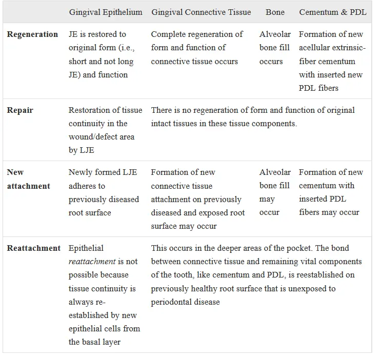
Basis for regeneration modalities
- SRP without additional graft or membrane
- biocompatible root surface, removal of diseased junctional and pocket epithelium by excision, wound stabilisation and clot protection
- can help in regenerating some bone and connective tissue at the base of defect
- new cementum or new functional PDL fibres not formed, healing by LJE formation (reparative healing after MWF procedure)
- root conditioning
- in theory
- surface detoxification, removal of smear layer, exposure of collagen fibrils that prevent epithelial migration over treated root surface, promoting fibroblast cell attachment to root surface
- this attempt yielded controversial results in humans, leaving it as adjunctive regen procedure
- bone grafts
- various materials for bony defects, result in decreased PPD and improved CAL
- bone grafting doesn't result in regeneration of all perio tissues, but acts as a space maintainer or scaffold for bone regeneration
- healing is by LJE when only bone grafts are used for defect fill
- still considered therapeutic success due to improvement in BOP, PPD, CAL
- barrier membranes GTR
- barriers of different types of to cover boen and PDL, temporarily separarting them from gingival epithelium and connective tissue
- excluding epithelium and gingival connective tissue from root surface during postsurgical healing phase prevents epithelial migration into wound and favours repopulation of are by cells from PDL and bone
- when sufficient time is given for deeper periodontal tissues to repopulate the clot under a membrane, healing by new periodontal connective tissue attachment and bone fill without LJE can potentially occur
- tissue egineering
- manipulation using one or more key elements
- signalling molecules (rhPDGF-BB, EMD)
- scaffold or supporting matrices (beta-TCP)
- cells
- three elements intertwine to stimulate regeneration using a different mechanism from bioexclusion principle, demonstrated with GTR membranes
- use of biologic agents can stimulate recruitment of stem cells to intrabony or furcation defect site, and their proliferation and differentiation into newly regenerated periodontal apparatus
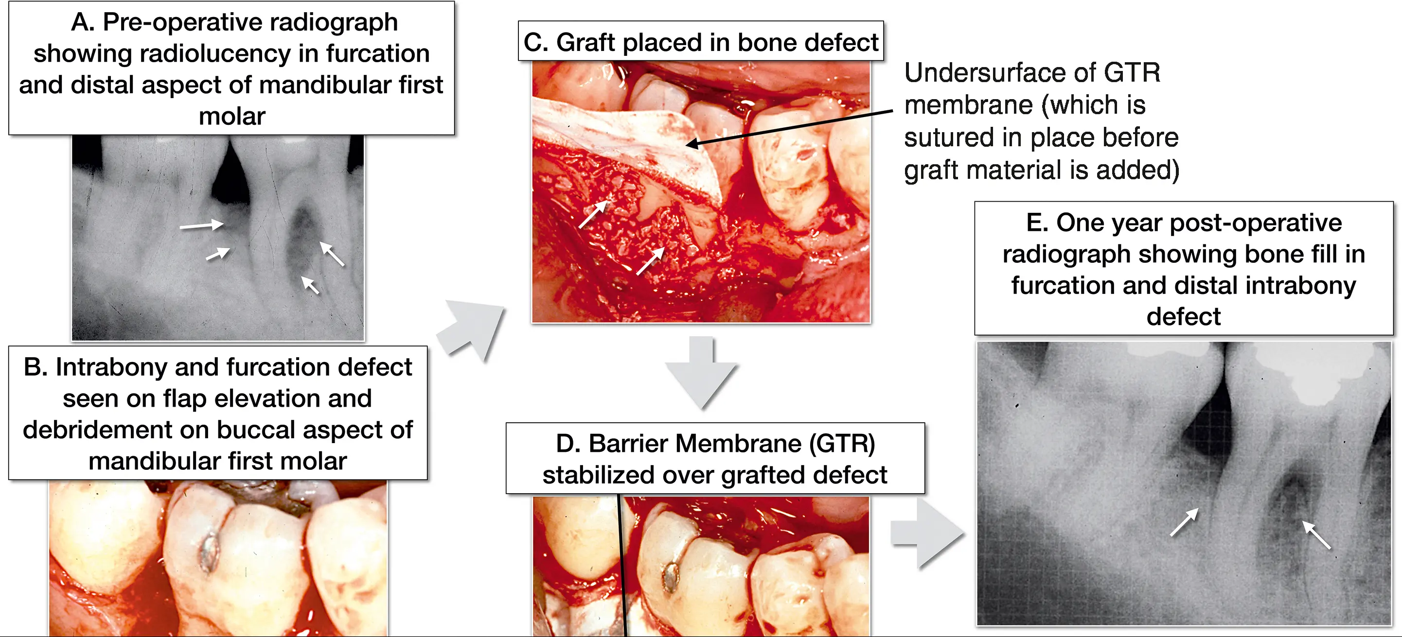
Guided Tissue Regeneration GTR
use
- prevent of epithelial migration along cemental wall and for maintaining space for clot stabilisation
- by preventing fast migrating epithelial and gingival connective tissues from prematurely occupying wound space under periodontal flap, GTR membrane guides precursor celsl from bone and ligament to preferentially occupy same wound space
- prevention of epithelial downgrowth into the wound and exclusion of gingival connective tissue is achieved using predominantly barrier techniques
- indications
- narrow two or three wall intrabony defects
- circumferential/moat shaped defects
- class II molar furcation
- recession defects
- contraindications
- width of attached gingiva <1mm
- generalised horizontal bone loss
- intrabony defects <4mm deep
Bone Grafting
- primarily a scaffold for healing
| Autograft | Allograft | Xenograft | Alloplast | |
|---|---|---|---|---|
| Description | Bone obtained from the same individual | Bone obtained from a different individual of the same species from commercial tissue banks | Bone obtained from a different species from which all cellular and protein material has been removed | Synthetic, biocompatible, and sometimes bioactive bone substitute |
| Example | Intraoral source: Osseous coagulum bone blend Extraoral source: Cancellous bone marrow from iliac crest |
Demineralized freeze-dried bone allograft (DFDBA) Freeze-dried bone allograft (FDBA) |
Bovine derived hydroxyapatite Porcine derived Equine derived |
Organic: Dentin, cementum, collagen, corals Inorganic: Plaster of Paris, bioceramics (hydroxyapatite, tricalcium phosphate), bioactive glass, polymers (PMMA/HEMA) |
| Role in bone regeneration | Osteogenic Osteoinductive Osteoconductive |
Osteoconductive (DFDBA–possibly osteoinductive) | Osteoconductive | Osteoconductive |
Difference between regeneration and bone fill
- bone fill only described clinical restoraiton of boen tissue in treated periodontal defect, but does not give any idea about presence or absence of epithelial or connective tissue new attachement or reattachment
- only histology can confirm regeneration vs repair
- bonefill can be confirmed clinically via surgical reentry or radiographically
Surgical Techniques
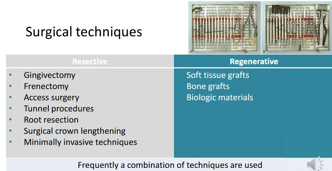
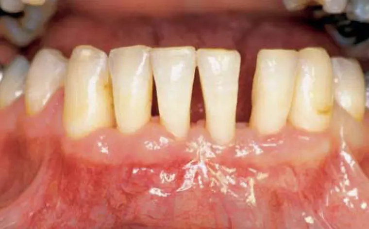
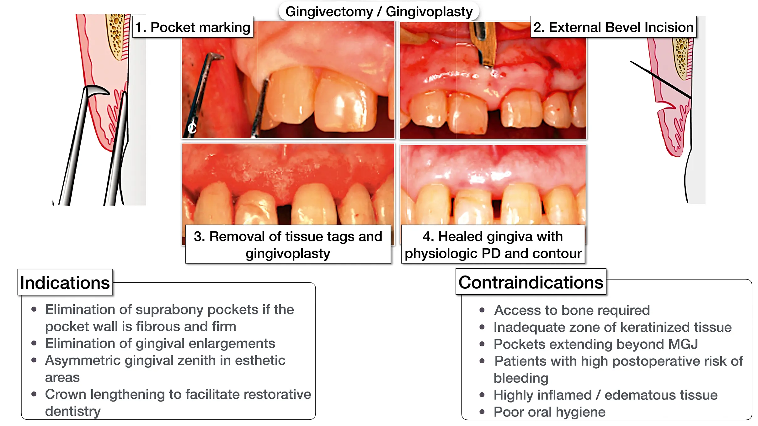
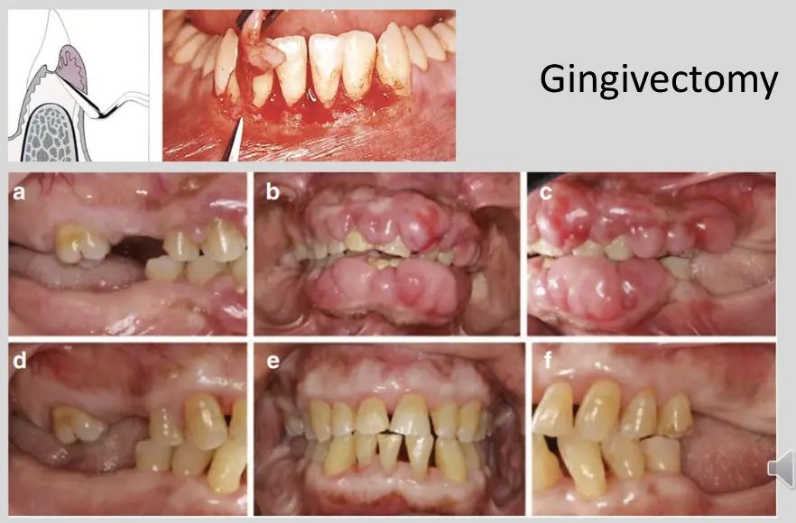
- gingivectomy for CCB induced gingival overgrowth
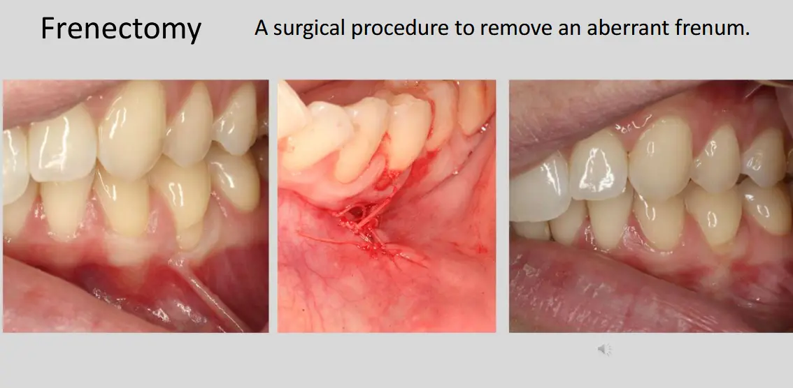
- frenectomy
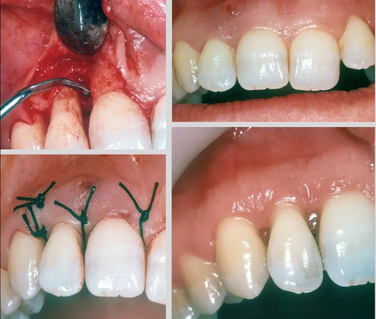
- access
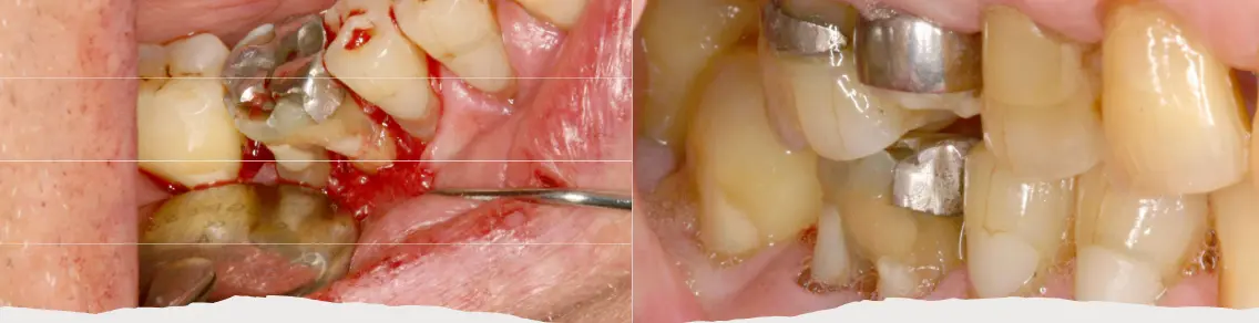
- tunnel
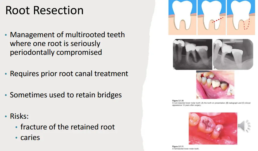
- root resection
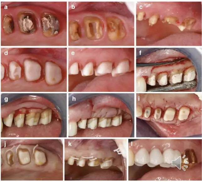
- surgical crown lengthening
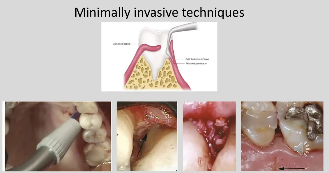
- minimally invasive split thickness flap
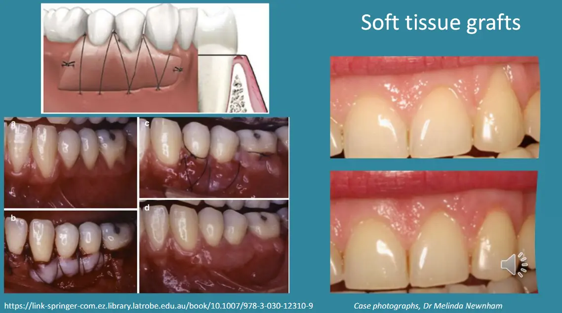
- soft tissue grafts
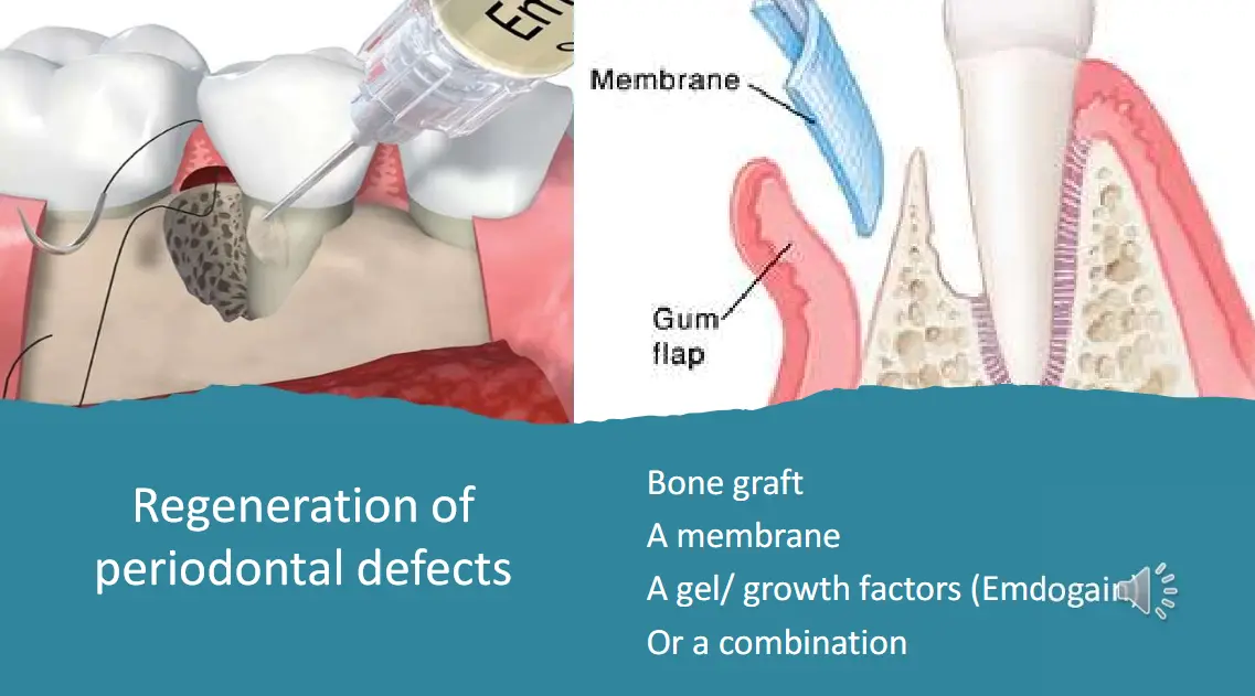
- regeneration of periodontal defects
Contraindications to periodontal surgery
Patient level
- systemic conditions
- bleeding dyscrasias, patient taking coagulation
- cardiac event within 6months
- unstable cardaic arryhthmias
- immunosuppression
- previous radiotherapy within the operating filed
- antiresorptive therapies exceeding three years
- patients with mental health conditions preventing them from coping with surgery
- other
- smoking
- oral dermatoses (erosive lichen planus, vesiculobullous conditions)
- poor plaque control
- severe crowding
Site level
- anatomical structures in close proximity at risk of damage
- mental foramen, lingual artery, neurovascular bundle within retromolar pad
- arterovenous malformations within surgical field
- tooth roots in close apposition preventing instrument access
- shallow sites with probing depths <4mm causing recession
- inflamed periodontal tissues
- teeth with hopeless prognosis