Pathological Tooth Resorption
Resorption
- loss of hard tissue by physiologic or pathologic means due to activiation of clastic cells
- may involve hard tissues of teeth or bone
- may be associated with
- trauma induced marginal alveolar bone recession
- marginal periodontitis - perio origin
- apical periodontitis - endo origin
**Physiologic mechanisms inhibiting tooth resorption
- Non-mineralized precursor tissues (e.g. Predentine) are resistant to resorption
- Cementum is also more resistant to resorption than bone or dentine
- Activation or continual triggering is required for resorption to proceed
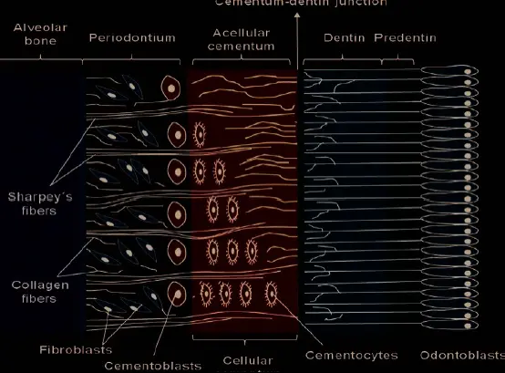
- Predentine does not resorb as needs calcified tissue for resorption to allow ostoeclasts to adhere
- osteoclasts do not adhere to nonmineralized collagen matrices
- Suggested the presence of a noncollagenous, organic component within dentin (odontoblast layer and predentin) prevents resorption of the root canal wall
- Similar to osteoclasts, odontoclasts might bind to mineralized extracellular matrix proteins containing the RGD binding peptides (arginine-glycine-aspartic acid) sequence of amino acids via integrins
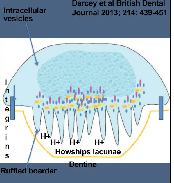
- resorption only occurs with various calcified connective tissue
- connective tissue composed largely of extracellular matrix in network of collagen fibres is dispersed within a gel like material, which itself is composed of a mixture of connective tissue polysaccharides
Cells associated with bone turn over
- MRONJ
- bisphosphonates reduce effect of osteoclasts
- altered bone surface needs combination of extracellular matrix proteins and mediators from osteoclast to activate odontoblast
- if disrupted bone not presented appropriately osteoblast may not be activated
- dentine and cementum are resorbed but enamel is undermined as no mineralised extracellular matrix in connective tissue for cells to attach
Classification
Andreasens Classification
- internal
- inflammatory
- external
- surface
- inflammatory
- replacement
**Linsdkog/Heithersay's Classification of pathologic tooth resorption
- trauma induced
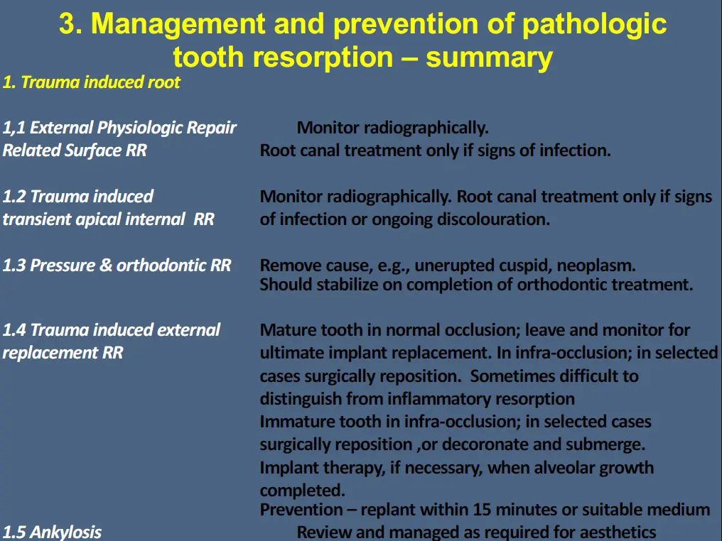
- trauma induced external physiologic repair related surface root resorption
- trauma induced transient apical internal root resorption
- pressure resorption and orthodontic apical root resorption
- trauma induced external replacement root resorption TIERRR
- Ankylosis (not formally separate)
- infection induced
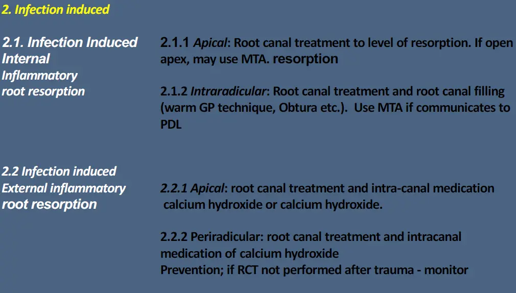
- Infection induced internal inflammatory root resorption IIIIRR
- infecti0n induced external inflammatory root resorption IIEIRR
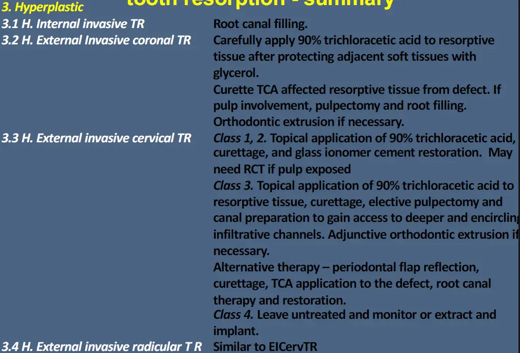
- **hyperplastic invasive
- Hyperplastic internal invasive tooth resorption
- Hyperplastic external invasive coronal tooth resorption
- Hyperplastic external invasive cervical tooth resorption
- Hyperplastic external invasive radicular tooth resorption
trauma induced external physiologic repair related surface root resorption
- physiologic - stable part of healing that arrests wit htime
- pathologic - unstable progressive response
- non-infective origin
- shallow resorption of cementum of underlying dentine
- self limiting
- transient
- may follow trauma or ortho
- associated with normal continuous bone and cementum remodelling
- physiologic
- heals uneventfully with reparative cementum
- usually not visible radiographically
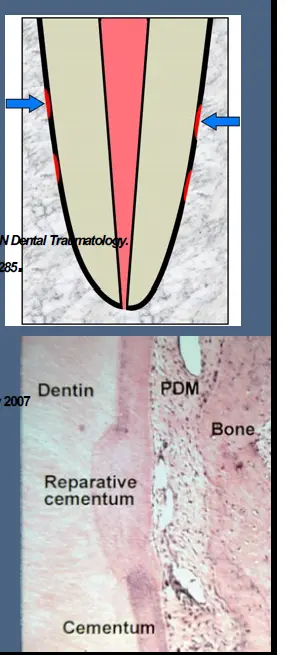
- aetiology, histopathology and management
- pathologic injury to cementum of PDL attracts clastic cells that phagocytose the damaged tissue
- without presence of infection in the root canal, resorption does not continue and repair with cementum like tissue is expected
- pulp remains vital, resorption is often not visible and is self limiting
trauma induced transient apical internal root resorption
- can follow subluxation or lateral luxation injuries
- may be associated with a transient apical breakdown
- internal apical resorption allows ingress of a greater vascular network to aid in the healing of a traumatised pulp
- may be an associated colour change due to intra pulpal haemorrhage and this may resolve spontaneously if revascularisation to the coronal pulp chamber occurs
- internally resorbed apex will close uneventfully
- often seen with trauma induced external physiological repair related surface root resorption
- last no more than 3 months
- purpose
- may occur in 10% of extruded or laterally luxated incisors with completed root formation
- within first months after injury
- both explained as space gaining manoeuvre to permit vascular ingrowth into root canal
- once repair reached remodelling phase, processes observed radiographically presumably resolves and is seen as reversal of radiographic condition
- may be associated with colour change
- colour change does not always resolve and more invasive approach may be warranted for aesthetics
pressure resorption and orthodontic apical root resorption
- pressure of a crypto of an unerupted erupting tooth or some neoplasms
- similar to TIEPRRSRR, usually due to low grade trauma and absence of infection
- more common during ortho with is limited once appliance are off
- repair and recontouring or root will occur as long as pulp remains vital
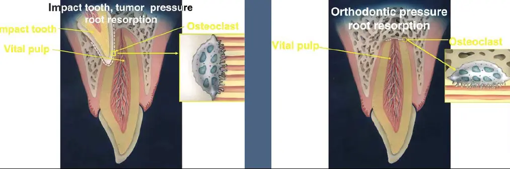
- severe in about 5-18% of cases
- possibly orthodontic pressure on roots damages PDL and initiates inflammatory response that results in apical root resorption
- cause of orthodontic RR due to bio and mechanical factors
- mechanical factors, extensive tooth movement, root torque and intrsuvie forces, movement type, ortho force magnitude, duration and type of force
- bio factors, genetic susceptibility, systemic factors, teeth agenesis, medication intake
- suggest management
- assess for apical root resorption after 6mo treatment
- if thought to have enhanced risk, radio review at 3 months
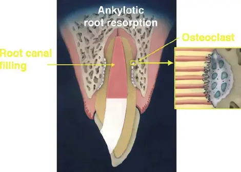
trauma induced external replacement root resorption
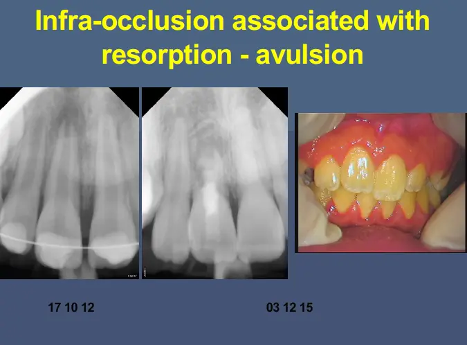
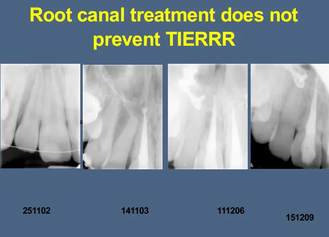
- progressive replacement of tooth structure by alveolar bone
- ultimately associated with tooth loss
- follows death of viable periodontal ligament cells and cementum due to compression (intrusive luxation) or drying of the PDL cells and cementum as in the case of delayed replantation of avulsed tooth
- usual response is that of development of an interface between bone and dentine
- total loss of mobility
- dull sound to percussion
- symptom free until tooth exfoliated
- total loss of image of PDL radiographically
- may progress slowly over years
- endo intervention no effect on progression
- sometimes may be present with Infection induced external inflammatory root resorption
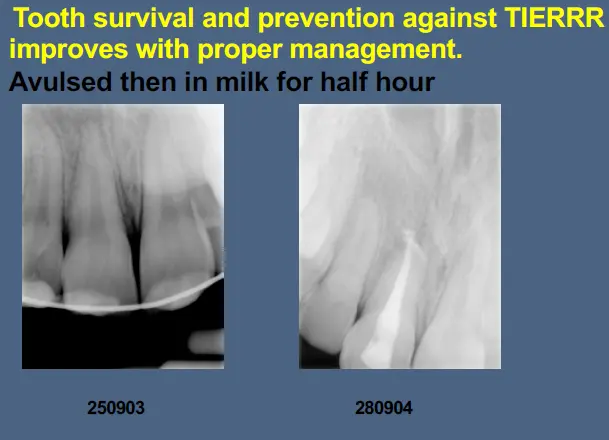
If you cant replant the tooth milk is a good storage medium
- milk has physiological osmolality (230-270mOsm/kg as is extra cellular fluid)
- okay for about 2 hour
- pH of milk is in physiological range
- can provide some nutrients to cells
- pasteurised milk has very low bacterial count
- milk must be fresh and refrigerated, sour milk is destructive
- fresh milk should be refrigerated and when the tooth is placed in fresh milk, keep contained chilled by surrounding with ice
- skim melk better because less fat content
- if no milk - use saliva until can get milk. After saliva, put saliva in milk
- baby formulas are ok
- some reconstitued powdered milk is ok
- yogurt has additives, only as a last resort
- gatorade and contact lens solution are hypertonic and not good
- soy milk is good
- normal saline can be used but results are variable
- coconut water too low pH or too much fat
- coconut milk is better than milk?
- water osmo 30 so no longer suggested
Avulsed permanent tooth
- replant before 5 minutes or in physiologic environment
- level of contamination of root before replantation
- no visible contamination best outcome with least resorption
- washed of visible contamination before replant next least resorption
- replanted with visible contamination still present most resorption
- extent of tooth formation - under 16 years less root formation has lower survivial rate
- comparable failure rates between studies initiating endo post op within and beyond 14 days
- infection related root resorption two times higher in studies initiating endo treatment beyond 14 days than within 14 days post op
- current guide advises if tooth is dirty, rinse is gentle in milk, saline or patients saliva and replant or return it to original position in the jaw
**After intrusive luxation
- in children, replacement resorption leads to loss of ankylosed teeth usually within 1-5 years
- in adults, replacement resorption occurs more slowly, allowing tooth to function for many years
- external inflammatory root resorption can occur rapidly in young teeth because dentinal tubules are wide and allow irritants to move freely to the external surface of the root
- speed of resorption not related toa ge if replaced within 15 minutes, if replanted after 60 minutes than age is relevant
Andreasen et al, prognosis about intrusive luxation
- stage of root development was most important factor in determining pulpal response and resorption
- both mature and immature teeth, about 50% underwent resorption
- half of all resorption were inflammatory (infection induced) and half replacement resorption (some combined)
- healing of resorption stabilises at 5 years
- recurrence of resorption may occur after 10 years and may lead to tooth loss
- 22% of teeth with open apex had pulpal survival, all these underwent pulp calcification
- teeth with immature root development wait for spontaneous reeruption
- all teeth with full root development had pulpal necrosis/infection (above age 12)
Costa et al Prognosis or luxation
- method did not affect resorption
- less marginal bone loss and necrosis with spontaneous re eruption than orthodontic or surgical
- more root resorption in full developed teeth compared to incomplete development
Ankylosis
- stiffening of a joint
- in teeth results in immobility of tooth
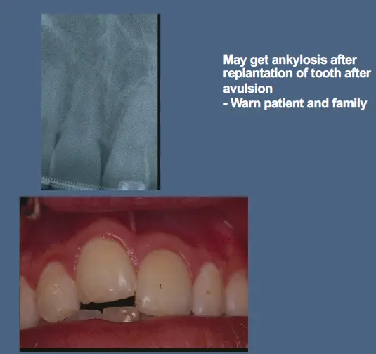
- interface and fusion/union between bone and tooth
- not always but usually associated with pulpal necrosis/infection
- usually associated with some truama, may be mild such as surgical exposure of a canine
- may be idopathic associated with eruption
- as trauma induced external replacement root resorption TIERRR initiates, on rare occasions intact cementum/cementoid layer acts as biological barrier so that ankylosis is not accompanied by progressive replacement resorption
- not all ankylosis associated with progressive trauma induced external replacement as resorption process may stop, but all TIERRR has element of ankylosis
- result in fusion of tooth and bone with loss of PDL
- may occur with TIERRR but not IIEIRR
- may need to distinguish idiopathic ankylosis from primary failure of eruption
- can result in immobile tooth and infraposition
- infraposition occur as local arrest of surrounding alveolar bone growth concomitant with continuous skeletal growth and development
- has been noted to be transient after truama
- may be spontaneous - not all ankylosis due to ETIRRR but all ETIRRR associated with ankylosis
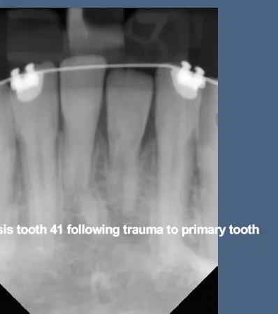
Infection induced internal inflammatory root resorption
- classified according to location as apical or intraradicular
- apical related to pulpal necrosis and infection with apical periodontitis - diseased
- intraradicular related to vital pulp with microbial overlay
Infection induced internal inflammatory root resorption - apical
- expect that resorption is due to granulation tissue of apical periodontitis diseased not pulp
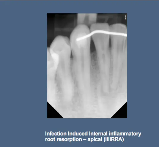
- file/flare and obturate to level of resorption
- need EAL to determine where invading PDL tissue is
- MTA for obturation
Infection induced internal inflammatory root resorption - intraradicular
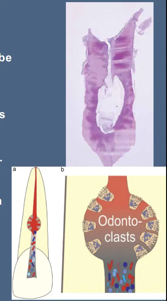
- apical pulp must be vital for resorption to be active
- apical pulp, although vital may be granulation tissue
- loss of odontoblast layer and predentine is required
- why odontoblasts are lost is uncertain
- bacterial overlay is located in coronal root canal and or dentinal tubules
- adjacent bone not affected until resorption has perforated
- usually related to traumatic injury or event that affects the pulp
- zimmets though
- history of trauma or caries may create a disturbance of odontoblast layer and microorganisms may enter dentien via enamel fracutres of caries and tubules are no longer filled with odontoblastic process facilitating infection stimulating the pulp
- Clinical features
- depends on location and extent
- usually asymptomatic but if active may be similar to pulpitis
- pinkish/red colour may be present centrally in crown if associated with pulpal changes
- sinus tract indicates perforation
- may be +ve or -nve pulp test
- progressive internal resorption associated with some pulpal infection
- bacteria may enter pulp canal thorugh dentine tubules, carious cavities, carcks fractures lateral canals
- for internal resorption to occur, pulp tissue apical to resorptive lesion must have viable blood supply to provide nutrient to clastic cells
- during initial pulp infection pulp may still be positive
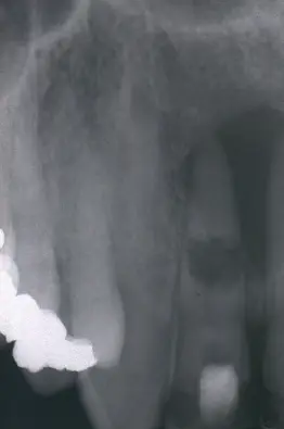
- radiographically
- uniform round or oval radiolucent enlargement of canal
- key feature is disappearance of original canal shape
- some cases CBCT may be useful
- Management
- most active lesions require prompt RCT
- bleeding from granulation tissue may cause difficulty in locating apical canal
- persistent bleeding may indicated perforation
- NaOCl use with passive ultrasonic irrigation is recommended
- short term dressing usually adequate, small defects may even be treated in single visit
- historically perforations required canals to be dressed long term with CaOH2
- short term dressing will subside the inflamed tissue at perforation site and repair with MTA will provide biocompatible seal
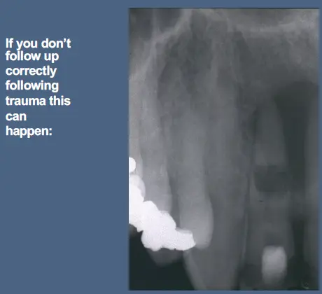
Infection induced external inflammatory root resorption
- apical or periradicular
- apical related to longstanding apical periodontitis - diseased
- periradicualr related to trauma
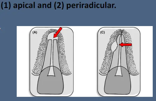
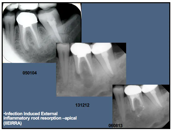
Infection induced external inflammatory root resorption - apical
- usually associated with pulpal necrosis and infection usually of long standing
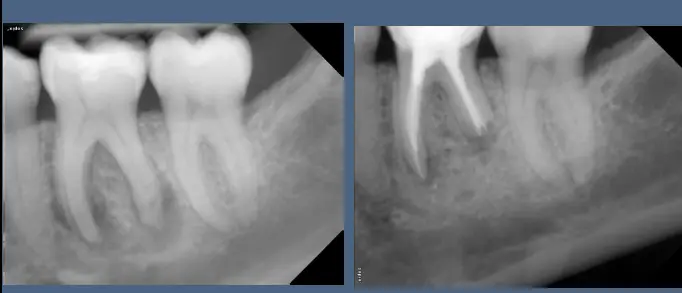
Infection induced external inflammatory root resorption - periradicular
- pulpal necrosis infection and trauma
- usually following replantation of avulsed tooth or luxation injury or intrusive luxation
- resolves with root canal treatment
- much more rapid than replacement resorption and therefore see large bowl shaped lesions
- due to damage to the root surface and an associated pulpal necrosis/infection
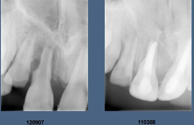
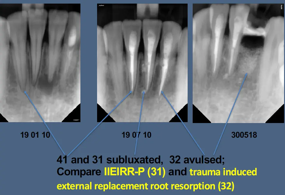
- tooth was sublux
- IIEIRRP occurs more quickly and usually assocaited with radiolucent lacunae and reversible if caught early enough, compared to TIERRR
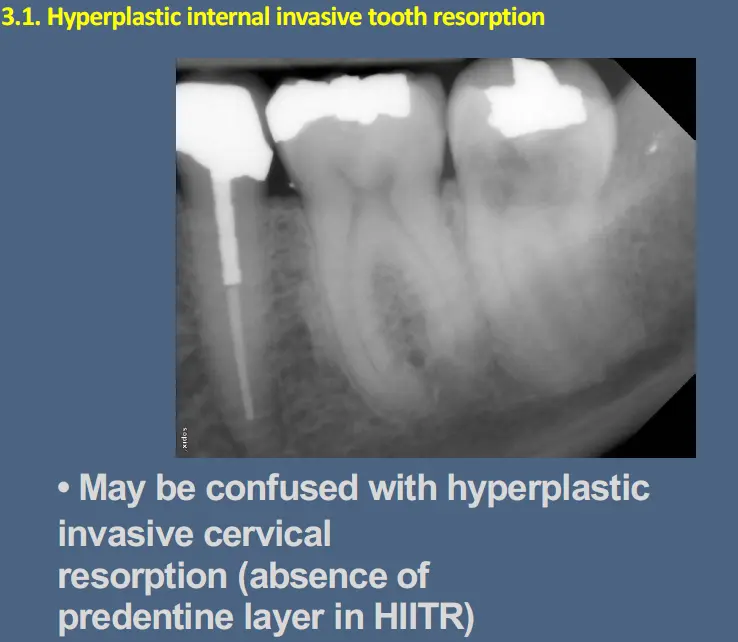
Hyperplastic internal invasive tooth resorption
- usually idiopathic and poorly understood
- resorbing tissue invades hard tissues of the tooth in a destructive and apparently uncontrolled fashion
- treatment involves total removal or inactivation of resorptive tissue is essential if recurrence/concurrence to be avoided
- concurrence indicates incomplete removal of resorptive tissue at time of treatment and recurrence is reestablishment of resorptive process
- may have pulpal or periodontal origin
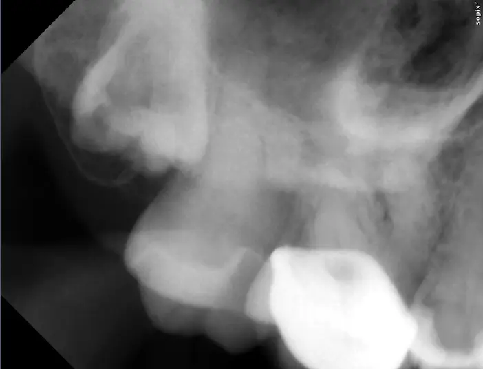
Hyperplastic external invasive coronal tooth resorption (HEICorTR)
- localised coronal enamel defect allows invasion of aggressive hyperplastic resorptive tissue in erupting tooth
- invasive coronal resorption has been observed in teeth which have been injured by the intrusive luxation of a primary tooth
- occurs in erupting teeth, hence thought to be due to tissue invade through developing external enamel epithelium of enamel organ
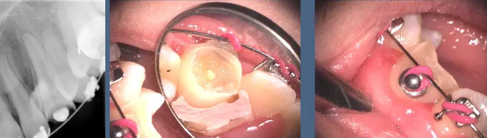
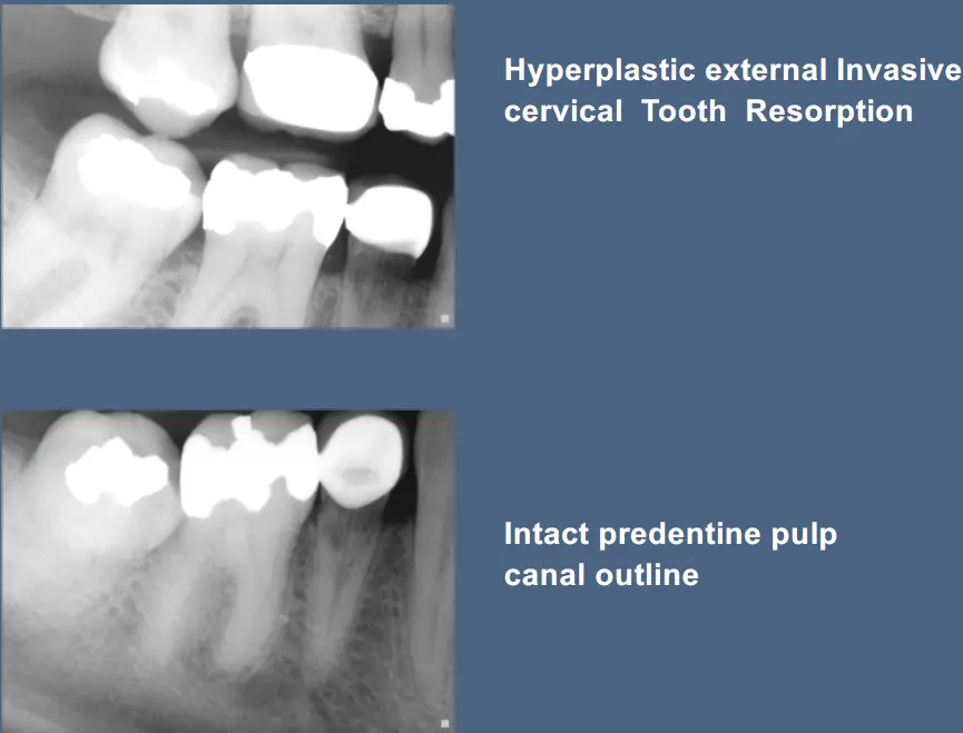
Hyperplastic external invasive cervical tooth resorption (HEICerTR)
- very obscure
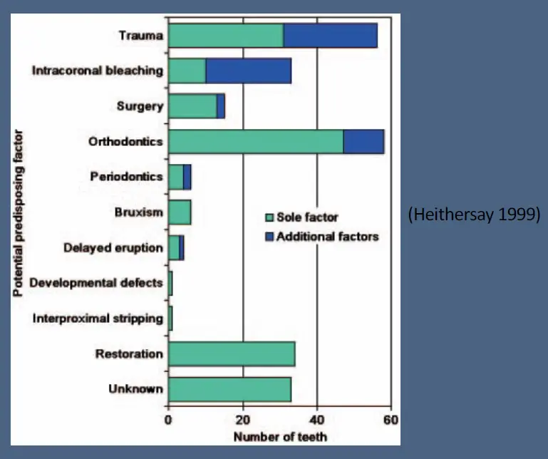
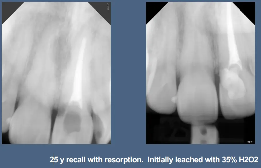
- potential predisposing factors have been identified and include trauma, ortho, and intracoronal bleaching as sole factors or in combo
- may be due to damage to cervical margin in association with exposed dentine at the CEJ
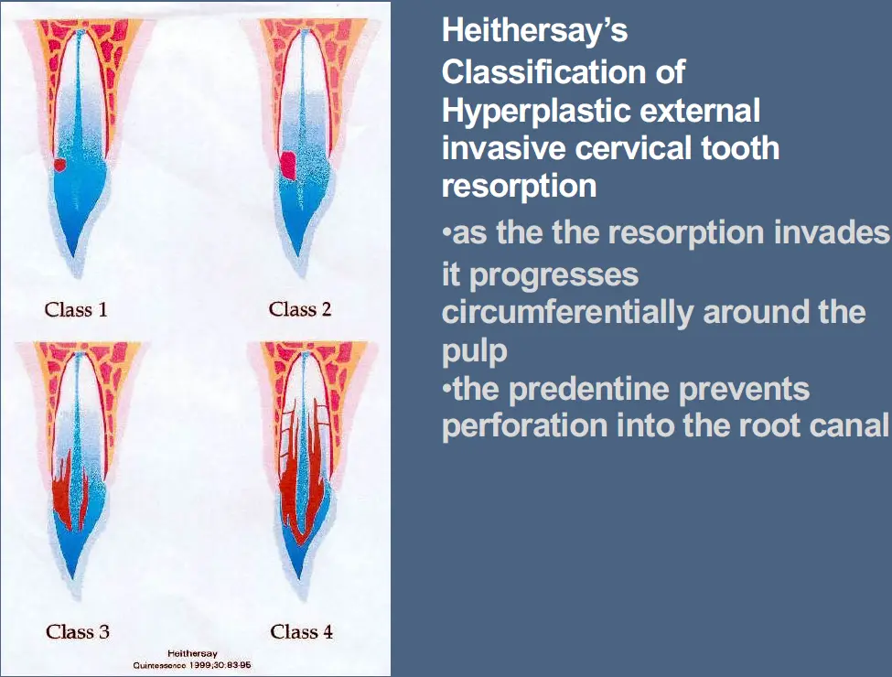
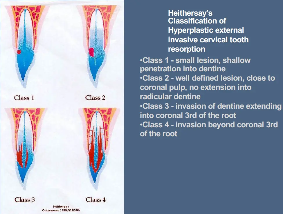
- treatment depends on position and extent of lesion
- often class 3 and 4 may not be treatable
- may use 90% trichloracetic acid (TCA) to inactivate fingers of resorption by coagulative necrosis
Hyperplastic external invasive radicular tooth resorption (HEIRTR)
- usually at level of where gingival margin attaches to root in region of marginal periodontitis with recession
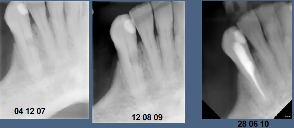
- progresses if do not remove the resorption
- check all teeth as me be multiple
Management of pathologic tooth resorption
- What type of tooth resorption is present? Diagnosis
- Is the tooth resorption external (periodontally derived), internal (pulpally derived) or communicating? Diagnosis
- Will the tooth resorptive process be self-limiting or transient and not require management other than careful monitoring of healing processes? Prognosis
- If the tooth resorptive process is progressive or self perpetuating will there be a favourable response to treatment and, if so, what is the appropriate therapy? Prognosis
- If treated what are the short and long-term Prognoses before and after treatment? (Endodontic, Marginal periodontal, Structural, Strategic)
- When is extraction and prosthetic therapy indicated?
Following trauma, prognosis relates to
- Tooth survival Structural
- Pulpal healing and pulpal necrosis – will the pulp be a source of infection? Endododontic
- Root development and disturbances in root growth with potential ankylosis. Strategic
- Periodontal healing and root resorption Marginal periodontal