Pulpal and Periradicular Pathosis
Aetiology of pulpal and periapical diseases
-
injury or irritation -> inflammation
- pulpitis - inflammation of the pulp
- apical periodontitis - inflammation of paical periodontium
-
reaction of pulp and periapical tissues to irritants dictated by type and duration of stimulus
-
irritants classified as
- non-microbial (mechanical, thermal, chemical)
- microbial
Mechanical irritants
- cavity preparatoins (dentine permeability increases closer to pulp
- deep scaler/curettage
- impact injuries
- mild/moderate trauma (extrusion) vs severe trauma (intrusion)
- immature vs closed apices
- impact trauma
- luxation/avulsion injuries cause direct PDL trauma
- occlusal trauma
- overinsturmentation of root canals
- root perforation
- overextension of root filling materials
- orthodontic forces beyond tolerance of PDL
Chemical irritants-pulpal
- cavity cleansers (alcohol or hydrogen peroxide)
- acids
- chemicals present in desensitisers, cavity liners, bases, temporary and permanent restorative materials
Chemical irritants-Periapical
- antibacterial irrigants
- intracanal medicaments
- some obturating materials
Microbial irritants - pulpal
- most significant cause of inflammation
- bacteria in dentine induce pulpal inflammation well before invading the pulp
- bacterial antigen diffuses through tubules
- localised inflammatory respopnse adjacent to affected tubules
- as caries progresses, intensity and character of infiltrate changes
- initial pulpal reaction mediated through innate immune response
- localised acute inflammatory response
- focal infiltration of PMNs and macrophages in sub odontoblastic tissue
- adaptive immune response develops (over time)
- bacteria antigen captured by dendritic cells
- focal accumulation of chronic inflammatory cells -> macrophages, lymphocytes, plasma cells
- pulpal response to bacteria constitutes first line of defence and is generally unable to clear the infection
- increased dentinal fluid flow (dilution and flushing effect)
- dentinal fluid components (IgG, albumin, fibrinogen, tubule occlusion)
- sclerosis of tubules (requires vital odontoblasts)
- production of tertiary dentine
- immunological reaction in the pulp
- pulp can survive a mild-moderate inflammatory episode, may cause death of odontoblasts, pulp may undergo fibrosis
- pulp may undergo eventual or rapid necrosis
- deeper damage (subodontoblastic zone) with microabscess formation is the point of no return (messer 2002)
- formatoin of small abscesses and necrotic foci progresses to total necrosis
- bacteria invade the entire root canal system
- what drives progression from pulpitis to necrosis
- dental pulp is unique connective tissue -> confined
- confined within rigid dentine walls
- self strangulation theory (old theory)
- pulp has mechanisms to oppose increased tissue pressure
- no collateral circulation
- effectiveness of inflammatory response depends on pulpal bloodflow
- progression to pulpal necrosis depends on
- number and virulence of microorganisms
- amount of circulation
- ability to avoid marked increase in intrapulpal pressure
- lymphatic drainage
- host resistance
- ultimately
- pulp is overwhelmed by increasing numbers of bacteria
- blood flow cannot meet demands for an effective response
- bacteria grow unopposed
- host response cause indirect tissue damage (PMNs release lysosomal contents -> liquefaction necrosis)
- microbial irritants from infected necrotic pulp diffuse from root canal to periapical tissues
- Apical Periodontitis - inflammatory response to infection of the root canal
- purpose is to prevent spread of infection from the root canal
Microbial irritants
-
bacteria play primary aetiological role in development or pulpal and periapical pathosis
- Kakehashi et al 1965 conventional vs germ free rats
- Sundqvist et al 1976 traumatised intact teeth with necrotic pulps, with and without apical pathosis
-
Routes of infection
- enamel and cementum are protective barriers
- happens via:
- caries
- fractures and cracks
- restorative procedures
- congenital anomalies of teeth
- scaling and root plaining
- loss of PDL attachment to apical foramen or lateral canal
- following pulpal necrosis bacteria invade the entire root canal system
-
infections classified as intra-radicular or extra radicular
- intraradicular
- primary
- secondary: m/o not present in primary infection but introducing during or after initial treatment
- persistent: m/o from primary infection resist intracanal antimicrobial procedures
- intraradicular
-
persistent and secondary
- responsible for clinical problems: persistent exudation, persistent symptoms, interappointment flare-ups, failure of treatment
- due to
- poor aseptic technique
- leaking restorations, caries not addressed
- untreated/inadequately cleaned canals
- inaccessible anatomy (isthmus etc.)
- procedural errors (ledging)
- inadequate obturation
- tooth/root fractures
- extra-radicular infections
- usually host defences in form of AP are effective at confining microorganisms to root canal
- situations where they do occur
- acute apical abscess
- periapical actinomycosis
- infected periapical cysts
- periapical biofilms
- displaced infected dentine pieces
host response to pulpal disease
- pulpal inflammatory response involves cellular and molecular changes
- degree of irritation stimulates a corresponding level of inflammation
- at some point inflammation changes from reversible to irreversible
- which is important distinct -> determines treatment
- cellular response
- increase in inflammatory cells (neutrophils, lymphocytes, macrophages, plasma cells, mast cells, dendritic cells)
- intensity of inflammatory response correlates to depth of carious lesion (Torabinejad et al 2021)
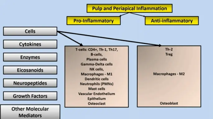
- Bacterial ingress into the pulp (severe pathosis) only occurred in cases diagnosed clinically as irreversible pulpitis (ricucci et al 2014)
- molecular mediators of pulpal inflammation
- some inflammatory mediators correlate with pulpal pain and diganosis of irreversible pulpitis
- PGE, neuropeptides, bradykinin, cytokines, chemokines, MMPs (mente et al 2016)
- increased mineralisation
- tertiary dentine, pulp stones
- pulp canal obliteration following dental trauma
- not considered pathologic in the absence of symptoms of apical pathosis
- internal resorption defects
- initiated by osteoclasts and odontoclasts (not normally present in a healthy pulp)
- arise from monocytes and are normally present in PDL and alveolar bone
- considered pathologic even in absence of symptoms
Classification of pulpal conditions
- pulpal diagnosis determined clinically by reliance on
- signs and symptoms of disease
- clinical tests
- radiographic imaging
- diagnosis must be obtained prior to contemplating endodontic procedure
- histologic examination of pulp at various stages of disease reveals that different areas of the pulp may have different stages of normalcy, inflammation or necrosis
- progression from reverible to irreversible to necrosis is gradual and occurs in small compartments of the pulp - can complicate diagnosis
Normal Pulp
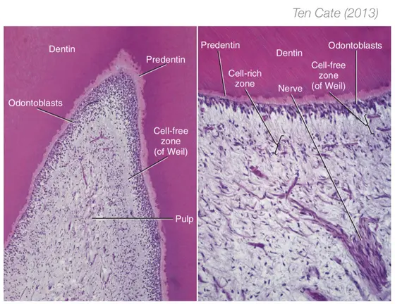
- histologic features
- intact odontoblast layer
- cell-free zone
- cell rich zone
- absence of inflammation or necrosis (sporadic inflammatory cells located in central pulp)
Reversible Pulpitis
- irritant
- inflammatory response in close proximity to area of irritation
- clinical features: no symptoms of mild hyperalgesia (sharp transient pain) in response to stimulus
- removal of irritant results in healing and resolution of symptoms (e.g. desensitisation of exposed dentine)
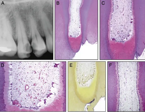
- histologically
- odontoblast layer may be reduced or flattened
- thick tertiary dentine, islands of calcification
- moderate chronic inflammation not disrupting architecture
- no necrosis, no bacterial invasion
Irreversible Pulpitis
- transition from reversible to irreversible pulpitis is gradual
- may or may not be associated with changes in symptoms
- clinical features
- may be associated with severe spontaneous and lingering pain, or milder symptoms or no pain
- inconsistent association of pain with irreversible pulpitis -> potentially for challenging diagnosis
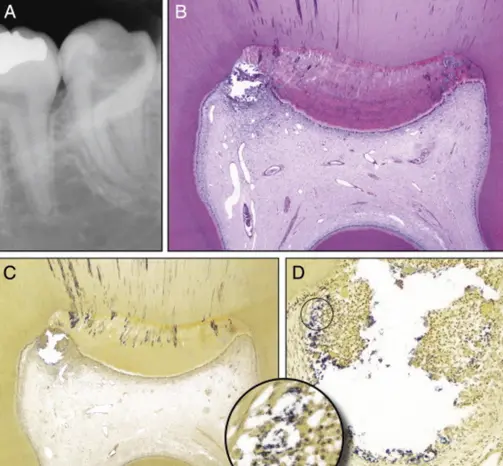
- histologically
- foci of pulpal necrosis (liquefaction) surrounded by severe inflammation
- bacterial invasion of the pulp
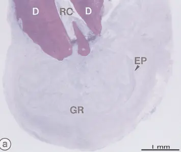
- additional conditions
- internal resorption with a vital (responsive) pulp
- hyperplastic pulpitis (pulp polyp)
- proliferative response occasionally seen in children
- pulp exposed to oral cavity and covered in desquamated oral epithelium
Pulp Necrosis
- pulp degenerates completely + infection with microbial biofilms
- 40% cases - irreversible le pulpitis transitions to necrosis without symptoms
- pulpal necrosis is asymptomatic but may be associated with apical conditions that are symptomatic
- why may a patient still feel instruments exploring a necrotic pulp space
- surviving pulpal nerve fibres
- fluid pressures that stimulate apical nociceptors
Periapical Disease
Host Response
- PA inflammatory response starts soon after initiation of pulpal inflammation
- PA inflammatory cell infiltrate + bone resorption occur in advance of total pulp necrosis, with vital pulp still resent in RC
- bone resorption occurs to accommodate formation of soft tissue lesion within which immunologic reactions occur to stop advancing infection
- microbial factors
- lead to development of periapical lesion
- LPS, lipteichoic acid, bacterial enzymes, adhesion factors, toxins etc.
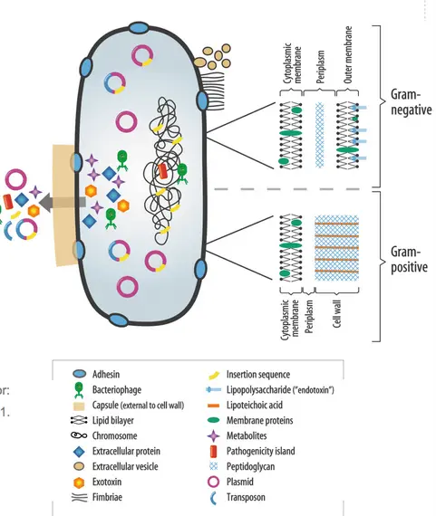
- bacterial components stimulate innate and adaptive immune responses, involving cells, molecular mediators, effector molecules and antibodies
- innate immune response
- dominates initial stages of pulpitis and AP
- complement activation (opsonisation, chemotaxis, inflammation)
- chemotaxis of neutrophils
- LPS stimulates endothelial cells adhesion molecules) and macrophages (produced IL-1 and TNF -> pro inflammatory effects)
- production of AA metabolites (leukotrienes, prostaglandins ->inflammation)
- neurogenic inflammation (neuropeptides)
- Adaptive immune response
- large load of virulent bacterial, critical in preventing disseminated infection
- activation of T and b lymphocytes bearing complementary antigen binding receptors
- proliferation and cytokine production by T cells
- T cell regulated macrophage activation
- proliferation of B cells with differentiation to plasma cells and antibody production
- bone resorption
- critical component of host response
- periapical bone loss correlates with degree of advancing microbial irritation and balance of pro inflammatory and anti-inflammatory factors
- modulation of bone resorption
- cytokines
- Activate resorption: IL-1, IL-6, IL-11, IL-17, TNF-alpha
- Limit resorption: IL-4, IL-10
- RANK-L / RANK / OPG interaction
- RANK-L (osteoclast differentiation factor) binds to its receptor (RANK)
- RANK-L / RANK Integration inhibited by OPG (osteoprotegerin)
- Levels of RANK-L peak at 2-3 weeks, tapering off at 4-8 weeks as OPG production increases, limiting bone loss.
- T-reg Lymphocytes
- Exert significant control over the size of periapical lesions
- cytokines
- PA host response is unable to eradicate the source of infection
- chronic inflammatory response usually effective at preventing microbial invasion into PA tissues
- mixed inflammatory cell infilitrate of T and B lymphocytes, PMNs, macrophages, plasma cells
- course of disease and structural components of the lesion depend largely on equilibrium between bacteria and host defence
Lesions and Histology
- PA lesions classified into
- granulomas
- cysts
- apical abscesses
Granuloma
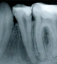
- chronic inflammatory cell infiltrate (lymphocytes, plasma cells, macrophages, mast cells) arranged in a granular manner
- multinucleate giant cells (osteoclasts) and foam cells (engorged macrophages)
Periapical cysts
- arise from long-standing granulomas
- proliferation of epithelial cell rests of malassez (remnants of embryonic hertwigs epithelial root sheath)
- contain fluid or semi solid material and may contain cholesterol crystals, lined by stratified squamous epithelium, surrounded by connective tissue containing all the elements of a PA granuloma
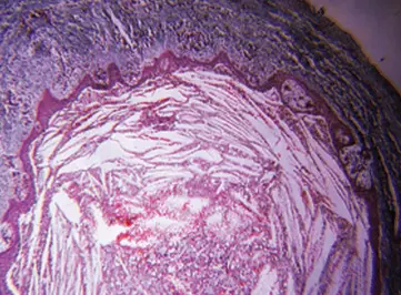
- less than 20% of all AP lesions (nair et al 2004)
- two types of apical cysts
- true cysts - do not communicate directly with apical foramen
- pocket cysts - root apex opens directly into cyst lumen
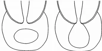
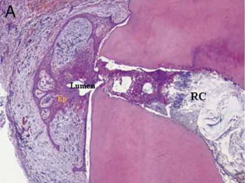
- was previous hypothesis that true cysts would not repsond to nonsurgical RCT and would require surgical enucleation (no direct evidence)
- differentiation of cysts and granulomas is not possible clinically
- large cyst-like periapical lesions can heal after non-surgical RCT
Periapical Abscess
- intense inflammatory response surrounding areas of tissue necrosis
- bacteria may be present in necrotic areas
- extra-radicular bacteria are rare in asymptomatic apical lesions, but may be more frequent in cases of persistent disease after RCT
- occasionally actinomyces spp. may form extra radicular bacterial colonies
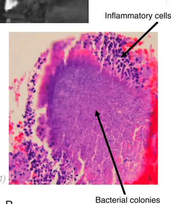
Clinical Classification of periapical conditions JOE 2009
-
normal periapex
- no evidence of apical pathosis clinically or radiographically
- asymptomatic to percussion and apical palpation
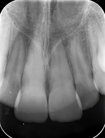
-
symptomatic apical periodontitis
- inflammation of apical periodontium producing symptoms
- tooth is tender to percussion and or palpation
- may be primary or secondary
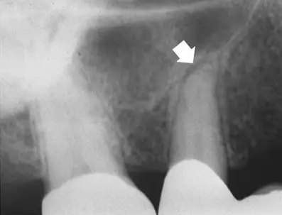
- primary
- initiated in a healthy periodontium
- etiology: inflammatory mediators from an irreversibly inflamed pulp, bacteria invading apical root canal, mechanical and chemical irritatnts
- histology: acute inflammation limited to apical PDL and adjacent cancellous bone, inflammatory exudate with infiltration of neutrophils
- clinically: pain, tooth elevation, TTP, pulp may be irreversibly inflamed or necrotic, slight widening of apical PDL space
- secondary
- acute inflammation which occurs in an already established chronic apical periodontitis lesion
- etiology: changed intracranial microbiota (increased load, virulent strains) or disruption of host response
- histology: focus of acute inflammation located within an otherwise chronic lesion
- Clinically: non vital, TTP, radiographic lesion, possibly rapid enlargement of the radiolucent area
-
asymptomatic apical periodontitis
- periapical radiolucent lesion
- necrotic pulp
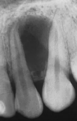
- etiology: bacterial infection of root canal contained by host response
- histologically: chronic inflammation - granulation tissue infiltrated with lymphocytes, plasma cells, and macrophages. May be epithelialised or non epithelialised. Nay be granulomas or cysts (cannot determine clinically)
- clinically: non vital tooth, periapical radiolucency, bone loss may occur at any portal of exit
-
acute apical abscess
- most serious clinicaly diagnosis in endo
- virulent bacteria from an infected root canal invade the periapical tissues causing pain and swelling
- subsides once the intracanal infection is treated
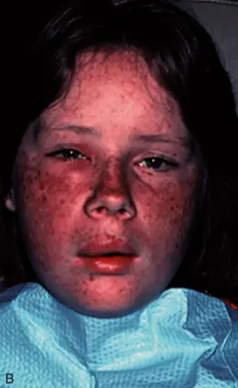
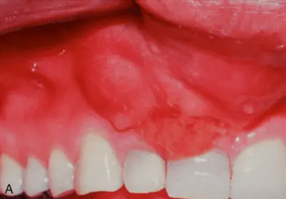
- histologically: intense PMN infiltrate, liquefaction necrosis surrounded by granulation tissue, rapid bone resorption may occur
- clinically: intense pain, swelling, TTpercussion TTpalpation, increased mobility, non-vital tooth.
- may range from small localised swelling to a large swelling that invades fascial spaces and may cause severe morbidity
- systemic signs include fever, malaise, lymphadenopathy, leucocytosis
- Radiographically widened PDL to obvious radiolucent lesion
-
chronic apical abscess
- virulent bacteria from an infected root canal invade the periapical tissues to cause purulent drainage through a sinus tract
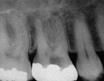
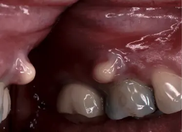
- sinus tract follows pathway of least resistance
- attached gingiva/oral mucosa
- via PDL space (isolated periodontal probing depth to root apex
- to skin of the face
- usually asymptomatic because drainage exists
- heals once the infected root canal system is treated
- histology: similar to asymptomatic apical periodontitis, sinus tract may be lined partialyl or totally by epithelium surrounded by granulation tissue
-
condensing osteitis
- variant of asymptomatic apical periodontitis resulting in an increase in trabecular bone
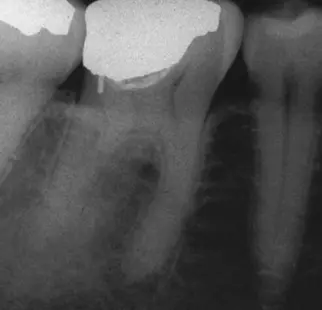
- etiology" chronic inflammation of pulp (reversible to irreversible) or necrotic infected pulp, result of low grade bone irritation
- histology: dense mass of bony trabeculae with few marrow spaces
- clinically: does not require treatment unless pulpal condition is irreversible, expected to resolve after endodontic treatment
-
non-endodontic pathosis
- other lesions of the jaws may mimic apical periodontitis
- ddx
- periodontal disease
- occlusal trauma
- benign and malignant lesions
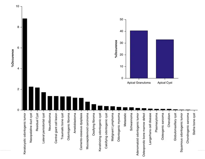
- usually (not always) lamina dura intact and pulp tests normal
- final diagnosis based on surgical biopsy and histopathologic examination