Examination and diagnosis or pulp, root canal and periradicular conditions
#D3/S2/W1
#Endo
Diagnostic process
- data gathering
- history
- clinical exam
- radiographic exam
- diagnosis
- treatment options
- treatment plan and informed consent
Diagnosis
- rule out other causes of pain
- TMD
- occlusal interferences
- non-odontogenic pain
- must perform pulp vitality tests
- if in doubt - do nothing irreversible
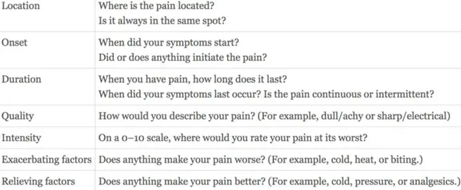
History
- presenting complaint
- history presenting complaint
- medical and dental history
Clinical Examination
eo
- swelling
- masticatory muscles
- TMJ
- lymph nodes
IO - soft tissue (inflammation, swelling, sinus tract)
- occlusion (traumatic occlusion, fremitus)
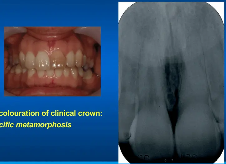
- teeth: caries, cracks, restoration, discolouration (calcific metamorphosis, attrition erosion abrasion, developmental abnormalities (dens evaginatus)
- tests
- percussion, palpation, probing, mobility
- probing
- discrete isolated pocket: VRF/crack or drainage via PDL (sinus tract)
- broad pocketing (periodontal disease)
- furcation involvement
Pulp testing
- EPT
- endofrost
- CO2 ice tests
- 2 purposes
- identify problem tooth by reproducing chief complaint
- determine a vital versus non vital pulp
- to test pulp vitality CO2 is considered most reliable
- false negative response
- dentinal scelroris (elderly)
- young patient
- trauma (inc ortho)
- EPT typically used to confirm a negative response to CO2
- false negative response
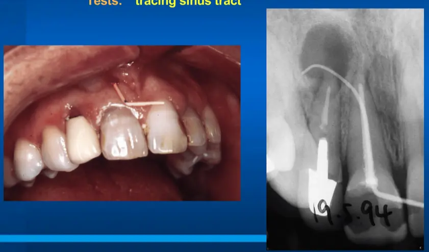
Tracing sinus tract
- sinus tract doesnt always arise beside tooth that is contributing to pathosis
Transillumination, staining (methylene blue), selective cuspal loading
- all undertaken for identification of cracked tooth particularly frac finder for recreating presenting complain (sharp transient pain on biting)
Selective anaesthesia
- unsure if pain is originating from what tooth
- elect to selectively anaesthetise and assess if pain has subsided or not
Radiographic examination
- must have good quality pre-op film
- periapical film + bitewing for all posterior teeth
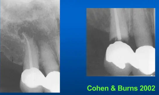
- CBCT imaging eliminates superimposition, useful as an aid in diffcult diagnosis
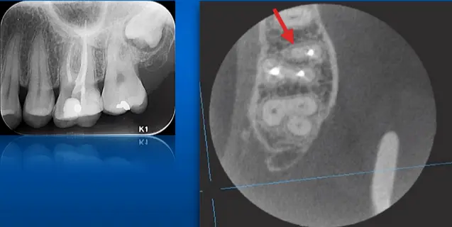
misdiagnosis case
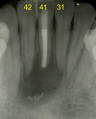
- referred to rct 31 and 42, retreat 41 for pain and swelling
- no tests performed, no diagnosis
- 31 and 42 CO2 positive
- advised no treatment for teeth 31 and 42
- option for revision vs surgery on tooth 41
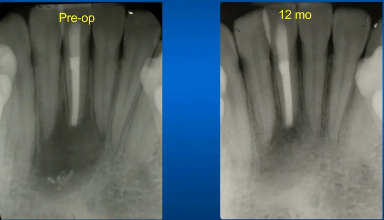
- positive CO2 always overrides radiographic appearance
- dont assume which teeth are involved based on size of radiolucency
Endodontic diagnosis
Pulpal diagnosis
- normal pulp
- symptom free, normally response to pulp testing
- reversible pulpitis
- vital inflamed pulp that may heal with vital pulp therapy
- e.g. slight hypersensitivity to cold
- symptomatic irreversible pulpitis
- vital inflamed pulp incapable of healing
- lingering thermal pain
- spontaneous unstimulated pain
- referred pain
- biologic basis
- pulp contains 2 types of nociceptors
- type A myelinated (preferentially located around pulp periphery)
- type C nonmyelinated and smaller (preferentially located in blood vessels in central area of pulp)
- C fibre activation indicates severe pulpal inflammation
- deep dull quality, poorly localised, lingering pain
- accumulation of inflammatory mediators: nociceptor sensitiation
- spontaneous pain, sleep disturbance
- TTpercussion + vital pulp: inflammation extends to periodontium
- referred pain indicated severe inflammation and irreversible pulpitis
- pulp contains 2 types of nociceptors
- asymptomatic irreversible pulpitis
- vital inflamed pulp incapable of healing
- no symptoms but irreversible inflammation still caused by carious exposure, caries excavation etc.
- e.g. routine bitewing shows large carious lesion in pulp but no symptoms
- pulpal necrosis
- negative pulp test (thermal/electric)
- previously treated
- endodontically treated and canals obturated
- previously initiated therapy
- partial endodontic therapy (e.g extirpation or pulpotomy)
Periapical diagnosis
- Normal apical tissues
- Symptomatic apical periodontitis (SAP)
- acute inflam of PA tissues
- TTPercussion, TTPalpation, pulp inflamed or necrotic, radiographically normal or apical radiolucency
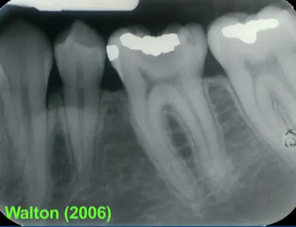
- example patient: strong persistent pain localised 36, exacerbated cold stimuli, affecting her sleep, painful to percussion
- DIAGNOSIS: symptomatic irreversible pulpitis and symptomatic apical periodontitis
- asymptomatic apical periodontitis (AAP)
- delicate equilibrium between microorganisms and host defence
- asymptomatic (not TTP TPP), necrotic pulp, PA radiolucency
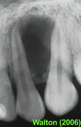
- no pain, 22 and 23 no TTpalpation or TTpercussion, radiograph shows PARL, 22 nonresponsive to cold test
- DAIGNOSIS: pulp necrosis + asymptomatic apical periodontitis
- cant determine histology from radiograph or clinically Nair et al 1996
- acute apical abscess (AAA)
- swelling, intense pain
- +- systemic signs
- necrotic pulp, TTP, increased mobility
- radiographically widened PDL or radiolucency
- two distinct presentations
- cellulitis
- initial presentation of infection
- spreading inflammatory exudate (no pus)
- large diffuse swelling, soft to palpate (initially)
- dangerous (host unable to lcoalise the lesion)
- abscess
- more chronic (days to weeks)
- pus surrounded by granulation tissue
- well-localised fluctuant swelling
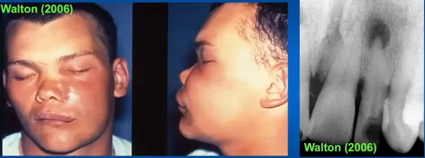
- acute facial swelling and PARL on 22
- DIAGNOSIS: Pulp necrosis + acute apical abscess
- chronic apical abscess (CAA)
- abscess that drains to a surface (oral mucosa, facial skin, gingival sulcus via PDL)
- sinus tract present, usually asymptomatic, necrotic pulp, periapical radiolucency
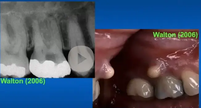
- negative response to cold test, abscess associated with MB root of tooth 16
- pulp necrosis and chronic apical abscess
- condensing osteitis (CO)
- variant of AAP resulting in increased trabecular bone
- pulp chronically inflamed or necrotic
- young and middle aged patients
- mandibular first molar
- diffuse radiopaque lesion at apex of a tooth
- expected to resolve after endodontic treatment
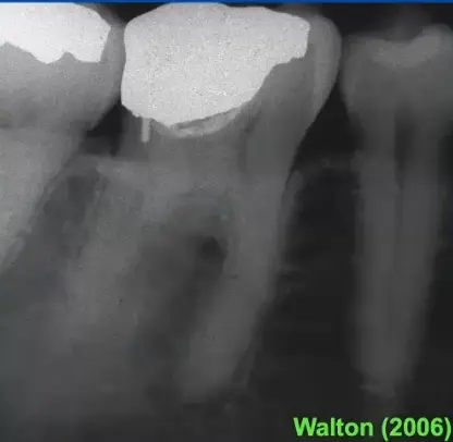
- negative response to sensibility testing, increased radiopacity with distal root of tooth
- necrotic pulp with condensing osteitis
- non endodontic pathosis (NEP)
case example
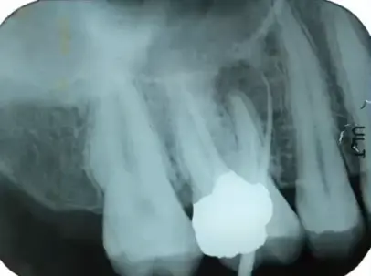
- draining sinus tract traces with GP point to apical radiolucency associated with MB root
- 16 is previously endodontically treated
- diagnosis: previously treated with chronic apical abscess