Endodontic procedural errors and their management
Important clinician questions
- how will the complication affect prognosis
- once complication has happened, how can the impact on prognosis be minimised
- how can the complication be avoided
Perforations
Why are they a problem
- create pathway for bacteria to reach periodontium and cause an inflammatory lesion
- can be difficult to disinfect and seal
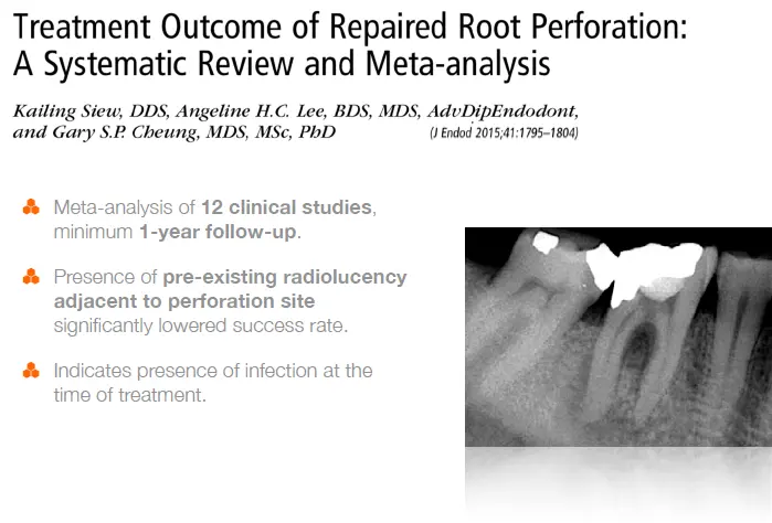
- prognosis depends on control of bacterial infection at perforation site
Size
- larger (>3mm) more likely to have pathological changes in adjacent perio tissue
- (Tsesis 2010, Mente 2014)
- more difficult to seal
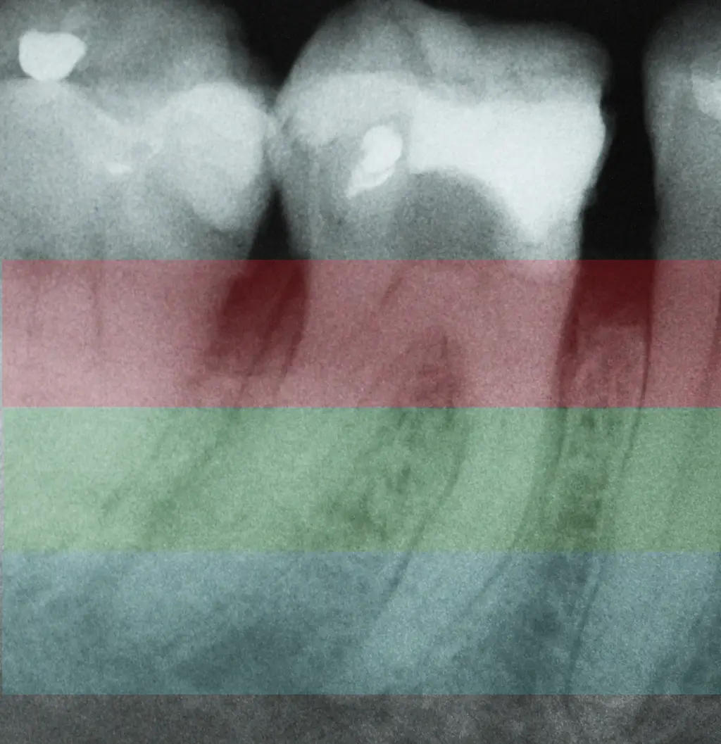
Time - older more likely to have established infection
- immediate repair has best prognosis
Location - crestal
- risk gingival epithelial downgrowth
- pocket formation
- bacterial ingress maintains inflammation
- periodontal destruction
Study
Perforation repair principles
- disinfect perforation site
- seal perforation with a material that prevents bacterial leakage
- repair material should be biocompatible and encourage regeneration of periodontal tissues
Mineral trioxide aggregate
- hydrophillic powder mixed with sterile water
- 75% portland cement, 20% bismuth oxide, 5% gypsum
- sets after 3 hours (Torabinejad 1995)
- calcium silicate cement (calcium crystals in an insoluble silicate matrix)
- release calcium hydroxide for >3month by surface hydrolysis
- promotes alkaline pH 11 -12 for >2month
- stimulate hard tissue formation
- superior sealing ability
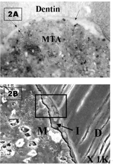
- forms mineralised interficial layer of hydroxyapatite crystals when placed against dentine
- adaptation and seal better than amalgam, zoe/irm, gic
- seal not affected by moisture or blood
- superior biocomptaibility
- stimulated regeneration of cementum and PDL
- biological seal
MTA disadvantage
- perforation with communicate with oral cavity/gingival sulcus
- slow setting time - freshly mixed MTA can wash out
- rough granular surface, promotes subgingival plaque formation
- RMGIC preferred
- can discolour teeth when used in cervical zone
- cost 170 / gram
- difficult to handle
Other bioactive endodontic cements
- biodentine, bioaggregate, endosequence, theracal LC
- calcium silicated based material with common bioactive properties
- release CaOH
- form interficial layer
- form apatie crystals on their surface in synthetic tissue fluid
- claim to have similar MTA properties without its drawback
- few studies to support, unknown long term efficacy
- use of MTA appears to give highest success rate
Furcation perforation repair
- non-surg approach with high mag
- decontaminate perforation site
- debride dentine walls - fine pulp burs or ultrasonics under maginification
- passive irrigation with sodium hypochlorite
- haemostasis - trichloroacetic acid TCA
- placement of mTA (protect canal orifices)
**Prevention of perforation
- intial radiographic assessment
- angulation of tooth
- location/depth of pulp chamber, measure distance from occlusal surface to pulp chamber floor
- during access
- remove all restoration
- cut access cavity to safe depth
- aim for any visible chamber/canal space
- expose a radiograph with a bur to check depth/direction
- identification
- cbct
Middle third perforations
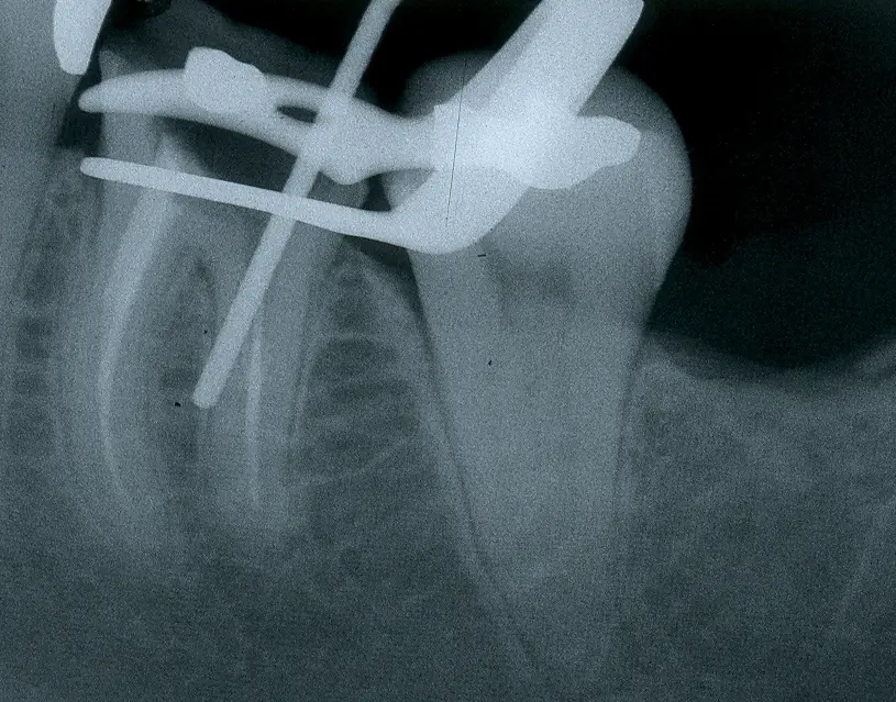
- misdirected posts
- strip perforation caused by endo files or gates glidden drills
- repaired via non-surgical approach
- prevention
- dentine on furcation aspect of roots can be very thin
- mean thickness of furcal wall in mesial root 0.7mm
- when using rotary NiTi, be aware of taper
- aim for mod taper 4% rather than larger taper 6%
Prevention of post space perforation
- distal canal of endodontically treated mandibular molars
- residual dentine thickness furcal wall <0.5mm in 17% of cases
- post space with # 4GG drill caused strip perf in 7% of cases
- post space should be limited to endodontically prepared canal
- safe post space prepartion
- immediately after root filling
- remove GP using heated instrument (+-# 2 gg drill = 0.7mm)
- use post drills by hand if active cutting is required
Apical third perforations
- often impossible to regain access to apical canal
- surgery required when tehse cases do not respond to conventional RCT
Clinical outcomes of perforation repair
- clinical studies show fair-good prognosis for repair of perforations using MTA in absence of periodontal pocketing
- healing rate 80-100%
- pocketing indicates poor prognosis
- prognosis reduced by
- inexperience operator
- post placement after perforation repair
Minimise impact on prognosis
- microscope required to adequately manage most perforations
- referral to endodontist is usually best
- if perforation detected
- place cavit over area as temp seal
- sound provisional restoraiton to prevent bacterial ingress
- inform patient
- refer patient and phone endo in advance to discuss
Fractured instruments
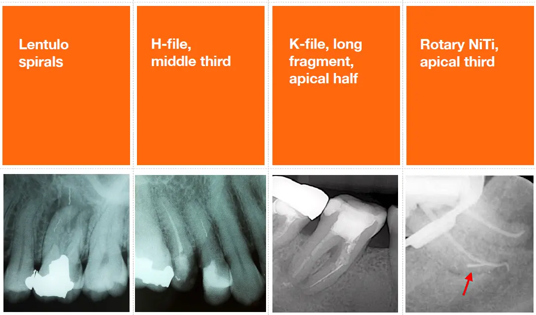
How common
- hand instrument 1.6%
- rotary niti 1.0%
Aetiology of instrument fracture
- Nickel titanium
- torsional fracture
- tip binds while shaft continues to rotate
- deformation precedes fracture (unwinding)
- usually due to excessive apical pressure or inadequate glide path
- **prevention
- adequate glide path prior to use
- clean file flutes regularly using endo sponge
- avoid excess apical pressure
- discard files that show distortion or significantly curved canal
- flexural fracture
- cyclic fatigue occurs when file rotates in curved canal
- work hardening and metal fatigue leads to initiation of surface cracks
- file breaks suddenly without distortion
- Prevention
- use touch retract action
- track file usage and discard after (up to) 4 uses
- follow manufacturer guidelines for motor speed/torque settings
- use heat treated NiTi files
- double curvature produce greater stress
- fatigue accumulates quickly and fracture may occur after very short time
- no correlation between number of uses and frequency
- single overloading event is most common mechanism
- torsional fracture
- see Root canal mechanical instrumentation 2, rotary canal preparation methods#Aetiology of NITi instrument fracture for rest of info
Does instrument fracture reduce prognosis
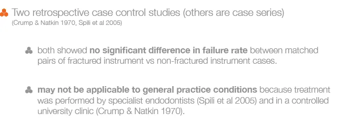
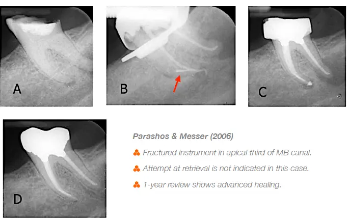
- Fractured instruments are a problem when they prevent cleaning and hsaping of an infected root canal
- prognosis will depend on
- presence of a PA lesion (indicates an infection) and
- stage of canal preparation when breakage occurs (extent to which microbial control is compromised)
- Breakage before completion of instrumentation in an infected root canal has a high risk of persistent disease
- breakage in final stages of cleaning and shaping has best prognosis
Treatment planning following instrument fracture
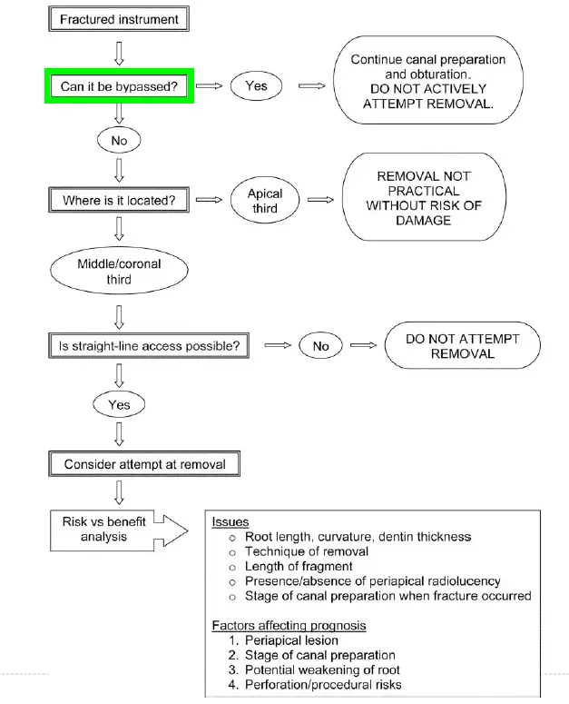
- bypassing broken files
- conserves tooth structure and still permits thorough cleaning and hsaping
- file incorporated into root filling
- 1 in 3 broken files could be bypassed
- Instrument removal - ultrasonic technique
- must be able to see broken file (microscope)
- modified GG drills used to create a staging platform
- ultrasonic tips trough around and expose the coronal part of the file
- ultrasonic tip wedged between file and canal wall to vibrate the file loose
- direct vision and straight line access to broke file are essential
- when coronal part of instrument lies in straight portion of the canal, safe removal was predictable
- instruments located around the curve could not be safely removed and attempts led to major canal damage
- platforming removes significant amount of tooth structure
- root fracture resistance declines the more apical the fragment
Loss of working length
Ledging and blockage
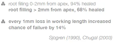
- instruments cant reach working length in a previously patent canal
- ledge
- iatrogenic gouging of the outer wall in a curved canal
- blockage
- compacted dentine mud, pulp tissue, foreign object (resto material)
- impact on prognosis
- prognosis is reduced when root canal is infected and cant be fully instrumented
- teeth with apical periodontitis
- Iatrogenic errors predisposing to ledging
- inadequate straight line access
- straight stainless steel files in curved canals
- incorrect length determination (short)
- false assessment of canal direction
- ledging with rotary NiTi
- inadequate glide path
- held stationary while rotating
- Bypassing blocks and ledges
- pre enlarge coronal canal
- precurved size #08 or #10 SS handfiles (21mm length)
- circumferential picking action to locate a catch
- advance file using watch-winding and short amplitude
- in-out strokes (keep file tip apical to the block)
- establish patency (electronic apex locator)
- circumferential short amplitude strokes to open path for precurved # 15-20 hand files before trying rotary NiTi
- precurve master GP cone+
- when unable to bypass blocks or ledges
- prognosis depends on
- presence of apical periodontitis
- distance between blockage and root apex
- stage of instrumentation when blockage occurred
- if disease persists, options are apical surgery or extraction (or intentional replantation)
- prognosis depends on