Endodontic Retreatment Case Selection & Procedures
Primary endo treatment
- clear diagnosis of endodontic disease
- point along contiuum of pulp irritation leading to pulpal necrosis and extension into periapical tissues
secondary endodontic treatment / retreatment
- assessment of chaos
Causes of primary treatment failure
RCT usually fails when treatment falls short of accepted standards
failure to control an intracana lbacterial infection
- aseptic technique
- coronal leakage (caries/restorations)
- missed canals
- inadequately cleaned canals
- procedural errors (blockage, ledge, transportation, perforation)
- inadequate obturation
persistence of disease in well treated teeth - persisting intracanal infection (inaccessible anatomy)
- extra radicualr infection
- radicular cyst
- foreign body reaction
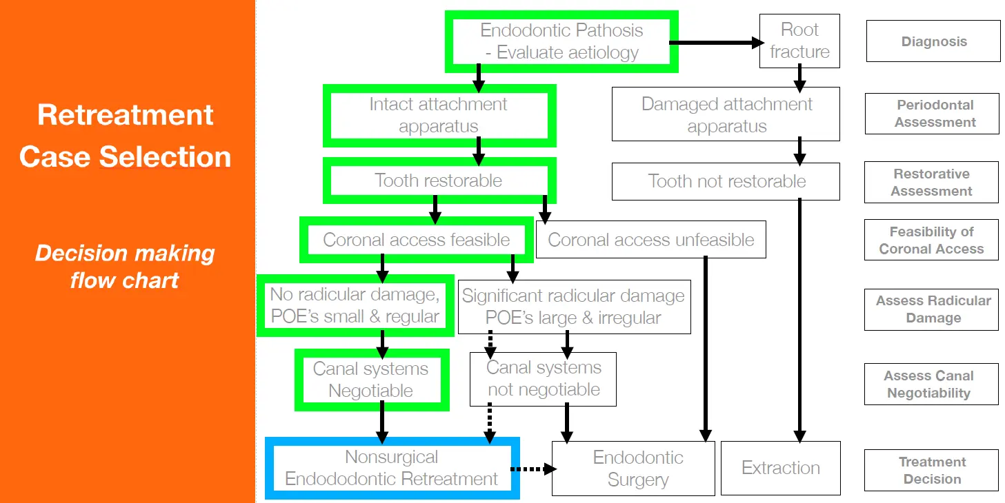
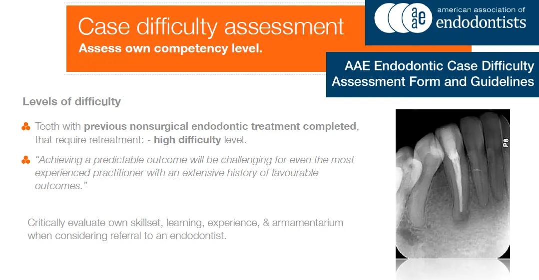
Retreatment case selection
Diagnosis
- can the reason for failure of the primary RCT be identified and corrected
- take CBCT
- identify additional untreated canals
- identify previous treatment complications
- exclude longitudinal tooth fracture
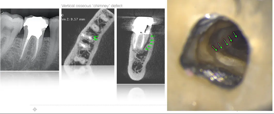
ROOT FRACTURE
detecting VRF in root filled teeth
- magnification
- transillumination
- isolated deep probing both sides of root
- probing + sinus tract (can be multiple)
- widened PDL along axial root wall
- bone loss pattern: vertical/crestal, lateral, frucation, J shaped, halo
- CBCT
Periodontal assessment- assess for attachment apparatus damage
restorative assessment - eliminate pathways for bacterial ingress
- detect and assess cracks
- assess amount of remaining tooth structure
- restorative plan
Feasability of coronal access
- posts can generally be removed
- consider amount of tooth structure likely to remain after disassembly
- time/cost of dismantling and reconstruction
assess radicualr damage
- stiff instruments to straighten
- over preparation of outer wall (apically) and inner wall (middle)
- compromised cleaning and filling
- ledging, perforation, weakening
- procedural errors that alter RC morphology will affect prognosis for endo retreat
- Gorni and gagliani 2004
- RC altered 47% healed
- RC respected 87% healed
Assess ability to negotiate canals
- calficiation
- blockage
- ledging
- fractured instruments
Outcomes
The toronto study (de Chevigny et al 2008)
- 4-6 year recall following NS retreatment of 229 teeth performed by supervised endo residents
- presence of pre op AP had important influence on outcome
- without ap 93% healed
- with AP 80% healed
Contemporary retreat techniques (he et al 2017)
- microscope, rotary niti, ultrasonic, irrigant activation
- 2 year recall following NS restreatment by endo resident
- 90.4% success
- during retreatment, negotiating entire canal length to patency doubled odds of success
Teeth exhibit high survival rates following endo retreatment
- Salehrabi et al 2010
- Ng et al 2011
- 4-5 year survival rate 89-95%
Disassembly techniques
Crown to remove or not
- if has defective margins of shows leakage/caries -> remove it
- PA and bitewing
- if accessing through crown, undertake internal assessment
Removal deep core material
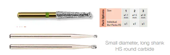
- magnification and illumination
- remove core material peripherally using HS burs, identify restorative dentine interface
- coarse ultrasonic tip to shatter thinned material at pulp chamber floor/orifices
Post removal
- piezon units with specialised tips at high power
- most posts can be safely removed
- negligible risk of root fracture
- can generate high temperatures
- prefabricated alloy posts
- adequate access outline, shorten post, remove surrounding core material
- fine US to remove bulk cemeent and open anatomic spaces
- coarse US to vibrate post back/forth, counterclockwise
- cast postcore
- remove surrounding core material HS burs
- fine US to trephine and remove bulk cement
- corase US to vibrate back forth counter clockwise
- Bonded fibre reinforced posts
- carbon fibre, glass, quartz fibres in composite matrix bonded into canal
- higher difficulty, requires microscope
- drilled out as far as possible using fine hS carbide burs
- US top to disintegrate apical part
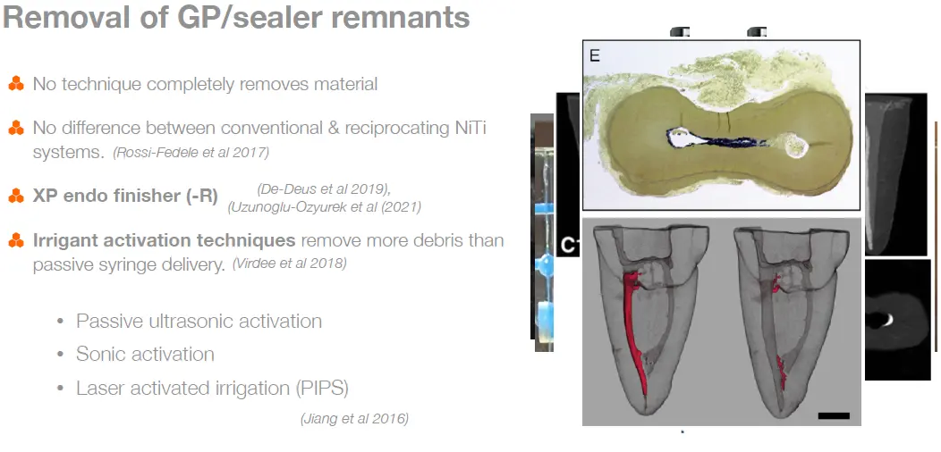
Gutta percha removal
- preferable to avoid/minimse use of solvents
- liquefied infected GP compacts into canal irregularities, hinders cleaning and can extrude
- retreatment without sovlent leaves canal walls and tubules cleaner
- depends on characteristics of root filling
- quality of condensation
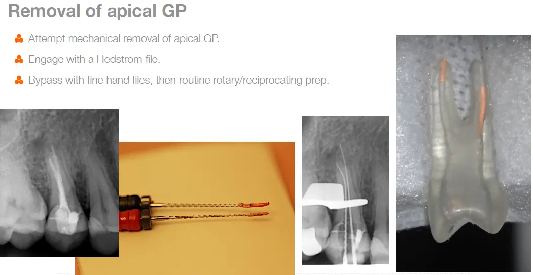
- poorly compacted - bypass and engaged
- bypass with find hand file, glide path
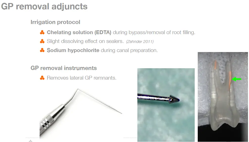
- engage with hedstrom file
- routine rotary/recip niti protocol
- braiding technique
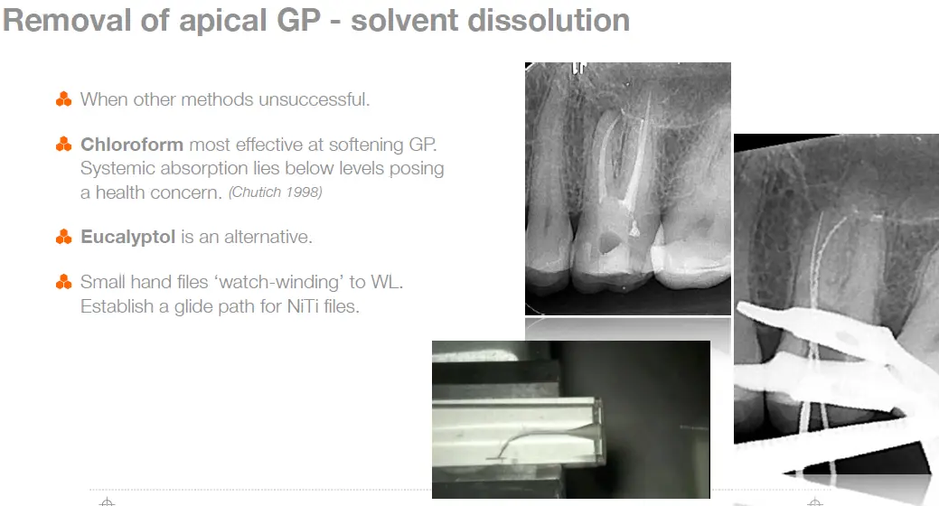
- well compacted - may require solvent
- no space available for instrument insertion
- remove as much GP as possible by mechanical means before using solvent
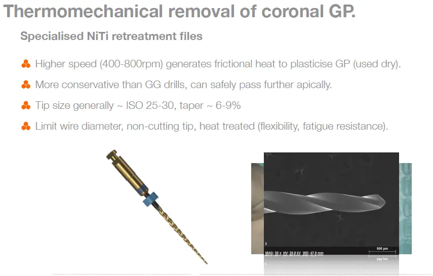
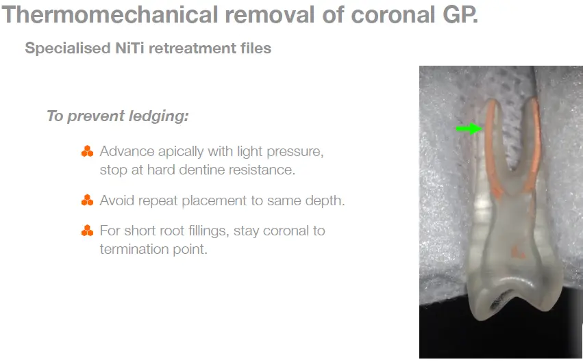
- Thermomechanical removal of coronal GP first, then apical GP removal with solvent if needed
- poorly compacted - bypass and engaged
- length of obturation
- under extended - ledge may be present, use solvent to help avoid further ledging
- overextended - carefully bypass and retrieve GP cone intact, avoid solvent
Thermal injury through heat transfer using ultrasonics
- root surface temp increase > 10 degrees -> necrosis of PDL and bone
- can occur within 5 minutes
- recommended protocol
- continuous water spray and suction, activate US for 15 second intervals
- if removal attemp reach 10min, allow 2 min rest
- refrigerant spray to cool post