Diagnosis and management of concurrent endodontic and periodontal diseases
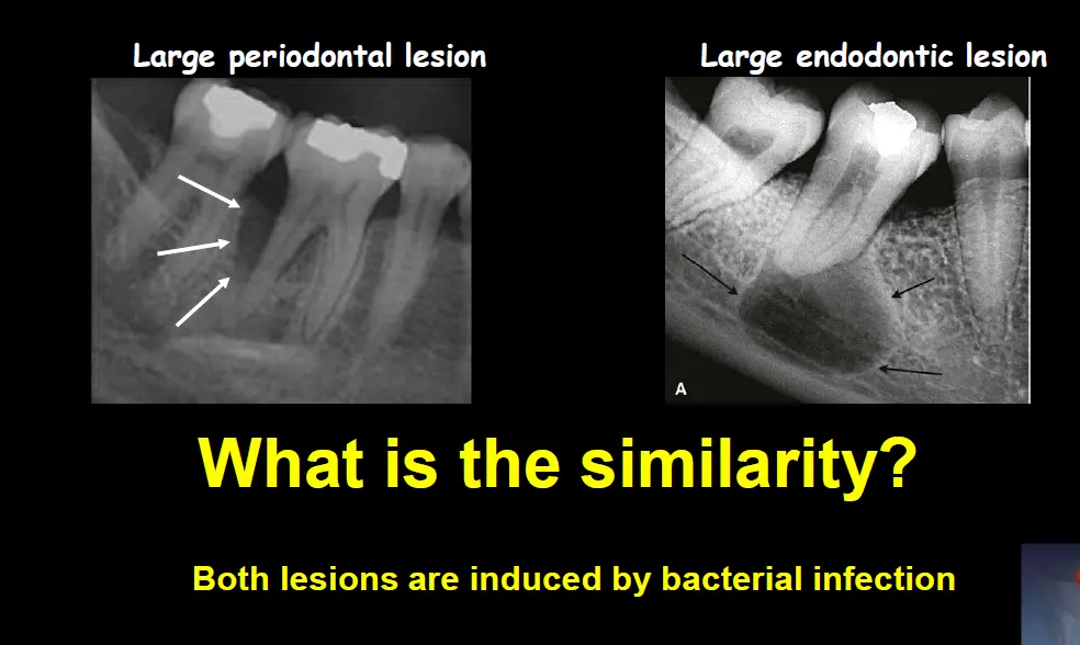
how to treat infection
-
eliminate the cause
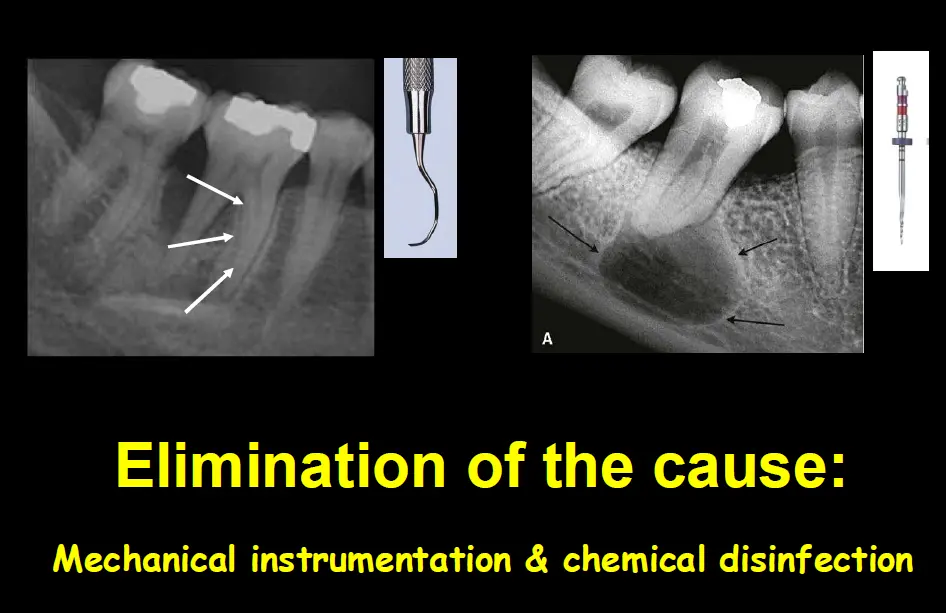
-
prevent reinfection
-
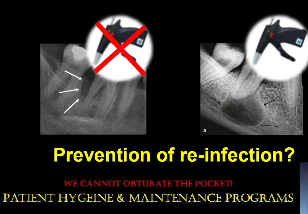
-
untreated disease in one can cause signs symptoms in the other
-
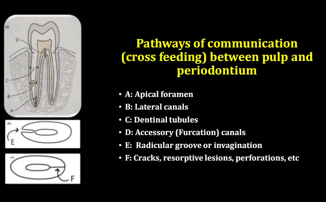
cross seeding of bacteria from one tissue to the other can also occur (in either direction)
Diagnosis
- most important
- history
- smoking, diabets, immunocompromised
- clinical examination
- radiographic examination
- perio probing
- vitality tests
Med history
- diabetes
- risk for perio
- delayed endo healing
- greater risk apical periodontitis
- Prolia
- increased risk external cervical resorption
Can moderate perio cause endo?
- maybe
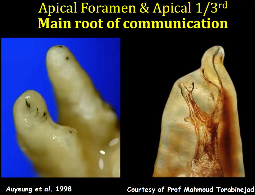
- accessory apical foramen exposed to oral environment
- perio treatment specially when cementum is removed
- canal calcification with respect to associated defect
- is the tooth vital with extensive perio?
Can severe perio cause endo
- yes in cases when disease envelopes apical and possible lateral/accessory foramina resulting in hypoxia and eventual necrosis
Can endo cause perio
- yes
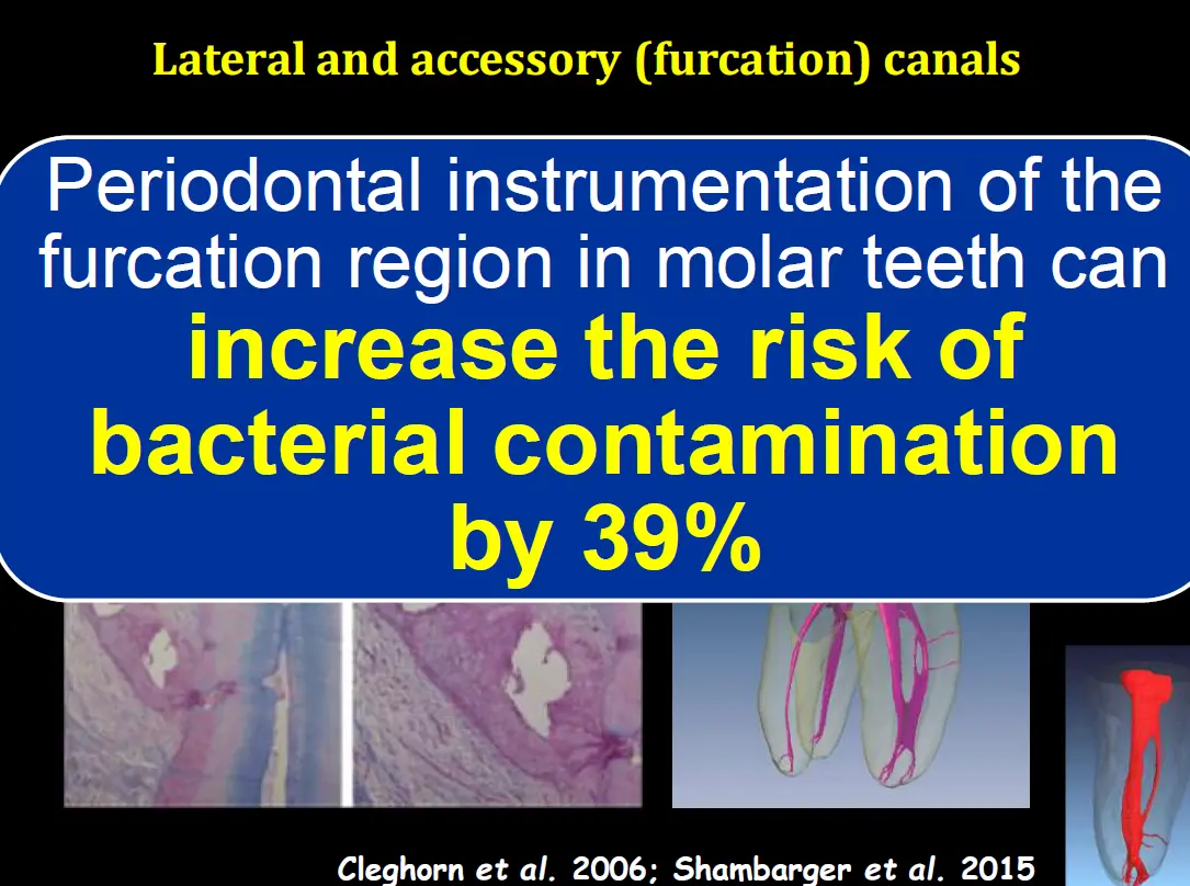
- through accessory and furcation canals
- PA lesion can drain through narrow pocket
- is tooth non vital with associated periodontal defect?
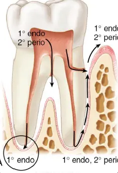
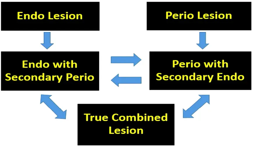
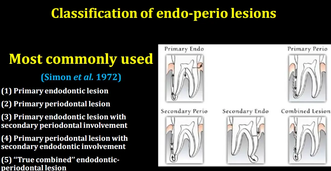
**Primary endodontic leisons (endodontic conditions with periodontal manifestations)
- localised collection of pus with draining sinus (CAA)
- drainage might be through gingival sulcus mimicking pocket
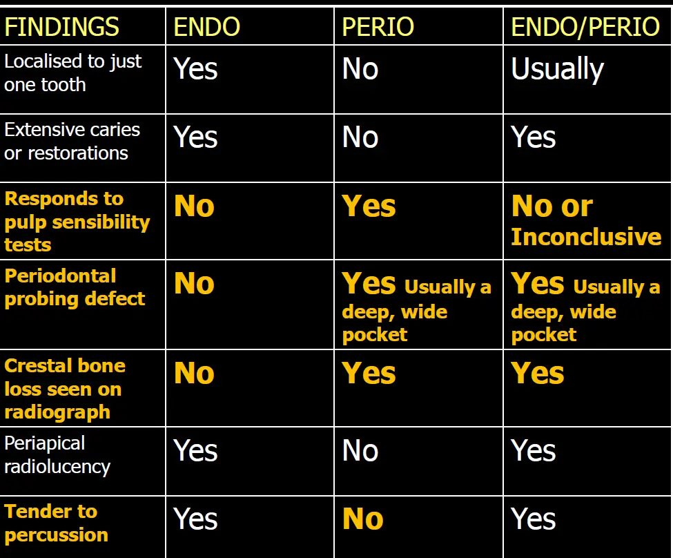
- narrow and deep pocket and lack of marginal bone loss
- minimal plaque and calc
- generally no response vitality tests and endo aetiology identified
Endo/perio lesions without communication
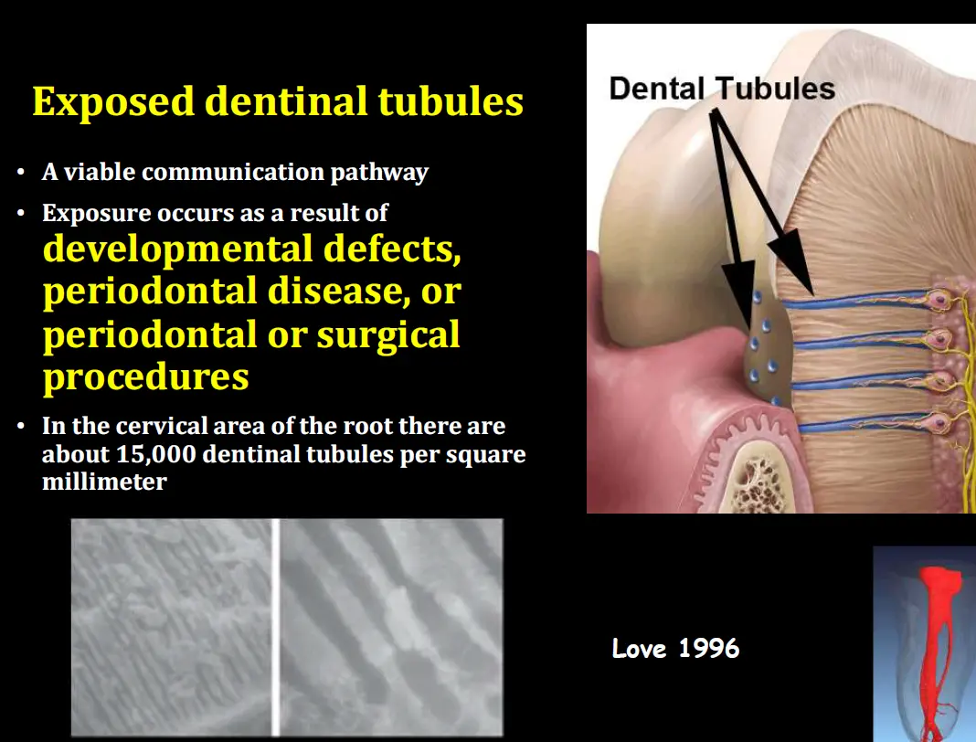
- endo infections can stimulate epithelial downgrowth along denuded dentine surfaces (after root planing) thus deepening pockets on instrumented root surfaces
- RCT first, then perio management
Primary Endodontic lesions with secondary periodontal involvement
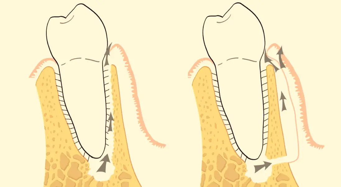
Primary periodontal lesions (periodontal conditions with endo manifestations)
- generally respond to vitality test despite apex radiolucency
- wide pockets, plaque/calculus, marginal bone loss
- other teeth with plaque calc and perio defect can help diagnosis
Primary endodontic lesion with secondary periodontal involvement
- tooth non vital and generally endodontic aetiology can be identified
- subgingival plaque/calculus present but mostly localised to affected tooth
- radiolucent area extending from apical/furcal region to crestal bone
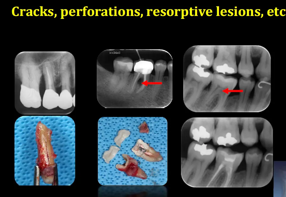
- pulp therapy does not resolve lesion completely
- endo perio with communication
- manage concurrently
- remove infections from both entities before placing final root filling
- remove pulp and dress canal with calcium hydroxide, complete periodontal management, then place final root filling
- prognosis
- would be better if no communicatoin
- home management need to be address
- exo always option
Primary periodontal lesoin with secondary endodontic involvement
- tooth may exhibit endodontic symptoms or may not respond to vitality tests, but generally endo aetiology cannot be identified
- subgingival plaque/calculus and perio bony defects present at other teeth too
- radiolucency involves crestal bone and may or may not reach apex
- perio treatment does not resolve lesion completely
True combined endodontic periodontal lesion
- non vital, can find endo aetiology
- subging plaque calc and perio defect at other teeth
- radiolucency involves crestal bone and generally reaches apex (J shaped)
- prognosis generally poorer, consider
- perio
- endo
- resto
- strategic value
- patient factors
- sequence of treatment
- pulp extirpation and intracanal dressing, deep double seal
- perio treatment once evidence of favourable endodontic healing observed
- NSPT if perio defect can be accessed without flap
- complete endo in rare cases
- surgical perio (consider GTR aswell)
- completion of endo once evidence of favourable perio healing